Gerald E. McDonnell - Antisepsis, Disinfection, and Sterilization
Здесь есть возможность читать онлайн «Gerald E. McDonnell - Antisepsis, Disinfection, and Sterilization» — ознакомительный отрывок электронной книги совершенно бесплатно, а после прочтения отрывка купить полную версию. В некоторых случаях можно слушать аудио, скачать через торрент в формате fb2 и присутствует краткое содержание. Жанр: unrecognised, на английском языке. Описание произведения, (предисловие) а так же отзывы посетителей доступны на портале библиотеки ЛибКат.
- Название:Antisepsis, Disinfection, and Sterilization
- Автор:
- Жанр:
- Год:неизвестен
- ISBN:нет данных
- Рейтинг книги:4 / 5. Голосов: 1
-
Избранное:Добавить в избранное
- Отзывы:
-
Ваша оценка:
Antisepsis, Disinfection, and Sterilization: краткое содержание, описание и аннотация
Предлагаем к чтению аннотацию, описание, краткое содержание или предисловие (зависит от того, что написал сам автор книги «Antisepsis, Disinfection, and Sterilization»). Если вы не нашли необходимую информацию о книге — напишите в комментариях, мы постараемся отыскать её.
is well suited as a textbook and is outstanding as a reference book for facilities managers and application engineers in manufacturing plants, hospitals, and food production facilities. It is also essential for public health officials, healthcare professionals, and infection control practitioners.
Antisepsis, Disinfection, and Sterilization — читать онлайн ознакомительный отрывок
Ниже представлен текст книги, разбитый по страницам. Система сохранения места последней прочитанной страницы, позволяет с удобством читать онлайн бесплатно книгу «Antisepsis, Disinfection, and Sterilization», без необходимости каждый раз заново искать на чём Вы остановились. Поставьте закладку, и сможете в любой момент перейти на страницу, на которой закончили чтение.
Интервал:
Закладка:
TABLE 1.6 Classification of protozoa, based on their motility mechanisms and microscopic morphologies
| Classification and organism | Disease(s) | Comments |
| Flagellates (motility by flagella) | 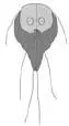 |
|
| Giardia lamblia | Giardiasis, including dysentery | Trophozoites (~20 μm; shown above) produce cysts, which can survive water chlorination |
| Trypanosoma gambiense | Sleeping sickness | Transferred in tsetse flies |
| Leishmania donovani | Leishmaniasis (kala-azar) | Transferred in sand flies |
| Amebas (motility by flowing cytoplasm, “pseudopodia”) |  |
|
| Entamoeba histolytica | Amebiasis, including dysentery and liver abscesses | The trophozoites reproduce asexually by binary division and can produce cysts, which can be transferred in contaminated food or water (surviving for up to 5 weeks at room temperature) |
| Acanthamoeba castellanii | Eye infections; associated with contaminated contact lenses | Commonly found free living in water, with two stages in life cycle (trophozoites and cysts) |
| Ciliates (motility using cilia) | 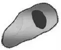 |
|
| Paramecium spp. | Dysentery | The trophozoite has two types of nuclei and can be up to 60 μm in length |
| Balantidium coli | Dysentery | Trophozoites can measure up to 150 μm, with transmission via cyst-contaminated meat |
| Sporozoans, apicomplexans (no specific motility extensions used) | 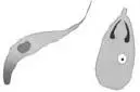 |
|
| Plasmodium falciparum | Malaria | Complicated life cycle; sporozoites transferred to humans by female mosquitoes |
| Cryptosporidium parvum | Severe diarrhea | Oocysts have marked resistance to biocides, surviving in water. When ingested, they hatch to release sporozoites. These forms invade cells of the intestine; they can reproduce asexually through two generations and then produce oocysts by sexual reproduction. |
| Toxoplasma gondii | Toxoplasmosis | The oocysts are formed in the cat intestine and transferred to other animals |
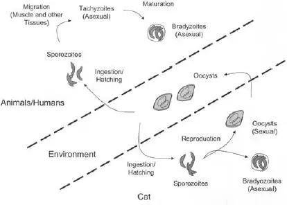
FIGURE 1.4 Life cycle of Toxoplasma gondii .
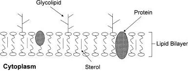
FIGURE 1.5 Simple representation of a mycoplasma cell surface structure.
Bacteria that contain cell walls can be simply classified based on their cell morphology and general reaction to a staining method known as the Gram stain. The Gram stain is used to differentiate between two types of cell wall structures: gram positive and gram negative. Microscopic examination of stained preparations allows further differentiation based on their shape ( Table 1.8). However, this is an oversimplification, as bacteria vary widely in their morphologies and staining characteristics; many other methods are used for further differentiation, including assays of oxygen requirements, growth characteristics, and lipid composition; immunoassays; and molecular biological procedures.
TABLE 1.7 Examples of pathogenic mycoplasmas
| Type | Example(s) | Significance |
| Spiroplasma | S. citri | Plant pathogens, insect parasites |
| Ureaplasma | U. urealyticum | Human parasites, genital tract diseases |
| Mycoplasma | M. genitalium, M. pneumoniae | Urethritis, atypical pneumonia |
The basic structure of cell wall-containing bacteria consists of an outer cell wall and an inner cell membrane surrounding the internal cytoplasm ( Fig. 1.6). The cell surface can also contain additional structures, such as pili, flagella, and capsules, depending on the bacterial species and its growth conditions.
The cell membrane is similar to that in mycoplasmas and consists of a phospholipid bilayer (without sterols) and associated proteins. Membrane proteins can be at the interface with the cytoplasm, embedded within the membrane, and/or associated with the external wall of the cell. Examples are some lipoproteins (proteins with lipid groups attached), in which the lipid component allows anchoring to the membrane. The overall structure is fluid but serves as a barrier to contain the cytoplasm and to restrict the passage of nutrients and ions into and out of the cell. Membrane proteins play vital roles in many cellular activities, including transport mechanisms, enzymatic reactions, cell signaling, energy generation, and cell wall synthesis. For this reason, damage to the cell membrane can render bacteria nonviable. The cell wall structures are less similar and can be considered as three basic types: gram-positive, gram-negative, and mycobacterial cell walls ( Fig. 1.7). Mycobacteria (not to be confused with the cell wall-free mycoplasmas) are considered separately due to their unique cell wall structure. The cell wall can play an important role in the resistance of bacteria to disinfection (see chapter 8).
TABLE 1.8 General differentiation of types of bacteria based on their microscopic morphologies and reactions to Gram staining
| Bacterial structure | Shape | Example(s) |
| Cocci |  |
Gram positive: Staphylococcus, Streptococcus Gram negative: Neisseria, Veillonella |
| Bacilli (rods) |  |
Gram positive: Bacillus, Listeria Gram negative: Escherichia, Pseudomonas |
| Spirals |  |
Gram negative: Treponema, Borrelia |
| Pleomorphic |  |
Gram negative: Bacteroides Cell-wall-free bacteria, e.g., Mycoplasma |
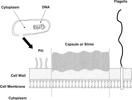
FIGURE 1.6 Basic structure of a bacterial cell, showing the cell surface in greater detail.
Читать дальшеИнтервал:
Закладка:
Похожие книги на «Antisepsis, Disinfection, and Sterilization»
Представляем Вашему вниманию похожие книги на «Antisepsis, Disinfection, and Sterilization» списком для выбора. Мы отобрали схожую по названию и смыслу литературу в надежде предоставить читателям больше вариантов отыскать новые, интересные, ещё непрочитанные произведения.


Обсуждение, отзывы о книге «Antisepsis, Disinfection, and Sterilization» и просто собственные мнения читателей. Оставьте ваши комментарии, напишите, что Вы думаете о произведении, его смысле или главных героях. Укажите что конкретно понравилось, а что нет, и почему Вы так считаете.