Gerald E. McDonnell - Antisepsis, Disinfection, and Sterilization
Здесь есть возможность читать онлайн «Gerald E. McDonnell - Antisepsis, Disinfection, and Sterilization» — ознакомительный отрывок электронной книги совершенно бесплатно, а после прочтения отрывка купить полную версию. В некоторых случаях можно слушать аудио, скачать через торрент в формате fb2 и присутствует краткое содержание. Жанр: unrecognised, на английском языке. Описание произведения, (предисловие) а так же отзывы посетителей доступны на портале библиотеки ЛибКат.
- Название:Antisepsis, Disinfection, and Sterilization
- Автор:
- Жанр:
- Год:неизвестен
- ISBN:нет данных
- Рейтинг книги:4 / 5. Голосов: 1
-
Избранное:Добавить в избранное
- Отзывы:
-
Ваша оценка:
Antisepsis, Disinfection, and Sterilization: краткое содержание, описание и аннотация
Предлагаем к чтению аннотацию, описание, краткое содержание или предисловие (зависит от того, что написал сам автор книги «Antisepsis, Disinfection, and Sterilization»). Если вы не нашли необходимую информацию о книге — напишите в комментариях, мы постараемся отыскать её.
is well suited as a textbook and is outstanding as a reference book for facilities managers and application engineers in manufacturing plants, hospitals, and food production facilities. It is also essential for public health officials, healthcare professionals, and infection control practitioners.
Antisepsis, Disinfection, and Sterilization — читать онлайн ознакомительный отрывок
Ниже представлен текст книги, разбитый по страницам. Система сохранения места последней прочитанной страницы, позволяет с удобством читать онлайн бесплатно книгу «Antisepsis, Disinfection, and Sterilization», без необходимости каждый раз заново искать на чём Вы остановились. Поставьте закладку, и сможете в любой момент перейти на страницу, на которой закончили чтение.
Интервал:
Закладка:
b NA, not applicable.
TABLE 1.2 Some advantages and disadvantages of microorganisms
| Advantage or disadvantage | Example(s) |
| Advantages | |
| Food and beverage production | Saccharomyces cerevisiae : bread and beer production Saccharomyces ellipsoideus : wine fermentation |
| Antibiotic production | Bacillus licheniformis : bacitracin Penicillium chrysogenum : penicillin |
| Vitamin metabolism | Pseudomonas spp.: vitamin B 12production Escherichia spp.: vitamin K synthesis in the gut |
| Genetic engineering | Agrobacterium tumefaciens : plasmids used for generating transgenic plants (e.g., herbicide or pathogen resistance) |
| Disease prevention | Bacteroides, Enterococcus spp.: prevention of pathogen colonization of the intestinal tract |
| Bioremediation | Desulfotomaculum spp.: arsenic detoxification |
| Disadvantages | |
| Animal/human diseases | Mycobacterium spp.: tuberculosis HIV:AIDS Plasmodium spp.: malaria |
| Plant diseases | Phytophthora : potato blight Corynebacterium : vegetable infections |
| Surface damage | Pseudomonas spp.: biofilm development and surface corrosion |
| Food spoilage | Rhizopus : bread mold Streptococcus : milk souring |
| Allergic reactions | Fungal spores, including Aspergillus spp. |
| General product contamination | Bacterial and fungal spores, including Bacillus spp. |
1.3.3.2 FUNGI
Fungi are eukaryotic cells, many of which can reproduce asexually (by cell division) or sexually (by the production of spores). A limited number of fungi have been implicated in plant and animal diseases (mycoses), but fungi are also widely used for bioremediation and biodegradation, product fermentation (e.g., beer, wine, and bread), and the production of biochemical products (e.g., antibiotics, enzymes, and vitamins). Due to their ubiquitous nature, they are often implicated in spoilage and as general contaminants. They are chemoheterotrophs (requiring organic nutrition), and many are saprophytes (living off dead organic matter), acquiring their food by absorption. They are generally classified as filamentous (molds) or unicellular ( Fig. 1.2).
Filamentous fungi multiply by cell division, but the cells do not separate and form long tubular structures known as hyphae (singular, hypha). The further development and branching of hyphae leads to the development of a mass of fungal growth on a surface known as a mycelium (plural, mycelia). Mycelia can often grow to such an extent that they are clearly visible to the naked eye on a surface (e.g., mold growth on bread). Fragments of hyphae can break off and allow the development of further mycelia. As the mycelia develop, a variety of fruiting bodies or other structures, which contain spores, are formed. Fungal spores can be present in a variety of shapes and sizes and can be asexual and/or sexual. The various molecular structures of fungal spores have not been studied in detail, but most are surrounded by a rigid wall distinguished by its low water content and low metabolic activity and which can contain lipids and pigments, as well as nutrient reserves; these are discussed further in section 8.10.
TABLE 1.3 Comparison of general prokaryotic and eukaryotic structures a
| Structure | Prokaryotes | Eukaryotes |
| Basic structure | 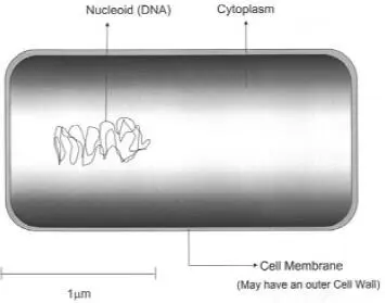 |
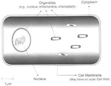 |
| Cytoplasmic membrane | + | + |
| Organelles, e.g., chloroplasts, mitochondria | − | + |
| Nucleus defined by a membrane | − | + |
| Ribosomes | 70S | 80S |
| Cell wall | +/− | +/− |
a Prokaryotic cells are simple, smaller structures, while eukaryotic cells are larger and more organized. Structural differences will vary, depending on the microorganism. For example, some prokaryotes (e.g., mycoplasmas) and eukaryotes (animal cells) do not have a cell wall.
TABLE 1.4 Helminths associated with disease
| Species | Disease | Comments |
| Nematodes (roundworms) |  |
|
| Wuchereria bancrofti | Elephantiasis (blood or lymphatic system blockage) | Transferred via mosquitoes; can grow up to 10 cm long |
| Onchocerca volvulus | River blindness | Transferred via blackflies |
| Ascaris lumbricoides | Generally asymptomatic, but can develop into ascariasis (pneumonitis and intestinal obstruction) | From contaminated water, food, or direct surface contact; worms can grow up to 30 cm long |
| Enterobius vermicularis | “Pinworms”; dysentery, intestinal blockage | From contaminated water, food, or direct surface contact; worms ~1 cm long |
| Cestodes (tapeworms) | 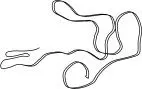 |
|
| Taenia saginata | Generally asymptomatic, but can cause mild intestinal complications (including abdominal pain and diarrhea) | Contaminated meat; worms can be very long (> 100 cm) |
| Trematodes (flukes) |  |
|
| Fasciola hepatica | Can be asymptomatic, with complications including liver abscesses | Contaminated grasses; snails are intermediate hosts |
| Schistosoma spp. | Schistomiasis; can cause many complications due to growth in the bloodstream and body tissues | Water contamination; snails are intermediate hosts |
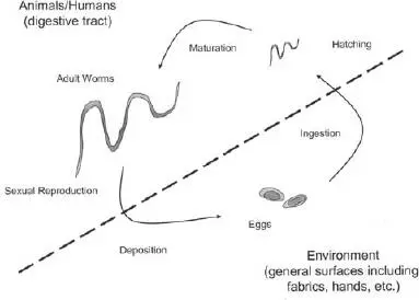
FIGURE 1.1 A typical helminth life cycle (example: Enterobius vermicularis ).
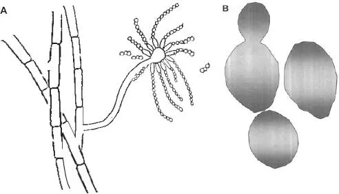
FIGURE 1.2 Typical fungal structures. (A) Filamentous fungus (mold). Hyphae are shown as long lines of unseparated cells, with the development of a fruiting body with attached spores. (B) Typical unicellular fungal (yeast) cells. The cells are generally polymorphic. In one case, a budding cell is shown.
Unicellular fungi (yeasts) do not generally form hyphae and produce growth that appears similar to bacteria (see section 1.3.4.1). Asexual reproduction of yeasts can occur by binary fission (e.g., in Schizosaccharomyces ), similar to bacterial fission, or by budding directly from the parent cell (e.g., in Saccharomyces [ Fig. 1.2]). In addition, some fungi are dimorphic, growing as either unicellular or hyphal (or pseudohyphal) forms. Common fungi are listed in Table 1.5.
Читать дальшеИнтервал:
Закладка:
Похожие книги на «Antisepsis, Disinfection, and Sterilization»
Представляем Вашему вниманию похожие книги на «Antisepsis, Disinfection, and Sterilization» списком для выбора. Мы отобрали схожую по названию и смыслу литературу в надежде предоставить читателям больше вариантов отыскать новые, интересные, ещё непрочитанные произведения.


Обсуждение, отзывы о книге «Antisepsis, Disinfection, and Sterilization» и просто собственные мнения читателей. Оставьте ваши комментарии, напишите, что Вы думаете о произведении, его смысле или главных героях. Укажите что конкретно понравилось, а что нет, и почему Вы так считаете.