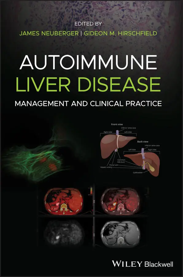
Autoimmune Liver Disease
Здесь есть возможность читать онлайн «Autoimmune Liver Disease» — ознакомительный отрывок электронной книги совершенно бесплатно, а после прочтения отрывка купить полную версию. В некоторых случаях можно слушать аудио, скачать через торрент в формате fb2 и присутствует краткое содержание. Жанр: unrecognised, на английском языке. Описание произведения, (предисловие) а так же отзывы посетителей доступны на портале библиотеки ЛибКат.
- Название:Autoimmune Liver Disease
- Автор:
- Жанр:
- Год:неизвестен
- ISBN:нет данных
- Рейтинг книги:5 / 5. Голосов: 1
-
Избранное:Добавить в избранное
- Отзывы:
-
Ваша оценка:
Autoimmune Liver Disease: краткое содержание, описание и аннотация
Предлагаем к чтению аннотацию, описание, краткое содержание или предисловие (зависит от того, что написал сам автор книги «Autoimmune Liver Disease»). Если вы не нашли необходимую информацию о книге — напишите в комментариях, мы постараемся отыскать её.
, practitioners will learn about the current state of autoimmune liver disease and how to focus on their diagnosis and treatment. The four-part book begins with a thorough investigation of current immunological thinking as it relates to the autoimmunity of the liver. It also covers the four major hepatic autoimmune liver diseases in both adults and children, their management and the role of liver transplantation, and learned approaches to patient management and empowerment.
Expert authors in the field have come together to provide a thorough examination of autoimmune liver disease to help support clinicians assisting patients. The text provides an in-depth look at topics including:
● The four major hepatic autoimmune liver diseases, their diagnosis, and potential disease management
● The use (and misuse) of autoantibodies in diagnosis and treatment
● The role and timing of liver transplantation and the impact of recurrent autoimmune liver disease as well as de novo autoimmune hepatitis
● Optimal approaches to managing patients and keeping care personalised
With breadth, depth and current-day relevance,
sheds light on recent developments in management of liver disease for practitioners, nurses, and health care professionals.
Autoimmune Liver Disease — читать онлайн ознакомительный отрывок
Ниже представлен текст книги, разбитый по страницам. Система сохранения места последней прочитанной страницы, позволяет с удобством читать онлайн бесплатно книгу «Autoimmune Liver Disease», без необходимости каждый раз заново искать на чём Вы остановились. Поставьте закладку, и сможете в любой момент перейти на страницу, на которой закончили чтение.
Интервал:
Закладка:
Carbohydrate Metabolism
The liver plays a key role in carbohydrate metabolism. It maintains carbohydrate stores by synthesizing glycogen and generating glucose from precursors such as lactate, pyruvate, and amino acids. Glycogen stored in the liver is the main source of rapidly available glucose for the whole organism. In the fed state, glycogen synthesis occurs preferentially in the perivenous hepatocytes whereas in the fasting state, glucose release via glycogenolysis and gluconeogenesis initially occurs in periportal hepatocytes. The liver glycogen stores contain up to a 2‐day supply of glucose, after which glucogenesis occurs mainly from lactate. In acute liver failure, the blood glucose level may drop whereas this is infrequent in chronic liver disease, where it is more common to observe hyperglycemia and insulin resistance. This may be related to decreased glucose uptake by muscle and reduced glycogen storage in the liver and muscle.
Lipid Metabolism
A major role of the liver in lipid metabolism is to synthesize large quantities of cholesterol and phospholipids, many of which are packaged with lipoproteins and made available to the rest of the body. Free cholesterol also derives from the uptake of chylomicron remnants and lipoproteins from the circulation. BAs are synthesized from free cholesterol, and both BA and cholesterol are secreted into bile. Bile provides a major route for cholesterol excretion.
The rate‐limiting step of cholesterol synthesis is the conversion of 3‐hydroxy‐3‐methylglutaryl‐CoA (HMG‐CoA) to mevalonate by the enzyme HMG‐CoA reductase, which is located almost exclusively in periportal cells. Synthesis is increased in biliary duct obstruction, terminal ileal resection, biliary or intestinal lymph fistula, and with medications such as colestyramine, corticosteroids and thyroid hormones. Cholesterol synthesis is inhibited by BAs, cholesterol feeding, fasting, and medications such as fibrates, nicotinic acid, and statins.
During chronic cholestasis, such as in primary biliary cholangitis and primary sclerosing cholangitis (PSC) but also in acute cholestasis, cholesterol serum levels are increased. This is mainly secondary to retention of cholesterol normally excreted in bile but also to increased hepatic synthesis of cholesterol, reduced plasma lecithin‐cholesterol acyltransferase activity, and regurgitation of biliary lecithin, which produces a shift of cholesterol from pre‐existing tissue cholesterol into the plasma. In patients with severe chronic cholestasis who are however malnourished, for example in premature ductopenic variant of primary biliary cholangitis (PBC) or in carcinomatous biliary obstruction, the serum cholesterol may be lower. Elevated cholesterol levels are clinically associated with skin xanthomas and xanthelasma. Hypercholesterolemia is not consistently associated with subclinical atherosclerosis in PBC, but should be treated if other risk factors for cardiovascular disease are also present.
In addition, the liver has other roles in lipid metabolism such as oxidizing triglycerides to produce energy; synthesizing lipoproteins; and converting excess carbohydrates and proteins into fatty acids and triglyceride, which are then exported and stored in adipose tissue.
Protein Metabolism
Amino acids are derived from the diet and from tissue breakdown and they reach the liver via the portal vein. Many critical aspects of protein metabolism occur in the liver, such as the deamination and transamination of amino acids, followed by conversion of the non‐nitrogenous parts of these molecules to glucose or lipids. Several of the enzymes used in these pathways, e.g. alanine and aspartate aminotransferases, are commonly measured in serum to assess liver damage.
The liver is also responsible for a number of vital metabolic processes, including the removal of ammonia (an important factor in the development of hepatic encephalopathy) via the synthesis of urea; the synthesis of non‐essential amino acids; and the synthesis of most plasma proteins such as albumin (the major plasma protein), fibrinogen, α 1‐antitrypsin, haptoglobin, ceruloplasmin, transferrin, and several coagulation factors.
Metabolic Zonation
The multiple functions of the liver are facilitated by a functional specialization of the liver parenchyma, known as metabolic zonation, where hepatocytes show different functional and structural characteristics according to their location in the liver acinus. Within each acinus, the functional unit in terms of blood flow, blood rich in nutrients and hormones enters at the portal triad through the portal vein, mixes with oxygen‐rich blood from the hepatic artery, flows through the sinusoids and eventually exits the lobule through the central vein. As the blood flows through the sinusoids there is free exchange of nutrients and metabolites between blood and hepatocytes. Functional variation is observed in hepatocytes based on their location along the portal–central axis. Hepatocytes exhibit a distinct gene expression based on their location within the acinus, which manifests as diverse availability of substrates and concentration of enzymes in different parts of the acinus. Based on this organization and heterogeneity of hepatocytes, the acinus comprises three geographical areas or zones: periportal or zone 1, midzonal or zone 2, and perivenous or zone 3. Hepatocytes in zone 3 contain the drug‐metabolizing P450 enzymes, have a reduced oxygen supply, receive a higher concentration of any toxic product of drug metabolism, and have a reduced glutathione concentration compared with zone 1. This makes them particularly susceptible to drug‐induced liver injury. Also, hepatocytes in zone 1 receive blood with a high bile salt concentration and are therefore particularly important in bile salt‐dependent bile formation, whereas hepatocytes in zone 3 are important in non‐bile salt‐dependent bile formation. Functions such as gluconeogenesis, glycolysis, and ketogenesis appear to be dependent on the direction of blood flow along the sinusoid. For others, such as cytochrome P450 activity, the gene transcription rate differs between perivenular and periportal hepatocytes. This functional zonation is often lost in liver diseases.
Hepatic Transport Systems
Hepatic uptake and efflux of electrolytes and solutes involved in bile formation are maintained by distinct transport systems expressed at the two surface domains of hepatocytes. After canalicular secretion, bile composition undergoes further modification in the bile canaliculi, involving reabsorption and secretion processes maintained by the apical and basolateral transport systems in cholangiocytes ( Figure 1.1).
Basolateral (Sinusoidal) Transporters
The uptake of exogenous and endogenous compounds from the portal circulation is facilitated by a number of basolaterally located, sodium‐dependent and sodium‐independent transporters. The sodium‐dependent transporters include the sodium taurocholate cotransporting polypeptide (NTCP), specific to conjugated bile salts and certain sulfated steroids. NTCP accounts for more than 80% of conjugated but less than half of unconjugated bile salt uptake. The sodium‐independent transporters include several members of the superfamily of organic anion‐transporting polypeptides (OATPs). With the exception of OATP2B1, the other OATPs exhibit overlapping transport activities for conjugated and unconjugated bile salts, neutral steroids, steroid sulfates and glucuronides, selected organic cations, and drugs including the antihistamine fexofenadine, opioid peptides, digoxin, the HMG‐CoA reductase inhibitor pravastatin, the angiotensin‐converting enzyme inhibitor enalapril, and the antimetabolite methotrexate. In addition, the basolateral hepatocyte membrane also localizes several ATP‐dependent efflux pumps belonging to the family of MRPs, multispecific transporters for different organic anions. These have been implicated in the cellular efflux of drugs conjugated with glutathione, glucuronic acid, and sulfate (MRP1); the efflux of bile salts (MRP3); and the transport of nucleoside analog drugs (MRP4).
Читать дальшеИнтервал:
Закладка:
Похожие книги на «Autoimmune Liver Disease»
Представляем Вашему вниманию похожие книги на «Autoimmune Liver Disease» списком для выбора. Мы отобрали схожую по названию и смыслу литературу в надежде предоставить читателям больше вариантов отыскать новые, интересные, ещё непрочитанные произведения.


Обсуждение, отзывы о книге «Autoimmune Liver Disease» и просто собственные мнения читателей. Оставьте ваши комментарии, напишите, что Вы думаете о произведении, его смысле или главных героях. Укажите что конкретно понравилось, а что нет, и почему Вы так считаете.