Michael Yudell - Welcome to the Genome
Здесь есть возможность читать онлайн «Michael Yudell - Welcome to the Genome» — ознакомительный отрывок электронной книги совершенно бесплатно, а после прочтения отрывка купить полную версию. В некоторых случаях можно слушать аудио, скачать через торрент в формате fb2 и присутствует краткое содержание. Жанр: unrecognised, на английском языке. Описание произведения, (предисловие) а так же отзывы посетителей доступны на портале библиотеки ЛибКат.
- Название:Welcome to the Genome
- Автор:
- Жанр:
- Год:неизвестен
- ISBN:нет данных
- Рейтинг книги:5 / 5. Голосов: 1
-
Избранное:Добавить в избранное
- Отзывы:
-
Ваша оценка:
Welcome to the Genome: краткое содержание, описание и аннотация
Предлагаем к чтению аннотацию, описание, краткое содержание или предисловие (зависит от того, что написал сам автор книги «Welcome to the Genome»). Если вы не нашли необходимую информацию о книге — напишите в комментариях, мы постараемся отыскать её.
offers substantial new and updated content to reflect recent major advances in genome-level sequencing and analysis, and demonstrates the vast increase in biological knowledge over the past decade. New sections cover next-generation technologies such as Illumina and PacBio sequencing, while expanded chapters discuss controversial ethical and philosophical issues raised by genomic technology, such as direct-to-consumer genetic testing. An essential resource for understanding the still-evolving genomic revolution, this book:
Introduces non-scientists to basic molecular principles and illustrates how they are shaping the genomic revolution in medicine, biology, and conservation biology Explores a wide range of topics within the field such as genetic diversity, genome structure, genetic cloning, forensic genetics, and more Includes full-color illustrations and topical examples Presents material in an accessible, user-friendly style, requiring no expertise in genomics Discusses past discoveries, current research, and future possibilities in the field Sponsored by the American Museum of Natural History,
is a must-read book for anyone interested in the scientific foundation for understanding the development and evolutionary heritage of all life.
Welcome to the Genome — читать онлайн ознакомительный отрывок
Ниже представлен текст книги, разбитый по страницам. Система сохранения места последней прочитанной страницы, позволяет с удобством читать онлайн бесплатно книгу «Welcome to the Genome», без необходимости каждый раз заново искать на чём Вы остановились. Поставьте закладку, и сможете в любой момент перейти на страницу, на которой закончили чтение.
Интервал:
Закладка:
It later turned out that Kornberg’s polymerase was not the key polymerase in DNA replication; DNA polymerase III was. Scientists who questioned the function of Kornberg’s polymerase in live organisms were only partially correct; polymerase I’s role was still found to be vital, playing a key role in chromosome replication and DNA repair. (25) Over the next two decades the approaches pioneered by Kornberg and his associates resulted in the discovery of a broad array of enzymes and other proteins important in the replication of DNA and the translation of proteins. An intriguing aspect of these discoveries is that polymerase enzymes do not need to be in cells to work. Biochemists used this feature of polymerase to develop methods to take proteins out of cells and coax them to activate in test tubes. The other enormously important result of Kornberg’s work was that scientists now had a laboratory reagent—the DNA polymerase itself—that could be used in a test tube to replicate DNA.
RESEARCH MILESTONE 4 : SEEING GENES
Sanger’s sequencing of insulin’s amino acids, the cracking of the genetic code, and Kornberg’s work on DNA polymerase were all technologies that would someday lead to the sequencing of a whole genome. But the ever‐increasing knowledge of the molecular basis of inheritance could not reach its full potential for both scientific and biomedical research without techniques to sequence genes quickly and accurately. So we now turn from deciphering the interiors of the cell to technologies that capitalized on these discoveries and enhanced our ability to see the most fundamental mechanisms of heredity. By the 1970s laboratories around the globe were focused on finding ways to better characterize, at the molecular level, genes and their component parts.
Oxford University biologist Edward Southern revolutionized molecular biology in 1975 with a method that came to be known as the Southern blot. (26) Southern blots allowed geneticists to locate and look at DNA and genes within a genome by capitalizing on the following characteristics of DNA. First, DNA is a negatively charged molecule; thus when electricity is present, it can hitch a ride on a current—it migrates to the positive terminal in an electric field.
Second, DNA molecules are small and can be separated by passing them through a porous gel made from either agarose (extracted from seaweed) or acrylamide (a synthetic polymer). The size of the DNA fragment, the strength of the current, and the concentration of acrylamide or agarose in the gel mixture dictate how fast molecules will pass through it. In fact, the concentration of an acrylamide gel can be adjusted to such a fine degree that DNA molecules of one base pair difference in length can be distinguished. Third, one fragment of DNA can be used to find another. This process, known as DNA hybridization, activates one strand of a double helix to search for the other strand, to reform hydrogen bonds and make a new double helix.
Hybridization doesn’t have to be perfect; only 60–70% of the two strands of a helix must match for the two strands to stick together.
Southern created a technique whereby a small piece of an organism’s genome can be arranged by size along an agarose gel. The technique then involves the transfer of DNA from gels onto nitrocellulose membranes. To detect where a specific gene is, a fragment of the gene of interest is labeled with radioactivity and then hybridized (attached) to the DNA on the nitrocellulose. Radioactive molecules hybridizing to the DNA on the filter emit particles that react with photographic film and can therefore be seen as a dark spot on the film. With his blot, Southern solved the problem of finding a genetic needle in a genomic haystack. Techniques were later developed to similarly isolate RNA molecules (jokingly named Northern blotting) and protein molecules (named Western blotting). (27)
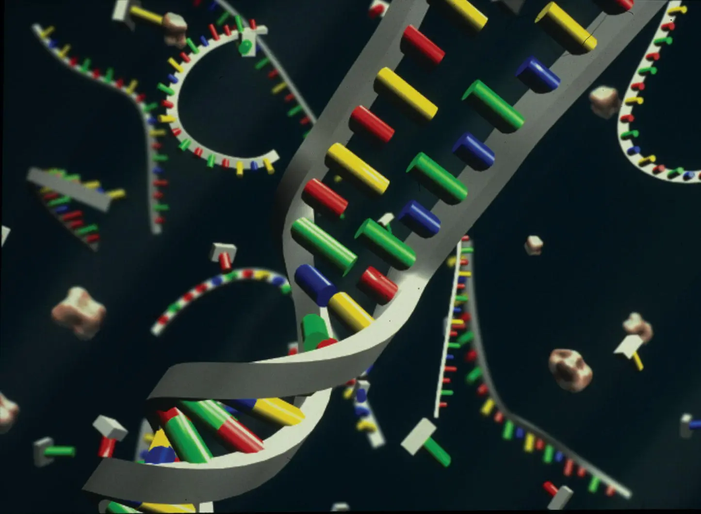
Figure 2.4DNA hybridization occurs when a single strand of a double helix finds a complementary strand to form a new double helix.
Credit: Exhibitions Department, American Museum of Natural History
RESEARCH MILESTONE 5 : COPYING DNA
During the 1970s scientists improved upon the Southern blot and other gel electrophoresis methods. Southern’s method required a tremendous amount of DNA and thus a tremendous amount of laboratory labor. It also lacked the precision to see the location of individual bases. To get around this shortcoming, scientists developed methods to amplify or clone (meaning simply to copy) DNA.
Found in bacteria, the small, circular extrachromosomal DNA molecule known as a plasmid often times carries genes that confer antibiotic resistance to bacteria. Under normal conditions a plasmid can facilitate genetic exchange between different bacterial strains: A gene fragment from one bacterium is carried to and inserted into a chromosome by a plasmid. In 1973 scientists discovered that if you biochemically insert a target stretch of DNA into a plasmid and put the plasmid into a bacterial cell, such a cell makes thousands and perhaps millions of copies of the plasmid and hence the attached DNA. (28) This procedure, incorporated into sequencing technology, made it easier to make large amounts of a desired stretch of DNA. There are, however, two serious shortcomings of the use of plasmids. First, bacterial plasmids must be cultured. This is time‐consuming. Second, plasmids can take up only a small piece of DNA efficiently. If the DNA stretch picked up by a plasmid is too large, the plasmid is unable to make accurate copies.
Since the discovery of plasmids or what might be termed bacterial copying machines, other vehicles have been created that can copy larger pieces of DNA. The average limiting size of a plasmid is about 5000 bases. Phages, a specific class of viruses that infect bacteria and can be stably replicated by them, can carry about 15,000 bases; cosmids, an artificial cloning vector with a phage gene, can carry about 35,000 bases; bacterial artificial chromosomes (also known as BACs) can take over 100,000 bases of sequence; and yeast artificial chromosomes (also known as YACs) can take approximately 1,000,000. Although these microbial methods remain an important component of DNA sequencing and were central to the effort to sequence the human genome, they are all arduous ways to copy DNA. BAC‐copied DNA was used in the sequencing of the human genome. (29)
RESEARCH MILESTONE 6 : SEQUENCING A VIRAL GENOME
By the 1970s advances in sequencing technology brought biology and genetics to the brink of the genomic revolution. The most important developments in sequencing technology occurred simultaneously in laboratories on opposite sides of the Atlantic. Two groups—biologist Walter Gilbert’s group at Harvard and Frederick Sanger’s group at Cambridge—exploited the chemistry of nucleic acids to come to the same brilliant idea. Unlike Edward Southern’s method, which revealed only the presence of DNA and genes, Gilbert’s and Sanger’s methods revealed the actual sequences of nucleotides along strands of DNA. The two methods had “complementary strengths,” and were used depending on what was to be sequenced. (30) The men shared the Nobel Prize in 1980 for this work. It was Sanger’s second Nobel. (31)
Sanger’s sequencing success rested on several premises. First, he knew that he could take a piece of DNA and synthesize its entire length with DNA polymerase. He was also aware of discoveries that showed that by using a class of nucleotides called chain terminators he could interrupt the synthesis of a DNA chain. These chain terminators come in four forms—terminator G, terminator C, terminator T, and terminator A—and when they were placed in a test tube with a DNA fragment and DNA polymerase and then placed on a gel, Sanger could determine the order of nucleotides in a given DNA fragment. He accomplished this by radioactively labeling the locations where the chain terminators stopped DNA synthesis at one of the four particular nucleotides. (32)
Читать дальшеИнтервал:
Закладка:
Похожие книги на «Welcome to the Genome»
Представляем Вашему вниманию похожие книги на «Welcome to the Genome» списком для выбора. Мы отобрали схожую по названию и смыслу литературу в надежде предоставить читателям больше вариантов отыскать новые, интересные, ещё непрочитанные произведения.


Обсуждение, отзывы о книге «Welcome to the Genome» и просто собственные мнения читателей. Оставьте ваши комментарии, напишите, что Вы думаете о произведении, его смысле или главных героях. Укажите что конкретно понравилось, а что нет, и почему Вы так считаете.