Michael Yudell - Welcome to the Genome
Здесь есть возможность читать онлайн «Michael Yudell - Welcome to the Genome» — ознакомительный отрывок электронной книги совершенно бесплатно, а после прочтения отрывка купить полную версию. В некоторых случаях можно слушать аудио, скачать через торрент в формате fb2 и присутствует краткое содержание. Жанр: unrecognised, на английском языке. Описание произведения, (предисловие) а так же отзывы посетителей доступны на портале библиотеки ЛибКат.
- Название:Welcome to the Genome
- Автор:
- Жанр:
- Год:неизвестен
- ISBN:нет данных
- Рейтинг книги:5 / 5. Голосов: 1
-
Избранное:Добавить в избранное
- Отзывы:
-
Ваша оценка:
Welcome to the Genome: краткое содержание, описание и аннотация
Предлагаем к чтению аннотацию, описание, краткое содержание или предисловие (зависит от того, что написал сам автор книги «Welcome to the Genome»). Если вы не нашли необходимую информацию о книге — напишите в комментариях, мы постараемся отыскать её.
offers substantial new and updated content to reflect recent major advances in genome-level sequencing and analysis, and demonstrates the vast increase in biological knowledge over the past decade. New sections cover next-generation technologies such as Illumina and PacBio sequencing, while expanded chapters discuss controversial ethical and philosophical issues raised by genomic technology, such as direct-to-consumer genetic testing. An essential resource for understanding the still-evolving genomic revolution, this book:
Introduces non-scientists to basic molecular principles and illustrates how they are shaping the genomic revolution in medicine, biology, and conservation biology Explores a wide range of topics within the field such as genetic diversity, genome structure, genetic cloning, forensic genetics, and more Includes full-color illustrations and topical examples Presents material in an accessible, user-friendly style, requiring no expertise in genomics Discusses past discoveries, current research, and future possibilities in the field Sponsored by the American Museum of Natural History,
is a must-read book for anyone interested in the scientific foundation for understanding the development and evolutionary heritage of all life.
Welcome to the Genome — читать онлайн ознакомительный отрывок
Ниже представлен текст книги, разбитый по страницам. Система сохранения места последней прочитанной страницы, позволяет с удобством читать онлайн бесплатно книгу «Welcome to the Genome», без необходимости каждый раз заново искать на чём Вы остановились. Поставьте закладку, и сможете в любой момент перейти на страницу, на которой закончили чтение.
Интервал:
Закладка:
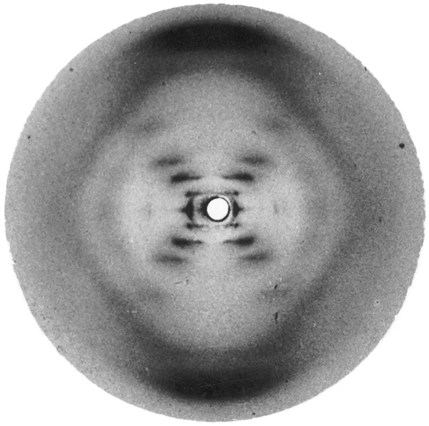
Figure 1.10Franklin’s X‐ray crystallography of DNA, shown to James Watson without her knowledge, helped Watson and Crick solve the puzzle of DNA. Franklin was renowned for her X‐ray crystallography talents.
Credit: Normal Collection for the History of Molecular Biology
Chargaff’s rules made Watson and Crick’s three‐dimensional model a reality. The great strength of Watson and Crick lay in their ability to reconcile their model with existing science. None of the other participants in this discovery put the pieces together quite as Watson and Crick had. And so they built their model.
There are three important chemical forces that hold together the DNA molecule. The first chemical bond, hydrogen bonds between a G and a C or an A and a T, connects the two strands of the helix. These bonds are relatively weak and can be broken apart by acids and/or heat. At approximately 90 °C the hydrogen bonds across a double helix can be broken, allowing the two strands of the double helix to separate.
The second kind of bond, the phosphodiester bond, keeps the Gs, As, Ts, and Cs together along a helix’s strand. These bonds can be made on both ends of a base and to any other base, resulting in long strands of Gs, As, Ts, and Cs. Phosphodiester bonds are the strongmen of the helix, withstanding high temperatures and even highly acidic conditions. It is the position of these bonds on the nucleotide carbon rings that gives DNA its helical twist, its third dimension. Molecular biology takes advantage of the characteristics of both hydrogen and phosphodiester bonds all the time. Because of the difference in relative strengths between these bonds, scientists would later figure out how to separate the two DNA strands. Why is this so important? Because to make copies of a double helix, you need to have both strands—let’s call them the Watson and Crick strands—as a template. If they are bonded in a double helix, they cannot be used to replicate themselves. By melting the weak hydrogen bonds between the two strands, the freed strands can now be copied.
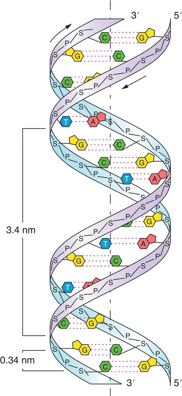
Figure 1.11This diagram shows the double helix structure of DNA. In the model you can see where hydrogen bonds bond the nucleic acids to one another and also the sugar‐phosphate backbone that holds the helix in place.
Credit: Wiley Publishers
The race to uncover the structure of DNA became the stuff of scientific legend after the publication in 1968 of James Watson’s The Double Helix . Watson’s telling of the DNA story drew ire from within the small community of scientists in which he himself worked. Facing strong objections from Francis Crick, Linus Pauling, Maurice Wilkins, and the family of Rosalind Franklin over the way in which Watson characterized all of the major players in the discovery, Harvard University Press dropped the book. (78) Of particular concern was Watson’s portrayal of Franklin, who was just 37 when she died in 1958 of ovarian cancer and whose role in the discovery was reduced in The Double Helix to that of an incompetent scientist and hot‐tempered woman. (79) Watson’s book, picked up by another publisher, went on to become a best‐seller, and for almost 30 years the story of the discovery of DNA was told by The Double Helix . It is only recently, with the publication of a new biography and with acknowledgments by Watson that Franklin’s work was “key” to their success, that Franklin’s image as a brilliant scientist was rehabilitated. Years after his own co‐discovery of the structure of the double helix, Francis Crick suggested that Franklin was just months away from solving the puzzle herself. (80)
As late as 1933, Thomas Hunt Morgan suggested that there is “no consensus opinion amongst geneticists as to what the genes are—whether they are real or purely fictitious.” (81) Working deductively, working on instinct, Morgan could never be sure that his gene maps or the work on genes conducted by his many colleagues amounted to anything. But beginning with Avery, McCarty, and MacLeod’s discovery in 1943 that DNA was the “stuff” of heredity, the gene became less an intellectual or theoretical entity and more a material reality. Watson and Crick’s discovery of the actual physical structure of DNA finally created a consensus among geneticists that genes were real and led genetics and molecular biology into a new and exciting realm. With the basics of heredity worked out, molecular biology became a driving force in science as the working characteristics of the gene came under scrutiny and study.
To complicate matters, research on and expansion of ideas about how phenotypes might be generated in a heritable fashion apart from genetics first formulated by C. H. Waddington and called epigenetics have made the biology of genes and inheritance even more complex and interesting. Epigenetics is what its name from the Greek implies—“beside genetics”. More precisely it concerns the expression of heritable phenotypes without changes in the DNA of the genome. In other words, epigenetic effects are heritable phenotypic effects without changes in genotype. The most famous case used to demonstrate this phenomenon is the Dutch Hunger Winter. A follow‐up study done on women who suffered through all or part of this 5‐month‐long famine that occurred near the end of World War II (the cause of the famine—which killed 20,000—was a Nazi blockade of food and fuel) revealed some important “heritable” problems. (82) It was reported that during the height of the famine, average caloric intake was less than 400 calories per day (the equivalent of say four pieces of toast). After the war a long‐term study of people who survived the famine was undertaken to determine the impact of famine on the offspring of women whose children were exposed prenatally to famine. As Laura C. Schultz puts it “the Dutch Hunger Winter study, from which results were first published in 1976, provides an almost perfectly designed, although tragic, human experiment in the effects of intrauterine deprivation on subsequent adult health.” (83)
The sad but remarkable results were that diabetes, obesity, microalbuminuria (a kidney malfunction), psychological and cognitive problems, and cardiovascular disease were seen in higher frequency in the offspring of women who lived through the famine than in the offspring of children of their siblings who were not exposed to famine. More remarkable was that women whose fetuses experienced the famine later in prenatal development were affected more severely than fetuses who experienced the famine earlier in their prenatal development (those fetuses that were conceived close to the end of the famine). Researchers could clearly show that this phenomenon was not due to DNA sequence changes. What then could cause this drastic change in the susceptibility to the offspring of women exposed to famine?
To understand this phenomenon completely we need first to describe the structure of DNA as it resides on our chromosomes. The DNA of our chromosomes is wrapped into what is called chromatin. First, the double helix is wrapped twice around a protein complex called a histone core. The histone cores have short parts of their proteins that “tail” off of the wrapped DNA. These “histone tails” are where the epigenetic action takes place, because these parts of the histone proteins can easily be modified by chemical reactions like the addition of methyl groups or acetyl groups. If a histone tail is methylated (or phosphorylated, acetylated, ubiquitylated, or sumoylated) this modification changes the shape of the histone core and disrupts the tightly wound chromatin altering the availability of the DNA in that region to transcription and hence gene expression of that region of the chromosome. Methylation can also occur on the DNA strand itself and this alters the availability of the region of DNA that is methylated to transcription.
Читать дальшеИнтервал:
Закладка:
Похожие книги на «Welcome to the Genome»
Представляем Вашему вниманию похожие книги на «Welcome to the Genome» списком для выбора. Мы отобрали схожую по названию и смыслу литературу в надежде предоставить читателям больше вариантов отыскать новые, интересные, ещё непрочитанные произведения.


Обсуждение, отзывы о книге «Welcome to the Genome» и просто собственные мнения читателей. Оставьте ваши комментарии, напишите, что Вы думаете о произведении, его смысле или главных героях. Укажите что конкретно понравилось, а что нет, и почему Вы так считаете.