Jane Flint - Principles of Virology
Здесь есть возможность читать онлайн «Jane Flint - Principles of Virology» — ознакомительный отрывок электронной книги совершенно бесплатно, а после прочтения отрывка купить полную версию. В некоторых случаях можно слушать аудио, скачать через торрент в формате fb2 и присутствует краткое содержание. Жанр: unrecognised, на английском языке. Описание произведения, (предисловие) а так же отзывы посетителей доступны на портале библиотеки ЛибКат.
- Название:Principles of Virology
- Автор:
- Жанр:
- Год:неизвестен
- ISBN:нет данных
- Рейтинг книги:3 / 5. Голосов: 1
-
Избранное:Добавить в избранное
- Отзывы:
-
Ваша оценка:
Principles of Virology: краткое содержание, описание и аннотация
Предлагаем к чтению аннотацию, описание, краткое содержание или предисловие (зависит от того, что написал сам автор книги «Principles of Virology»). Если вы не нашли необходимую информацию о книге — напишите в комментариях, мы постараемся отыскать её.
Volume I: Molecular Biology
Volume II: Pathogenesis and Control
Principles of Virology, Fifth Edition
Principles of Virology — читать онлайн ознакомительный отрывок
Ниже представлен текст книги, разбитый по страницам. Система сохранения места последней прочитанной страницы, позволяет с удобством читать онлайн бесплатно книгу «Principles of Virology», без необходимости каждый раз заново искать на чём Вы остановились. Поставьте закладку, и сможете в любой момент перейти на страницу, на которой закончили чтение.
Интервал:
Закладка:
Virus-Induced Signaling via Cell Receptors
Binding of virus particles to cell receptors not only concentrates the particles on the cell surface, but also activates signaling pathways that facilitate virus entry and movement within the cell or produce cellular responses that enhance virus propagation and/or affect pathogenesis. Binding of virus particles may lead to activation of protein kinases that trigger cascades of responses at the plasma membrane, cytoplasm, and nucleus ( Chapter 14). Second messengers that participate in signaling include phosphatidylinositides, diacylglycerides, and calcium. Regulators of membrane trafficking and actin dynamics also contribute to signaling. Additionally, virus-receptor interactions can stimulate antiviral responses, such as synthesis of type I interferons (Volume II, Chapter 3).
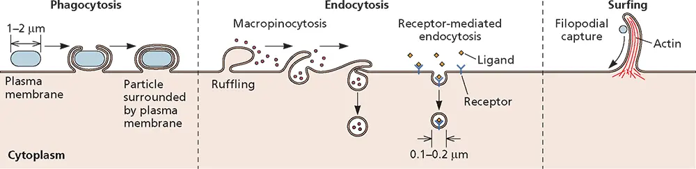
Figure 5.10 Mechanisms for the uptake of macromolecules from extracellular fluid. During phagocytosis, large particles such as bacteria or cell fragments that come in contact with the cell surface are engulfed by extensions of the plasma membrane. Phagosomes ultimately fuse with lysosomes, resulting in degradation of the material within the vesicle. Endocytosis comprises the invagination and pinching off of small regions of the plasma membrane either by macropinocytosis or by receptor-mediated endocytosis. Macropinocytosis mediates nonspecific uptake of fluids and small molecules. It is triggered by ligands and dependent on actin and signaling pathways different from those required for phagocytosis. Receptor-mediated endocytosis results in the specific uptake of molecules bound to cell surface receptors. Filopodia are actin-rich protrusions that sample the environment and participate in many cellular processes such as migration. Movement along filopodia occurs by an actin-dependent mechanism.
Signaling is essential for the entry of some viruses, such as simian virus 40, into cells. Binding of this virus particle to its glycolipid cell receptor, GM1 ganglioside, causes activation of tyrosine kinases. The signaling that ensues induces reorganization of actin filaments, internalization of the virus in caveolae, and transport of the caveolar vesicles to the endoplasmic reticulum ( Fig. 5.6). The activities of more than 50 cellular protein kinases regulate the entry of this virus into cells.
Routes of Entry
A wide range of ligands, fluid, membrane proteins, and lipids are taken into cells from the extracellular milieu by various processes depending on their size ( Fig. 5.10). Phagocytosis and endocytosis mediate the uptake of larger and smaller, respectively, molecules and particles. Endocytosis is the mechanism of entry of many viruses ( Fig. 5.10and 5.11). Three pathways of endocytosis have been identified: clathrin-dependent, caveolin-dependent, and clathrin- and caveolin-independent. Clathrin-coated pits can comprise as much as 2% of the surface area of a cell, and some receptors are clustered therein even in the absence of their ligands, whereas others require ligand binding to cluster. Successive transport and fusion with late endosomes exposes the contents of the vesicles to increasingly acidic pH—6.5 to 6.0 (early) and 6.0 to 5.0 (late)—while in lysosomes they are exposed to multiple degradative enzymes. Viral fusion proteins with a high pH threshold for fusion, such as that of vesicular stomatitis virus, mediate entry from early endosomes; but most mediate entry into the cytoplasm from late endosomes, and a few from lysosomes.
The caveolin-dependent pathway participates in transcytosis, signal transduction, and uptake of membrane components and extracellular ligands. Binding of a virus particle to the cell surface activates signal transduction pathways required for pinching off caveolae, which then are transported within the cytoplasm. These vesicles ultimately fuse with the caveosome, a larger membranous organelle that contains caveolin ( Fig. 5.11). In contrast to endosomes, the pH of the caveosome lumen is neutral. Some viruses, like echovirus type 1, penetrate the cytoplasm from the caveosome.
Although in cell culture virus particles can enter cells preferentially by one pathway, many viruses appear indiscriminate and enter via multiple pathways. For example, herpes simplex virus can enter cells by three different routes and simian virus 40 is taken up by both caveolin-dependent and clathrin-/caveolin-independent pathways. Influenza A virus can enter cells by both clathrin-dependent endocytosis and macropinocytosis, a process by which extracellular fluid is taken into cells via large vacuoles. Many virus particles are taken up by this pathway, including vaccinia virus, ebolaviruses, and herpesviruses (even though the latter can also fuse at the plasma membrane). Upon receptor binding, viruses that enter cells via macropinocytosis trigger a signaling cascade that leads to changes in cortical actin and ruffling of the plasma membrane ( Fig. 5.10). When these plasma membrane extensions retract, the viruses are brought into macropinosomes and eventually leave these vesicles via membrane fusion. A more dramatic rearrangement of actin filaments leads to the formation of filopodia, thin extensions of the plasma membrane. Virus particles of polyomaviruses can be visualized moving laterally on the plasma membrane on filopodia, and filopodia bridges participate in cell-to-cell spread of retrovirus particles in cells in culture ( Chapter 13).
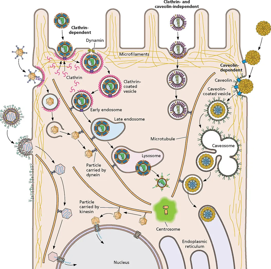
Figure 5.11 Virus entry and movement in the cytoplasm. Examples of various routes of virus entry are shown. Fusion at the plasma membrane releases the nucleocapsid or subviral components (lower left side of the cell) that can travel on actin filaments and microtubules. Uptake of virus particles by endocytosis can be either clathrin dependent, caveolin dependent, or clathrin and caveolin independent. Clathrin-dependent endocytosis (top left side of the cell) commences with binding to a specific cell surface receptor followed by diffusion into an invagination of the plasma membrane coated with the protein clathrin on the cytosolic side (clathrin-coated pits). The coated pit further invaginates and pinches off, a process that is facilitated by the GTPase dynamin. Within a few seconds, the clathrin coat is lost and the vesicles fuse with small, smooth-walled vesicles located near the cell surface, called early endosomes. The lumen of early endosomes is mildly acidic (pH 6.5 to 6.0), a result of energy-dependent transport of protons into the interior of the vesicles by a membrane proton pump. The contents of the early endosome are then transported via endosomal carrier vesicles to late endosomes located closer to the nucleus. The lumen of late endosomes is more acidic (pH 6.0 to 5.0). Late endosomes in turn fuse with lysosomes, which are vesicles containing a variety of enzymes that degrade sugars, proteins, nucleic acids, and lipids. Particle uncoating usually occurs from early or late endosomes. Virus particles may enter cells by a dynamin- and caveolin-dependent endocytic pathway (right side of the cell). Caveolae are distinguished from clathrin-coated vesicles by their flask-like shape, their smaller size, the absence of a clathrin coat, and the presence of a marker protein called caveolin. Three types of caveolar endocytosis have been identified. Dynamin 2-dependent endocytosis by caveolin 1-containing caveolaeis observed in cells infected with simian virus 40 and polyomavirus. Dynamin 2-dependent, noncaveolar, lipid raft-mediated endocytosis occurs during echovirus and rotavirus infection, while dynamin-independent, noncaveolar, raft-mediated endocytosis is also observed during simian virus 40 and polyomavirus infection. This pathway brings virions to the endoplasmic reticulum via the caveosome, a pH-neutral compartment. Clathrin- and caveolin-independent endocytic pathways of viral entry have also been described (top center-right of cell). Movement of endocytic vesicles within cells occurs on actin filaments or microtubules, the components of the cytoskeleton. Actin filaments are two-stranded helical polymers of actin. They are dispersed throughout the cell but are most highly concentrated beneath the plasma membrane, where they are connected via integrins and other proteins to the extra-cellular matrix. Transport along actin filaments is accomplished by myosin motors. Microtubules are 25-nm hollow cylinders made of tubulin. They radiate from the centrosometo the cell periphery. Movement on microtubules is mediated by kinesin and dynein motors.
Читать дальшеИнтервал:
Закладка:
Похожие книги на «Principles of Virology»
Представляем Вашему вниманию похожие книги на «Principles of Virology» списком для выбора. Мы отобрали схожую по названию и смыслу литературу в надежде предоставить читателям больше вариантов отыскать новые, интересные, ещё непрочитанные произведения.


Обсуждение, отзывы о книге «Principles of Virology» и просто собственные мнения читателей. Оставьте ваши комментарии, напишите, что Вы думаете о произведении, его смысле или главных героях. Укажите что конкретно понравилось, а что нет, и почему Вы так считаете.