Jane Flint - Principles of Virology
Здесь есть возможность читать онлайн «Jane Flint - Principles of Virology» — ознакомительный отрывок электронной книги совершенно бесплатно, а после прочтения отрывка купить полную версию. В некоторых случаях можно слушать аудио, скачать через торрент в формате fb2 и присутствует краткое содержание. Жанр: unrecognised, на английском языке. Описание произведения, (предисловие) а так же отзывы посетителей доступны на портале библиотеки ЛибКат.
- Название:Principles of Virology
- Автор:
- Жанр:
- Год:неизвестен
- ISBN:нет данных
- Рейтинг книги:3 / 5. Голосов: 1
-
Избранное:Добавить в избранное
- Отзывы:
-
Ваша оценка:
Principles of Virology: краткое содержание, описание и аннотация
Предлагаем к чтению аннотацию, описание, краткое содержание или предисловие (зависит от того, что написал сам автор книги «Principles of Virology»). Если вы не нашли необходимую информацию о книге — напишите в комментариях, мы постараемся отыскать её.
Volume I: Molecular Biology
Volume II: Pathogenesis and Control
Principles of Virology, Fifth Edition
Principles of Virology — читать онлайн ознакомительный отрывок
Ниже представлен текст книги, разбитый по страницам. Система сохранения места последней прочитанной страницы, позволяет с удобством читать онлайн бесплатно книгу «Principles of Virology», без необходимости каждый раз заново искать на чём Вы остановились. Поставьте закладку, и сможете в любой момент перейти на страницу, на которой закончили чтение.
Интервал:
Закладка:
Fluorescent Proteins
The discovery of green fluorescent protein revolutionized the study of the cell biology of virus infection. This protein, isolated from the jellyfish Aequorea victoria , is a convenient reporter for monitoring gene expression, because it is directly visible in living cells without the need for fixation, substrates, or coenzymes. Mutagenesis of the gene encoding this protein has led to the development of new fluorescent probes ranging in color from blue to yellow ( Fig. 2.15A). Additional fluorescent proteins emitting in the red, deep red, cyan, green, yellow, and orange spectral regions have been isolated from other marine species. Codon optimization for maximum translation in specific cell types and improved stability and brightness are other modifications that have broadened the utility of these proteins.
Fluorescence Microscopy
Fluorescence microscopy allows virologists to study all steps of virus reproduction, including cell surface attachment, cell entry, trafficking, replication, assembly, and egress. Single virus particle tracking can be achieved by inserting the coding sequence for a fluorescent protein into the viral genome, often fused to the coding region of a viral protein. The fusion protein is incorporated into the viral particle, which is visible in cells by fluorescence microscopy ( Fig. 2.15B). An alternative approach is to attach small-molecule fluorophores to viral capsid proteins. Light microscopy has a resolution in the range of 200 to 500 nm, whereas most viruses are between 20 and 400 nm in size and are therefore below the difraction limit. However, when the virus particle emits a high fluorescent signal in a low background, it is possible to use a computational point tracking algorithm to locate the particle with greater precision than the diffraction limit of the light microscope. This technique allows single particle tracking with accuracy in the range of low tens of nanometers.
Recent improvements in microscopy technology and computational image manipulation have led to unprecedented levels of resolution and contrast and an ability to reconstruct three-dimensional structures from captured images. The first advance was confocal microscopy, which utilizes a scanning point of light instead of full-sample illumination. In a conventional light microscope, light can penetrate the specimen only to a fixed depth. In a confocal microscope, a small beam of light is focused to multiple narrow depths. By capturing multiple two-dimensional images at different depths, it is possible to reconstruct high-resolution three-dimensional structures, a process known as optical sectioning.
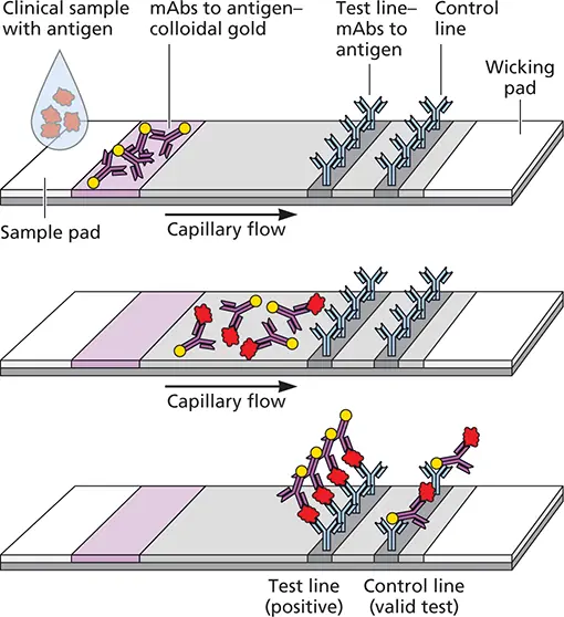
Figure 2.14 Lateral flow immunochromatographic assay. A slide or “dipstick” covered with a membrane is used to assay for the presence of viral antigens. The clinical specimen is placed on an absorbent pad at one end and is drawn across the slide by capillary action. Antigens in the sample react with a virus-specific antibody, which is linked to an indicator, in this example, colloidal gold. The antigen-antibody complexes move across the membrane until they are captured by a second virus-specific antibody in a test line. If viral antigen is present in the sample, an indicator line becomes visible in the test line. Accumulation of the indicator-containing antibody at the control line provides validation that the assay is functioning.
Superresolution microscopycombines the advantages of fluorescent imaging (multicolor labeling and live-cell imaging) while breaking the resolution limit of light microscopy. Different formats include single molecule localization microscopy, in which only a subset of fluorophores are turned on during each imaging cycle, thus allowing position determination with nanometer accuracy. Fluorophore positions from a series of images are then used to reconstruct the final image. Structured illumination microscopyutilizes standing waves formed by interference in laser illumination to create an excitation field that allows optical sectioning at very high resolution. These approaches can achieve resolution below 1 nm, well below the limit of light microscopy. This resolution is achieved by combining sequential acquisition of images with random switching of fluorophores on and off. From several hundred to thousands of images are collected and processed to generate a superresolution data set that can resolve cellular ultrastructure.
These superresolution microscopy methods are well suited for providing high-resolution images of static sections. Because these methods acquire images slowly, are phototoxic, and require computationally intensive image processing, their use for time-lapse imaging of live cells is impractical.
Fluorescence resonance energy transfer (FRET)microscopy can be used to examine protein-protein and protein-DNA or RNA interactions and conformational changes in these molecules. FRET solves the problem encountered in conventional fluorescence microscopy, which is of insufficient resolution to determine if molecules interact. The method is based on the principle that fluorescent emissions of one wavelength can excite a second distinct fluorophore at a distance of approximately 10 nm. For example, if two proteins are thought to interact under certain conditions, one can be labeled with a donor fluorophore that will emit light of a certain wavelength. If the two proteins are farther apart than 10 nm, only the donor color will be observed. However, if the two proteins are in close contact, then fluorescence of the second protein, which is linked to an acceptor fluorophore, will take place.
Another commonly used fluorescent microscopy technique in virology is fluorescence recovery after photo-bleaching (FRAP), a method for determining the kinetics of diffusion in cells. A viral or cellular protein is labeled with a fluorescent molecule, a portion of the cell is photobleached to eliminate fluorescence, and then recovery of fluorescence is observed over time. Fluorescence in the bleached area recovers as bleached fluorophore-linked proteins are replaced with unbleached molecules from a different part of the cell.
Detection of Viral Nucleic Acids
The detection of viruses in cell cultures is being increasingly supplanted by molecular methods such as the polymerase chain reaction and high-throughput sequencing, especially for discovery of new viruses associated with human diseases. These methods can be used to identify viruses that cannot be propagated in cell culture, offering new ways to fulfill Koch’s postulates ( Box 1.4).
Polymerase chain reaction.In this technique, specific oligonucleotides are used to amplify viral DNA sequences from infected cells or clinical specimens. Amplification is done in cycles, using a thermostable DNA polymerase ( Fig. 2.16). Each cycle consists of thermal denaturation, primer annealing, and extension, carried out by automated cycler machines. The result is exponential amplification (a 2 n -fold increase after n cycles of amplification) of the target sequence that is located between the two DNA primers.
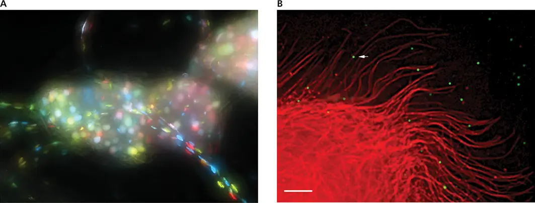
Figure 2.15 Using fluorescent proteins to study virus particles and virus-infected cells. (A)Submandibular ganglia after infection of the salivary gland with three recombinant pseudorabies viruses, each expressing a different color fluorescent protein. Courtesy of Lynn Enquist, Princeton University. (B)Single-virus-particle imaging with green fluorescent protein illustrates microtubule-dependent movement of human immunodeficiency virus type 1 particles in cells. The cells were infected with virus particles that contain a fusion of green fluorescent protein with a viral protein. Rhodamine-tubulin was injected into cells to label microtubules (red). Virus particles can be seen as green dots (white arrow). Bar, 5 μm. Courtesy of David McDonald, University of Illinois.
Читать дальшеИнтервал:
Закладка:
Похожие книги на «Principles of Virology»
Представляем Вашему вниманию похожие книги на «Principles of Virology» списком для выбора. Мы отобрали схожую по названию и смыслу литературу в надежде предоставить читателям больше вариантов отыскать новые, интересные, ещё непрочитанные произведения.


Обсуждение, отзывы о книге «Principles of Virology» и просто собственные мнения читателей. Оставьте ваши комментарии, напишите, что Вы думаете о произведении, его смысле или главных героях. Укажите что конкретно понравилось, а что нет, и почему Вы так считаете.