Jane Flint - Principles of Virology
Здесь есть возможность читать онлайн «Jane Flint - Principles of Virology» — ознакомительный отрывок электронной книги совершенно бесплатно, а после прочтения отрывка купить полную версию. В некоторых случаях можно слушать аудио, скачать через торрент в формате fb2 и присутствует краткое содержание. Жанр: unrecognised, на английском языке. Описание произведения, (предисловие) а так же отзывы посетителей доступны на портале библиотеки ЛибКат.
- Название:Principles of Virology
- Автор:
- Жанр:
- Год:неизвестен
- ISBN:нет данных
- Рейтинг книги:3 / 5. Голосов: 1
-
Избранное:Добавить в избранное
- Отзывы:
-
Ваша оценка:
Principles of Virology: краткое содержание, описание и аннотация
Предлагаем к чтению аннотацию, описание, краткое содержание или предисловие (зависит от того, что написал сам автор книги «Principles of Virology»). Если вы не нашли необходимую информацию о книге — напишите в комментариях, мы постараемся отыскать её.
Volume I: Molecular Biology
Volume II: Pathogenesis and Control
Principles of Virology, Fifth Edition
Principles of Virology — читать онлайн ознакомительный отрывок
Ниже представлен текст книги, разбитый по страницам. Система сохранения места последней прочитанной страницы, позволяет с удобством читать онлайн бесплатно книгу «Principles of Virology», без необходимости каждый раз заново искать на чём Вы остановились. Поставьте закладку, и сможете в любой момент перейти на страницу, на которой закончили чтение.
Интервал:
Закладка:
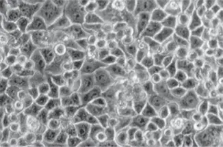
Despite the wide utility of monolayer and suspension cell cultures in virology, they are not without limitations, including the finite life span of primary cell cultures and the abnormal phenotype of continuous cell lines, such as immortality. These problems can be overcome by the use of induced pluripotent stem cells (iPSCs), which are adult cells that have been reprogrammed genetically to an embryonic stem-cell like state by the introduction of four genes ( Oct4 , Sox2 , Kif4 , and cMyc ). They are most commonly made from human fibroblasts, although other cell types have been used. Such iPSCs can be differentiated into many different cell types, such as cardiomyocytes, neurons, and hepatocytes, by treatment with specific growth factors. Viral reproduction can be studied in specific human cell types using cells derived from iPSCs.
BOX 2.2
BACKGROUND
The cells of Henrietta Lacks
The most widely used continuous cell line in virology, the HeLa cell line, was derived from Henrietta Lacks. In 1951, the 31-year-old mother of five visited a physician at Johns Hopkins Hospital in Baltimore and was found to have a malignant tumor of the cervix. A sample of the tumor was taken and given to George Gey, head of tissue culture research at Hopkins. Gey had been attempting for years, without success, to produce a line of human cells that would live indefinitely. When placed in culture, Henrietta Lacks’ cells propagated as no other cells had before.
On the day in October that Henrietta Lacks died, Gey appeared on national television with a vial of her cells, which he called HeLa cells. He said, “It is possible that, from a fundamental study such as this, we will be able to learn a way by which cancer can be completely wiped out.” Soon after, HeLa cells were used to propagate poliovirus, which was causing poliomyelitis throughout the world, and they played an important role in the development of poliovirus vaccines. Henrietta Lacks’ HeLa cells started a medical revolution: not only was it possible to propagate many different viruses in these cells, but the work set a precedent for producing continuous cell lines from many human tissues. However, the family of Henrietta Lacks did not learn about HeLa cells, or the revolution they started, until 24 years after her death. Her family members were shocked that cells from Henrietta lived in so many laboratories and that they had not been told that any cells had been taken from her.
The story of HeLa cells is an indictment of the lack of informed consent that pervaded medical research in the 1950s. Since then, biomedical ethics have changed, and there are now strict regulations in clinical research: physicians may not take samples for research from patients without permission. Nevertheless, in early 2013, HeLa cells generated more controversy when a research group published the cells’ genome sequence. The Lacks family objected to the publication, claiming that the information could reveal private medical information about surviving family members. As a result, the sequence was withdrawn from public databases. Months later, a second HeLa cell genome sequence was published, but this time the authors were bound by an agreement brokered by the National Institutes of Health, which required an application process for any individual wishing to view the sequence.
Adey A, Burton JN, Kitzman JO, Hiatt JB, Lewis AP, Martin BK, Qiu R, Lee C, Shendure J. 2013. The haplotype-resolved genome and epigenome of the aneuploid HeLa cancer cell line. Nature 500:207–211.
Callaway E. 2013. Deal done over HeLa cell line. Nature 500:132–133.
Callaway E. 23 March 2013. HeLa publication brews bioethical storm. Nature. https://www.nature.com/news/hela-publication-brews-bioethical-storm-1.12689.
Skloot R. April 2000. Henrietta’s dance. Johns Hopkins Magazine. http://pages.jh.edu/∼jhumag/0400web/01.html.
Skloot R. 2011. The Immortal Life of Henrietta Lacks. Broadway Books, New York, NY.
BOX 2.3
EXPERIMENTS
Zika virus blocks the neuronal road
Zika virus infection during pregnancy is a cause of the human birth defect called microcephaly. Babies born with this defect have smaller heads than expected for their age and smaller brains that do not develop normally. Organotypic brain slice cultures from embryonic mice have been used to study the effect of Zika virus on brain development.
To produce organotypic embryonic brain slice cultures, fetal mouse brains were removed, embedded in low-melting-point agarose, and thinly sliced with a vibratome. The slices were placed in cortical culture medium and then infected with Zika virus.
When first- and second-trimester brain slice cultures were infected with different isolates of Zika virus from 1947 to 2016, reproduction was observed as determined by plaque assay. These findings demonstrate that neurotropism of Zika virus is not a recently acquired phenotype.
The small heads observed in microcephalic children reflect a physically smaller brain—specifically, the neocortex is thinner than in a normal brain. The neocortex, the largest part of the cerebral cortex of the brain, is composed of six distinct layers of neurons, which are established during embryonic development. First, glial cells originating from progenitor cells in the ventricular zone extend their processes throughout the cortex and anchor at the pia, the outer surface of the brain. These long fibers provide a scaffold on which neurons, produced from the same progenitor cells, migrate outwards to establish the six layers of the cortex.
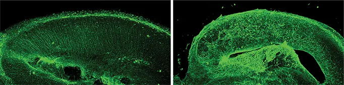
Neuronal migration is impaired during Zika virus infection.Brain slice cultures from embryonic day 15 mice were infected with 10 5PFU of Zika virus and at 4 dpi, were fixed and stained with antibody against vimentin to mark the radial glia progenitor (RGP) basal processes, which are the fibers upon which bipolar neurons migrate. ZIKV infection perturbed the RGP scaffold compared with control slices.
Glial fibers are visible as parallel tracks in the mouse embryonic brain slice cultures stained with an antibody to vimentin, a protein component of the fibers (image, left panel). When embryonic brain slice cultures were infected with Zika virus, the structure of the glial tracks was altered. Instead of parallel tracks, the fibers assumed a twisted morphology that would not allow neurons to travel from the ventricular zone to the developing neocortex (image, right panel). Disruption of glial fibers was observed after infection with Zika viruses isolated from 1947 to 2016.
These results suggest that Zika virus-mediated disruption of glial fibers during embryonic development contributes to microcephaly: if neurons cannot migrate to the pial surface, the neocortex will be thinner.
Rosenfeld AB, Doobin DJ, Warren AL, Racaniello VR, Vallee RB. 2017. Replication of early and recent Zika virus isolates throughout mouse brain development. Proc Natl Acad Sci U S A 114:12273–12278.
Monolayer and suspension cell cultures do not reproduce the cell type diversity and architecture typical of tissues and organs. One way to overcome this limitation is by the use of organotypic slice cultures, which can be produced from a variety of organs, including brain, liver, and kidney. These cultures are prepared by slicing embryonic or postnatal rodent organs into 100- to 400-micrometer slices. They are placed on substrates, such as porous or semiporous membranes, and bathed in cell culture medium. Such cultures remain viable for 1 to 2 weeks. The effect of Zika virus infection on neuronal migration has been examined in organotypic brain slice cultures derived from embryonic mice ( Box 2.3).
Читать дальшеИнтервал:
Закладка:
Похожие книги на «Principles of Virology»
Представляем Вашему вниманию похожие книги на «Principles of Virology» списком для выбора. Мы отобрали схожую по названию и смыслу литературу в надежде предоставить читателям больше вариантов отыскать новые, интересные, ещё непрочитанные произведения.


Обсуждение, отзывы о книге «Principles of Virology» и просто собственные мнения читателей. Оставьте ваши комментарии, напишите, что Вы думаете о произведении, его смысле или главных героях. Укажите что конкретно понравилось, а что нет, и почему Вы так считаете.