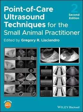
Point-of-Care Ultrasound Techniques for the Small Animal Practitioner

Здесь есть возможность читать онлайн «Point-of-Care Ultrasound Techniques for the Small Animal Practitioner» — ознакомительный отрывок электронной книги совершенно бесплатно, а после прочтения отрывка купить полную версию. В некоторых случаях можно слушать аудио, скачать через торрент в формате fb2 и присутствует краткое содержание. Жанр: unrecognised, на английском языке. Описание произведения, (предисловие) а так же отзывы посетителей доступны на портале библиотеки ЛибКат.

Point-of-Care Ultrasound Techniques for the Small Animal Practitioner: краткое содержание, описание и аннотация

Предлагаем к чтению аннотацию, описание, краткое содержание или предисловие (зависит от того, что написал сам автор книги «Point-of-Care Ultrasound Techniques for the Small Animal Practitioner»). Если вы не нашли необходимую информацию о книге — напишите в комментариях, мы постараемся отыскать её.

This book offers a thorough revision and update to the first landmark book that presented a standardized approach to focused point-of-care ultrasound exams of the abdomen, thorax, musculoskeletal and eye in veterinary practice. Now incorporating new applications for focused ultrasound exams and additional species, this Second Edition continues to be a state-of-the-art reference for using abbreviated ultrasound exams in clinical practice. A companion website features supplementary video clips of these point-of-care techniques depicting actual ultrasound exams for comparison and comprehension. 
New chapters in
cover ultrasound-guided nerve blocks, musculoskeletal, brain imaging, and applications of focused ultrasound techniques in cats, exotics and marine mammals—making it an essential purchase for veterinarians wanting to incorporate point-of-care ultrasound techniques into their veterinary practices. 
Presents a standardized approach to point-of-care ultrasound as an extension of the physical exam, including trauma, non-trauma, and monitoring applications Includes coverage of new techniques for focused ultrasound exams, including lung, anesthesia and ultrasound guided nerve blocks, transcranial brain imaging, musculoskeletal, volume status evaluation, and rapid assessment for treatable forms of shock Adds cats, exotic and wildlife mammals, and marine mammals to the existing canine coverage Emphasizes the integration of point-of-care ultrasound techniques for optimizing patient care and accurate patient assessment Offers access to a companion website with supplementary video clips showing many clinically relevant didactic examples The second edition of
is an excellent resource for veterinary practitioners, ranging from the general practitioner to nearly all clinical specialists, including internal medicine, oncology, cardiology, emergency and critical care, anesthesiology, ophthalmology, exotics, and zoo medicine specialists, and veterinary students.
Point-of-Care Ultrasound Techniques for the Small Animal Practitioner — читать онлайн ознакомительный отрывок

Ниже представлен текст книги, разбитый по страницам. Система сохранения места последней прочитанной страницы, позволяет с удобством читать онлайн бесплатно книгу «Point-of-Care Ultrasound Techniques for the Small Animal Practitioner», без необходимости каждый раз заново искать на чём Вы остановились. Поставьте закладку, и сможете в любой момент перейти на страницу, на которой закончили чтение.

Тёмная тема
Сбросить

Интервал:

Закладка:

Сделать

18 Chapter 18Figure 18.1. Dry aerated normal lung and pneumothorax. Shown in the top row ...Figure 18.2. Correlation of line drawings and B‐mode still image for B‐lines...Figure 18.3. Correlating images with pseudo B‐lines and overlays. In t...Figure 18.4. Pseudo pleural effusion from the gastric wall. Shown in (A) and...Figure 18.5. The “lung point” on a cross‐section of the thorax....Figure 18.6. The “lung point” with the finding of “lung sliding....Figure 18.7. Differences in positioning and the effect on the location of th...Figure 18.8. The “parking garage ‐ skyscraper effect” for the “lung point....Figure 18.9. Pneumothorax and the TFAST PTX 1/3s Rule. (A) Performing Vet BL...Figure 18.10. M‐mode findings correlated to line drawings and B‐mode images....Figure 18.11. Power Doppler for “lung sliding.” Shown in (A) is the us...Figure 18.12. Subcutaneous emphysema and probe pressure technique. In (A) is...Figure 18.13. Subcutaneous emphysema and its B‐mode appearance. (A) Ca...Figure 18.14. The “step sign.” The “step sign” is the loss of the expe...Figure 18.15. Anatomy of the heart and its right TFAST pericardial short‐axi...Figure 18.16. Right pericardial short‐axis views and their danger zone....Figure 18.17. Most reliable echo views for pericardial effusion. (A–D) show ...Figure 18.18. Imaging strategy for PCE is toward the muscular apex of the he...Figure 18.19. “One view is no view” – composite of multiple views for the ac...Figure 18.20. Integration of information in PCE and the presence and absence...Figure 18.21. Composite of images in a dog with a left atrial tear secondary...Figure 18.22. The “cardiac gallbladder” in a case series of dogs....Figure 18.23. The “cardiac gallbladder” in a cat. In rows (A) th...Figure 18.24. Composite of pleural effusion in a cat. Same images are shown ...Figure 18.25. Pitfall of pericardial FAST at the TFAST PCS views. In (A) and...Figure 18.26. Pitfall of pericardial fat. Shown is from the left TFAST PCS t...Figure 18.27. Hemothorax in a bluntly traumatized dog occult on thoracic rad...Figure 18.28. TFAST is better for patient care than the “flash” approach....Figure 18.29. Peritoneopericardial hernia detected during TFAST. The cat pre...Figure 18.30. Diaphragmatic hernia in a dog. The same concept in the cat in ...Figure 18.31. Composite of echo views from the right TFAST PCS view. The top...Figure 18.32. Composite for how TFAST echo views are a screening test for va...Figure 18.33. Composite of the TFAST echo views and expected normals and abn...Figure 18.34. TFAST goal‐directed template.

19 Chapter 19Figure 19.1. Dog on an echo table in right lateral recumbency with the sonog...Figure 19.2. A cat in sternal recumbency with a rolled‐up towel under the fo...Figure 19.3. Dog with transducer probe positioned on the right recumbent sid...Figure 19.4. Normal right parasternal short‐axis views at heart base optimiz...Figure 19.5. Normal M‐mode views of the left heart. (A) Normal M‐mode ...Figure 19.6. Dog with transducer probe positioned on the recumbent right sid...Figure 19.7. Dog with the probe positioned on the left hemithorax (nonrecumb...Figure 19.8. Normal two‐dimensional image of the left ventricular outflow tr...Figure 19.9. Dog positioned for subcostal views with the probe positioned ju...Figure 19.10. Subcostal view of fluctuations of caudal vena cava diameter du...

20 Chapter 20Figure 20.1. Right parasternal long‐axis view of severe left atrial enlargem...Figure 20.2. Right parasternal short‐axis view of severe left atrial enlarge...Figure 20.3. Right parasternal long‐axis view of the left ventricular outflo...Figure 20.4. Canine dilated cardiomyopathy. (A) Right parasternal long‐axis ...Figure 20.5. Feline hypertrophic cardiomyopathy. Right parasternal short‐axi...Figure 20.6. Feline hypertrophic cardiomyopathy and its complications. Right...Figure 20.7. Large right atrial tumor and pericardial effusion in a dog. Rig...Figure 20.8. Large heart base tumor, likely a chemodectoma, in a dog with pe...Figure 20.9. Canine pulmonary hypertension. (A) Right parasternal short axis...Figure 20.10. Subcostal view of severe caudal vena caval and hepatic venous ...Figure 20.11. Subcostal view of fluctuations of the caudal vena caval diamet...

21 Chapter 21Figure 21.1. Right parasternal short‐axis view of pericardial effusion in a ...Figure 21.2. Subxiphoid (or diaphragmatic hepatic) view of pericardial effus...Figure 21.3. Cardiac tamponade, right atrial tumor, and heart base tumor. (A...Figure 21.4. Right atrial mass (probable hemangiosarcoma) in a dog with peri...Figure 21.5. Left atrial tear in a dog with advanced degenerative mitral val...Figure 21.6. Ultrasound‐assisted pericardiocentesis in a dog. (A) Afte...

22 Chapter 22Figure 22.1. Vet BLUE performed in standing showing correlation with lung re...Figure 22.2. Gator sign orientation externally on a dog. Shown in (A) is the...Figure 22.3. Gator sign orientation on B‐mode. The “gator sign” orient...Figure 22.4. Depth and proportionality. Depth is generally set at 4–8 cm (up...Figure 22.5. Difference in appearance of longitudinal versus transverse orie...Figure 22.6. Optimizing the image – gain, depth, and focus cursor. Maj...Figure 22.7. The effect of angle of insonation and lung ultrasound artifacts...Figure 22.8. Imaging the “lung line” and angle of insonation. (A...Figure 22.9. Caudodorsal transition zone depicted radiographically. All imag...Figure 22.10. Caudodorsal transition zone on B‐mode and “curtain sign....Figure 22.11. Left‐sided Vet BLUE views. The lateral thoracic radiogra...Figure 22.12. Right‐sided Vet BLUE views. The lateral thoracic radiogr...Figure 22.13. The cranial transition zone. The top image shows Vet BLUE on a...Figure 22.14. Vet BLUE DH view. The view is shown without (A) and with (B) t...Figure 22.15. Determining and following the Vet BLUE line. In (A) and (C) th...Figure 22.16. Vet BLUE protocol and its three intercostal spaces. In (A) and...Figure 22.17. Boundaries of lung. The boundaries of lung are correlated to t...Figure 22.18. Ribs versus costochondral junction. (A–C) Images from the same...Figure 22.19. Subcutaneous emphysema. Shown in (A) is radiographically evide...Figure 22.20. Wet Versus dry lung. Shown in (A) and (B) are a line drawing o...Figure 22.21. B‐lines previously referred to as ultrasound lung rockets....Figure 22.22. Vet BLUE B‐line scoring system. Numbers of B‐lines have ...Figure 22.23. Composite of three different published lung ultrasound formats...

23 Chapter 23Figure 23.1. Lung pathology must touch the lung line to be detected. Simple ...Figure 23.2. Vet BLUE six lung ultrasound signs. The Vet BLUE visual lung la...Figure 23.3. “Wet” versus “dry lung.” (A,B) Line drawings ...Figure 23.4. B‐lines also referred to as ultrasound lung rockets. Exam...Figure 23.5. “Dry lung” – A‐lines with “lung sliding....Figure 23.6. Pneumothorax – A‐lines with absent “lung sliding....Figure 23.7. “Wet lung” – B‐lines. Correlative image...Figure 23.8. Shred sign – air bronchogram, consolidation with aeration....Figure 23.9. Tissue sign – hepatization, consolidation without aeration....Figure 23.10. Nodule sign. Correlative images of (A) line drawing of the nod...Figure 23.11. Wedge sign. Correlative images of (A) line drawing of the wedg...Figure 23.12. “Dry lung” – A‐lines and “lung sliding....Figure 23.13. “Wet lung” – B‐lines most commonly a result of fluid next to a...Figure 23.14. Vet BLUE B‐line scoring system. Numbers of B‐lines corre...Figure 23.15. Examples of how to score each regional Vet BLUE view by maximu...Figure 23.16. Vet BLUE B‐line scoring system. Line drawings in row (A)...Figure 23.17. Vet BLUE B‐line scoring system compared to the Volpicelli Stro...Figure 23.18. Example of an abnormal Vet BLUE considered negative by the Vol...Figure 23.19. Line Drawings and overlays of the six Vet BLUE lung ultrasound...Figure 23.20. Caudodorsal transition zone (CdTZ). The curtain sign is the te...Figure 23.21. Lung pathology during Vet BLUE at the DH view. (A–C) Various d...Figure 23.22. How the lung ultrasound line system lacks illustrative purpose...Figure 23.23. Hitches and blips. Lung ultrasound is extremely sensitive for ...Figure 23.24. Vet BLUE profiles using its regional pattern‐based approach....Figure 23.25. “Dry lung” All Vet BLUE Views also called Absent B‐lines All (...Figure 23.26. Examples of Vet BLUE left‐sided congestive heart failure profi...Figure 23.27. Use of the Vet BLUE B‐line scoring system for guiding loop diu...Figure 23.28. Examples of Vet BLUE noncardiogenic lung edema profile. Latera...Figure 23.29. Examples of Vet BLUE pneumonia profile. Lateral thoracic radio...Figure 23.30. Vet BLUE severity scoring of lung contusions. As with assessin...Figure 23.31. Comparative imaging of Vet BLUE, TXR, and CT. Shown in the top...Figure 23.32. Coughing dog post anesthesia event. This heart disease (class ...Figure 23.33. Respiratory distress feline. (A) The Vet BLUE profile is “Dry ...Figure 23.34. Aspiration pneumonia detected on Vet BLUE over thoracic radiog...Figure 23.35. Old dog lungs. The “old dog lungs” on three‐view radiography (...Figure 23.36. Fungal granulomas easily detected on Vet BLUE missed on thorac...Figure 23.37. Primary lung lobe mass versus disseminated lung disease. The t...

Читать дальше
Тёмная тема
Сбросить

Интервал:

Закладка:

Сделать

Похожие книги на «Point-of-Care Ultrasound Techniques for the Small Animal Practitioner»

Представляем Вашему вниманию похожие книги на «Point-of-Care Ultrasound Techniques for the Small Animal Practitioner» списком для выбора. Мы отобрали схожую по названию и смыслу литературу в надежде предоставить читателям больше вариантов отыскать новые, интересные, ещё непрочитанные произведения.


Отзывы о книге «Point-of-Care Ultrasound Techniques for the Small Animal Practitioner»

Обсуждение, отзывы о книге «Point-of-Care Ultrasound Techniques for the Small Animal Practitioner» и просто собственные мнения читателей. Оставьте ваши комментарии, напишите, что Вы думаете о произведении, его смысле или главных героях. Укажите что конкретно понравилось, а что нет, и почему Вы так считаете.

x