Crispian Scully - Clinical Guide to Oral Diseases
Здесь есть возможность читать онлайн «Crispian Scully - Clinical Guide to Oral Diseases» — ознакомительный отрывок электронной книги совершенно бесплатно, а после прочтения отрывка купить полную версию. В некоторых случаях можно слушать аудио, скачать через торрент в формате fb2 и присутствует краткое содержание. Жанр: unrecognised, на английском языке. Описание произведения, (предисловие) а так же отзывы посетителей доступны на портале библиотеки ЛибКат.
- Название:Clinical Guide to Oral Diseases
- Автор:
- Жанр:
- Год:неизвестен
- ISBN:нет данных
- Рейтинг книги:4 / 5. Голосов: 1
-
Избранное:Добавить в избранное
- Отзывы:
-
Ваша оценка:
Clinical Guide to Oral Diseases: краткое содержание, описание и аннотация
Предлагаем к чтению аннотацию, описание, краткое содержание или предисловие (зависит от того, что написал сам автор книги «Clinical Guide to Oral Diseases»). Если вы не нашли необходимую информацию о книге — напишите в комментариях, мы постараемся отыскать её.
Provides nearly 300 high-quality clinical photos and relevant questions to help lead readers to the proper diagnosis of common oral diseases Contains concise tables relevant to each chapter with a list of common oral lesions and conditions Offers MCQs of varying levels of difficulty to help readers test their knowledge in Oral Medicine Includes clinical flow charts according to the location and duration of oral lesions Incorporates the ICD-10 Codes of oral lesions and diseases
is a valuable reference for general dental and medical practitioners, undergraduate dental students, and postgraduate trainees in oral and maxillofacial surgery, oral medicine, oral pathology, periodontology as well as general pathology, dermatology or head and neck oncology.
Clinical Guide to Oral Diseases — читать онлайн ознакомительный отрывок
Ниже представлен текст книги, разбитый по страницам. Система сохранения места последней прочитанной страницы, позволяет с удобством читать онлайн бесплатно книгу «Clinical Guide to Oral Diseases», без необходимости каждый раз заново искать на чём Вы остановились. Поставьте закладку, и сможете в любой момент перейти на страницу, на которой закончили чтение.
Интервал:
Закладка:
Case 3.9
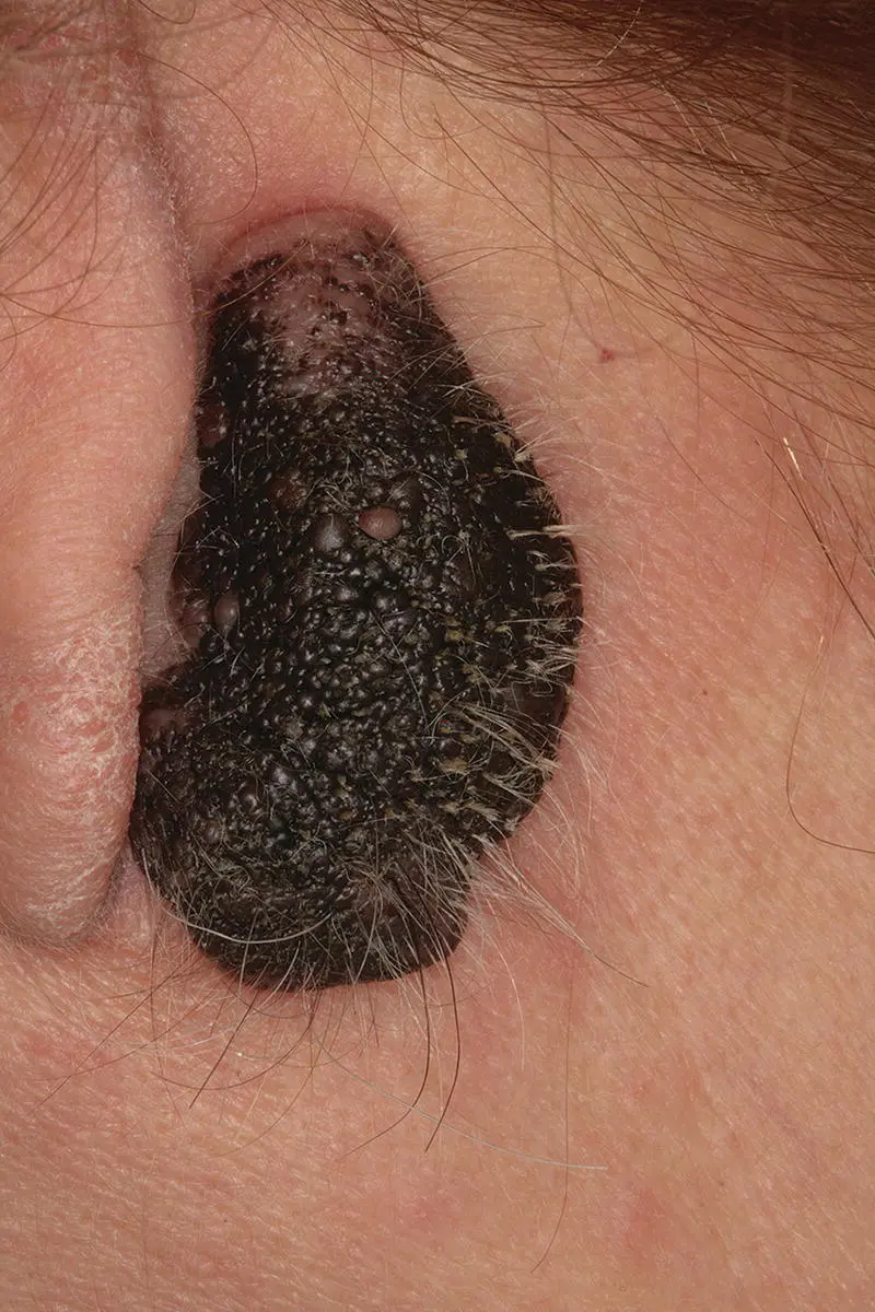
Figure 3.9
CO: A 56‐year‐old woman was presented for a routine medical–dental check‐up and during her examination a dark brown lesion behind her right ear lobe was found.
HPC: The lesion had been present since childhood, but increased in size and color during puberty and had remained unchanged since then.
PMH: This middle‐aged post‐menopausal woman had a history of mild hypertension, dislipidemia, and depression which were treated with an ACE inhibitor, fluvastatin plus diet and psychological counseling respectively. She had a basal cell carcinoma on the skin of her nose that was removed in surgery two years ago. She used to smoke two to five cigarettes on a daily basis and had no history of alcohol use. None of her close relatives had similar brown lesions.
OE: Clinical examination revealed a dark brown oval lesion on the skin behind her right ear lobe with a larger dimension of 3.5 cm. The lesion was well‐defined, soft with a nodular surface, unique color and contained few hairs ( Figure 3.9). No other similar lesions were found apart from a few small moles on her back.
Q1What is the diagnosis?
1 Pigmented basal cell carcinoma
2 Melanocanthoma
3 Congenital melanotic nevus
4 Melanoma
5 Neurocutaneous melanosis
Answers:
1 No
2 No
3 Congenital melanotic nevus is the cause and is characterized by a well‐defined, dark pigment lesion on the skin of the face with a nodular appearance, containing hairs. Congenital nevus is characterized by its large size (>3.5 cm in diameter) and early onset (from birth). Acquired melanotic nevi are usually numerous, of small size (<1 cm) and develop later in puberty or adolescence.
4 No
5 No
Comments: Although the lesion is congenital, the absence of other melanocytic tumors in the leptomeninges of the central nervous system (CNS) excludes neurocutaneous melanosis from the differential diagnosis. The lack of induration or bleeding at palpation as well asymmetry and color variation together with the satellite new lesion formation ruled out melanoma from diagnosis. Another benign pigmented lesion that mainly affects female skin, especially the face, is melanoacanthoma; it differs in that it affects older women with dark skin (>30 years of age) and is characterized histologically by proliferation of epithelial and nevus cells.
Q2What are the differences between congenital and acquired nevi apart from the year of onset?
1 Size
2 Skin distribution
3 Color
4 Risk for melanoma transformation
5 Location of nevi cells
Answers:
1 The acquired nevi are smaller than the congenital ones. Congenital nevi can be small (<2 cm); intermediate (>2 cm but <20 cm) or giant covering a whole part of the body such as the face, back, or legs.
2 No
3 No
4 No
5 The congenital nevi cells are usually found deeper into the dermis, within neurovascular bundles.
Comments: Both types of nevi have been observed in the skin and oral mucosa as benign pigmented lesions with color ranging from brown to dark black. Both are asymptomatic and do not show any indication of melanoma following the asymmetry, border, color, diameter, evolving (ABCDE) rule. Only the giant congenital nevus has an increased risk of developing melanoma and that is why it should be closely monitored.
Q3Which other treatment options are available apart from surgical excision for congenital melanocytic nevi?
1 Phototherapy
2 Corticosteroid creams
3 Chemical peeling
4 Hyperbaric oxygen
5 Dermabrasion
Answers:
1 No
2 No
3 Chemical peeling with trichloracetic acid or phenol solutions lighten the color of nevus but cause local skin irritation.
4 No
5 Dermabrasion involves the partial removal of a large congenital nevus causing its color lighting, although it can be scarring.
Comments: Phototherapy involves the exposure of skin to ultraviolet light. On a regular basis, this is used for the treatment of psoriasis, and not for nevus disappearance. This therapy may stimulate nevi cells and produce melanin. Hyperbaric oxygen is used to treat wrinkles induced by ultraviolet radiation, and steroids are used for the eczema around the nevus, but not for the nevus.
Case 3.10
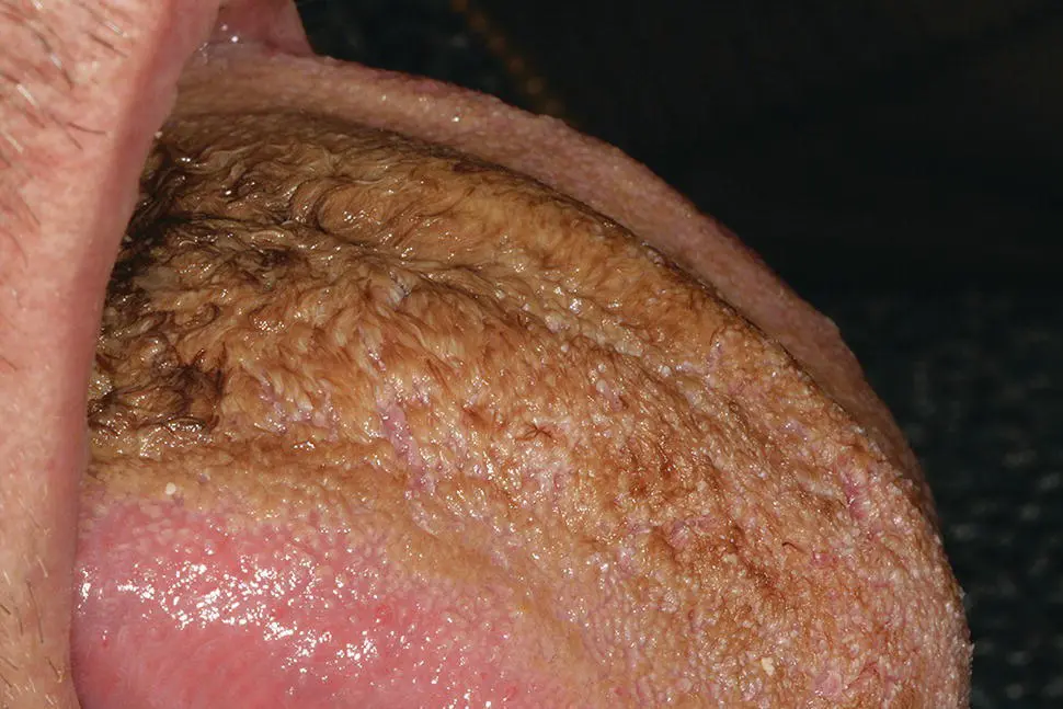
Figure 3.10a
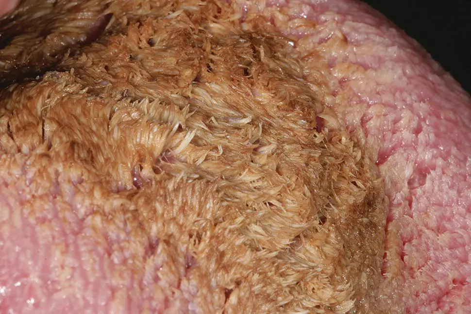
Figure 3.10b
CO: A 22‐year‐old student was evaluated for a brown discoloration of his tongue.
HPC: The tongue became brown after a course of antibiotics for pericoronitis on his lower left wisdom tooth two weeks ago.
PMH: His medical history is clear from any serious diseases except for a few episodes of aphthous stomatitis, two to three times a year. He is not on any medicines apart from antibiotics and chlorhexidine mouthwash recently. He is a chronic smoker of 10 to 15 cigars per day and chews mint flavor chewing gum on a regular basis.
OE: The oral examination revealed a brown discoloration on his dorsum of his tongue ( Figure 3.10a) associated with hypertrophy of filiform papillae ( Figure 3.10b).This discoloration covered the middle of the area, but was more prominent on the posterior part of the tongue and was related to mild halitosis. This discoloration was partially wiped off by scrubbing his tongue with a wooden spatula, and it had no other connection with similar lesions on the oral and other mucosae.
Q1What is the cause?
1 Pseudomembranous candidiasis
2 Brown hairy tongue
3 Hairy leukoplakia
4 Mint‐induced stomatitis
5 Smoking melanosis
Answers:
1 No
2 No
3 Brown hairy tongue is a variation of hairy tongue and is presented with an abnormal keratin coating of the elongated filiform papillae on the dorsum of the tongue. It is commonly found among young anxious patients with poor oral hygiene, who smoke, take antibiotics – metronidazole in particular – or use strong mouthwashes on a regular basis, as in the case of this young man.
4 No
5 No
Comments: The lesions in pseudomembranous candidiasis are white creamy lesions that are spread all over the oral mucosa apart from the tongue, and can be easily removed with a spatula, leaving only an erythema underneath. Hairy leukoplakia lesions appear as white fixed lesions on the lateral margins and not on the dorsum of tongue in immuno‐compromised patients, while the mint induced stomatitis lesions are not only restricted to the area of tongue (lateral margins) but can be also be seen in other parts that are exposed to mint flavor. Cigar smoking may contribute to the brown discoloration through the accumulation of nicotine stains within filiform papillae in hairy tongue, but also by stimulating the gingival melanocytes to produce melanin, therefore causing a gingival melanosis.
Q2The diagnosis of this lesion is based mainly on:
1 Intra‐oral examination
2 History
3 Culture
4 Biopsy
5 Allergic tests
Читать дальшеИнтервал:
Закладка:
Похожие книги на «Clinical Guide to Oral Diseases»
Представляем Вашему вниманию похожие книги на «Clinical Guide to Oral Diseases» списком для выбора. Мы отобрали схожую по названию и смыслу литературу в надежде предоставить читателям больше вариантов отыскать новые, интересные, ещё непрочитанные произведения.


Обсуждение, отзывы о книге «Clinical Guide to Oral Diseases» и просто собственные мнения читателей. Оставьте ваши комментарии, напишите, что Вы думаете о произведении, его смысле или главных героях. Укажите что конкретно понравилось, а что нет, и почему Вы так считаете.