Biological Mechanisms of Tooth Movement
Здесь есть возможность читать онлайн «Biological Mechanisms of Tooth Movement» — ознакомительный отрывок электронной книги совершенно бесплатно, а после прочтения отрывка купить полную версию. В некоторых случаях можно слушать аудио, скачать через торрент в формате fb2 и присутствует краткое содержание. Жанр: unrecognised, на английском языке. Описание произведения, (предисловие) а так же отзывы посетителей доступны на портале библиотеки ЛибКат.
- Название:Biological Mechanisms of Tooth Movement
- Автор:
- Жанр:
- Год:неизвестен
- ISBN:нет данных
- Рейтинг книги:5 / 5. Голосов: 1
-
Избранное:Добавить в избранное
- Отзывы:
-
Ваша оценка:
Biological Mechanisms of Tooth Movement: краткое содержание, описание и аннотация
Предлагаем к чтению аннотацию, описание, краткое содержание или предисловие (зависит от того, что написал сам автор книги «Biological Mechanisms of Tooth Movement»). Если вы не нашли необходимую информацию о книге — напишите в комментариях, мы постараемся отыскать её.
delivers a comprehensive reference for orthodontic trainees and specialists.
It is fully updated to include new chapters on personalized orthodontics as well as the inflammatory process occurring in the dental and paradental tissues. It is heavily illustrated throughout, making it easier for readers to understand and retain the information discussed within. The topics covered range from bone biology, the effects of mechanical loading on tissues and cells, genetics, tissue remodeling, and the effects of diet, drugs, and systemic diseases.
The Third Edition of
features seven sections that cover subjects such as:
The development of biological concepts in orthodontics, including the cellular and molecular biology behind orthodontic tooth movement Mechanics meets biology, including the effects of mechanical loading on hard and soft tissues and cells, and biological reactions to temporary anchorage devices Inflammation and orthodontics, including markers for tissue remodeling in the gingival crevicular fluid and saliva Personalized diagnosis and treatment based on genomic criteria, including the genetic influences on orthodontic tooth movement Rapid orthodontics, including methods to accelerate or decelerate orthodontic tooth movement Perfect for residents and PhD students of orthodontic and periodontal programs,
is also useful to academics, clinicians, bone biologists, and researchers with an interest in the mechanics and biology of tooth movement.
Biological Mechanisms of Tooth Movement — читать онлайн ознакомительный отрывок
Ниже представлен текст книги, разбитый по страницам. Система сохранения места последней прочитанной страницы, позволяет с удобством читать онлайн бесплатно книгу «Biological Mechanisms of Tooth Movement», без необходимости каждый раз заново искать на чём Вы остановились. Поставьте закладку, и сможете в любой момент перейти на страницу, на которой закончили чтение.
Интервал:
Закладка:
very high forces, capable of crushing the tissues, causing irreparable damage.
He concluded that an optimal force is smaller in magnitude than that capable of occluding PDL capillaries. Occlusion of these blood vessels, he reasoned, would lead to necrosis of surrounding tissues, which would be harmful, and would slow down the velocity of tooth movement.
The proposed optimal orthodontic force concept by Schwartz was supported by Reitan ( Figure 1.11), who conducted thorough histological examinations of paradental tissues incidental to tooth movement. Reitan’s studies were conducted on a variety of species, including rodents, canines, primates, and humans, and the results were published during the period from the 1940s to the 1970s. Figure 1.12displays the appearance of an unstressed PDL of a cat maxillary canine. The cells are equally distributed along the ligament, surrounding small blood vessels. Both the alveolar bone and the canine appear intact. In contrast, the compressed PDL of a cat maxillary canine that had been tipped distally for 28 days, with an 80 g force ( Figure 1.13), appears very stormy. The PDL near the root is necrotic, but the alveolar bone and PDL at the edge of the hyalinized zone are being invaded by cells that appear to remove the necrotic tissue, as evidenced by a large area where undermining resorption has taken place. Figure 1.14shows the mesial side of the same root, where tension prevails in the PDL. Here the cells appear busy producing new trabeculae arising from the alveolar bone surface, in an effort to keep pace with the moving root. To achieve this type of tissue and cellular responses to orthodontic loads, Reitan favored the use of light intermittent forces, because they cause minimal amounts of tissue damage and cell death. He noted that the nature of tissue response differs from species to species, reducing the value of extrapolations.
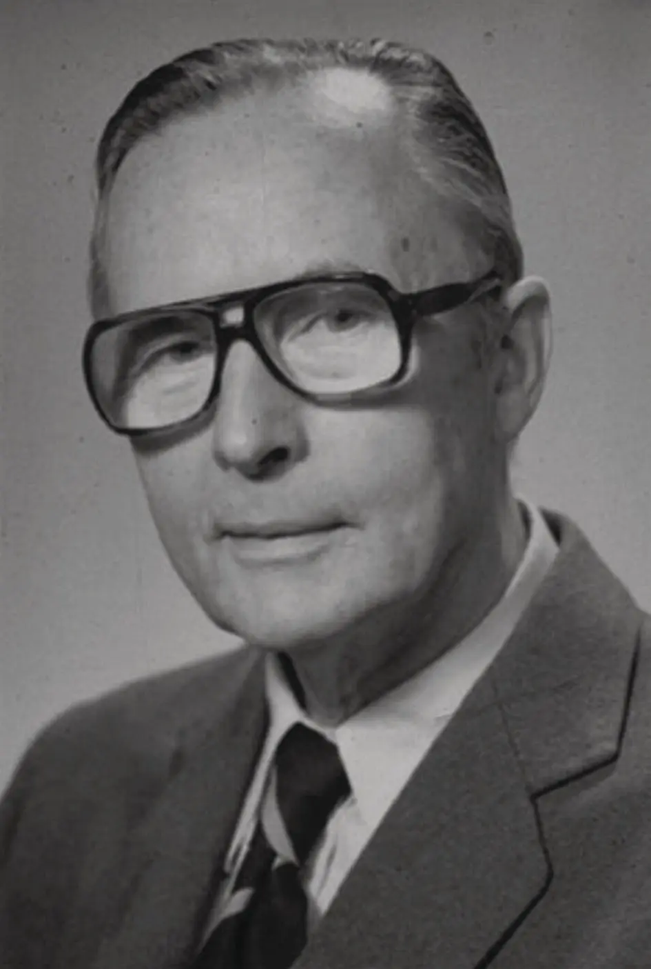
Figure 1.11 Kaare Reitan (1903–2000), who conducted thorough histological examinations of paradental tissues.
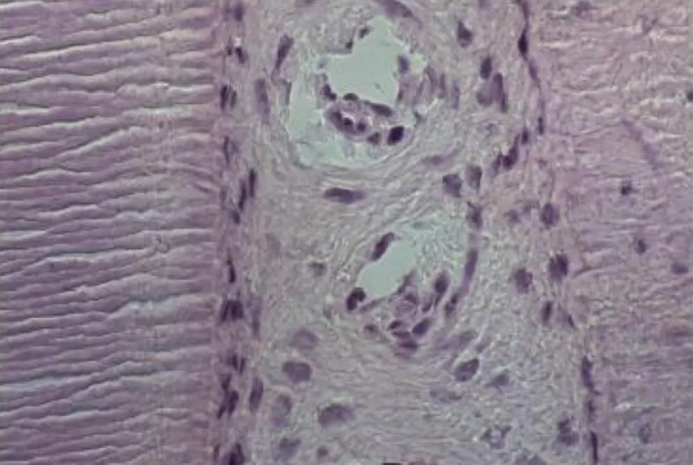
Figure 1.12 A 6 μm sagittal section of a frozen, unfixed, nondemineralized cat maxillary canine, stained with hematoxylin and eosin. This canine was not treated orthodontically (control). The PDL is situated between the canine root (left) and the alveolar bone (right). Most cells appear to have an ovoid shape.
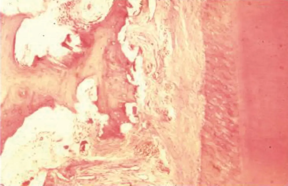
Figure 1.13 A 6 μm sagittal section of a cat maxillary canine, after 28 days of application of 80 g force. The maxilla was fixed and demineralized. The canine root (right) appears to be intact, but the adjacent alveolar bone is undergoing extensive resorption, and the compressed, hyalinized PDL is being invaded by cells from neighboring viable tissues (fibroblasts and immune cells). H & E staining.
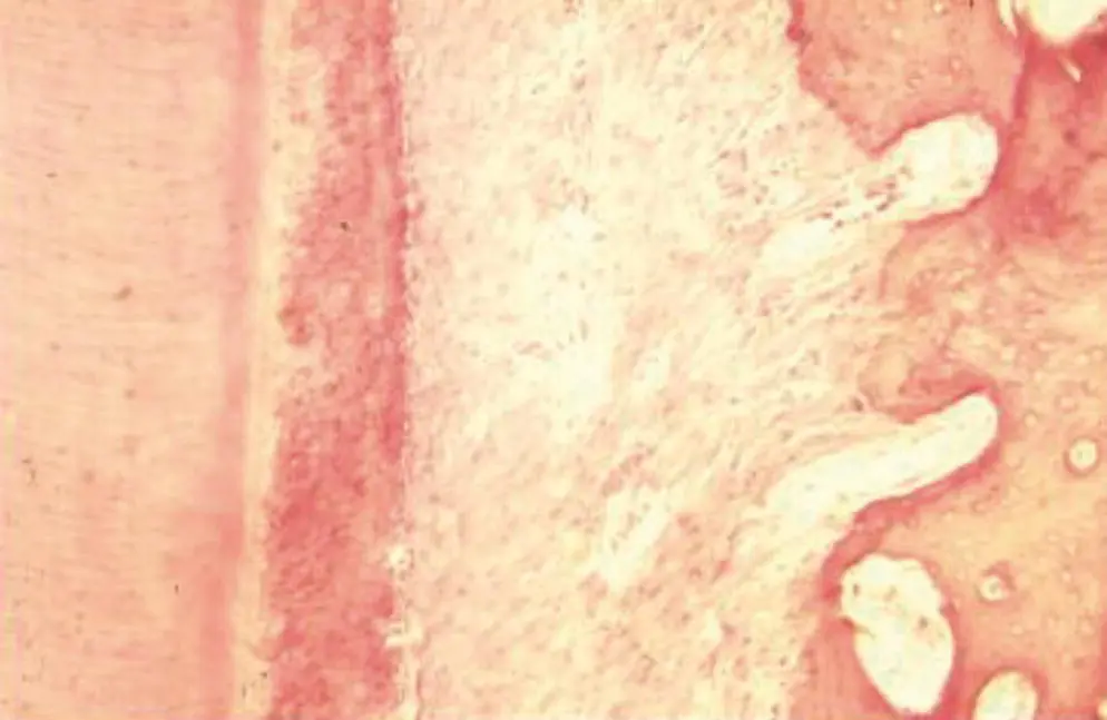
Figure 1.14 The mesial (PDL tension) side of the tooth shown in Figure 1.13. Here, new trabeculae protrude from the alveolar bone surface, apparently growing towards the distal‐moving root. H & E staining.
With experiments on human teeth, Reitan observed that tissue reactions can vary, depending upon the type of force application, the nature of the mechanical design, and the physiological constrains of the individual patient. He observed the appearance of hyalinized areas in the compressed PDL almost immediately after continuous force application and the removal of those hyalinized areas after 2–4 weeks. Furthermore, Reitan reported that in dogs, the PDL of rotated incisors assumes a normal appearance after 28 days of retention, while the supracrestal collagen fibers remain stretched even after a retention period of 232 days. Consequently, he recommended severing the latter fibers surgically. He also called attention to the role of factors such as gender, age, and type of alveolar bone, in determining the nature of the clinical response to orthodontic forces. He also reported that 50 g of force is ideal for movement of human premolars, resulting from direct resorption of the alveolar bone.
Another outlook on differential orthodontic forces was proposed by Storey (1973). Based upon experiments in rodents, he classified orthodontic forces as being bioelastic, bioplastic, and biodisruptive, moving from light to heavy. He also reported that in all categories, some tissue damage must occur in order to promote a cellular response, and that inflammation starts in paradental tissues right after the application of orthodontic forces.
Continuing the legacy of Sandstedt, Kvam and Rygh studied cellular reactions in the compression side of the PDL. Rygh (1974, 1976) reported on ultrastructural changes in blood vessels in both human and rat material as packing of erythrocytes in dilated blood vessels within 30 minutes, fragmentation of erythrocytes after 2–3 hours, and disintegration of blood vessel walls and extravasation of their contents after 1–7 days. He also observed necrotic changes in PDL fibroblasts, including dilatation of the endoplasmic reticulum and mitochondrial swelling within 30 minutes, followed by rupture of the cell membrane and nuclear fragmentation after 2 hours; cellular and nuclear fragments remained within hyalinized zones for several days. Root resorption associated with the removal of the hyalinized tissue was reported by Kvam and Rygh. This occurrence was confirmed by a scanning electron microscopic study of premolar root surfaces after application of a 50 g force in a lateral direction (Kvam, 1972). Using transmission electron microscopy (TEM), the participation of blood‐borne cells in the remodeling of the mechanically stressed PDL was confirmed by Rygh and Selvig (1973), and Rygh (1974, 1976). In rodents, they detected macrophages at the edge of the hyalinized zone, invading the necrotic PDL, phagocytizing its cellular debris and strained matrix.
After direct measurements of teeth subjected to intrusive forces, Bien (1966) hypothesized that there are three distinct but interacting fluid systems involved in the response of the PDL to mechanical loading: the fluids in the vascular network, in the cells and fibers, and the interstitial fluid. Mechanical loading moves fluids into the vascular reservoir of the marrow space through the many minute perforations in the tooth alveolar wall. The hydrodynamic damping coefficient ( Figure 1.15) is time dependent, and therefore the damping rate is determined by the size and number of these perforations. As a momentary effect, the fluid that is trapped between the tooth and the socket tends to move to the boundaries of the film at the neck of the tooth and the apex, while acting to cushion the load and is referred to as the “squeeze film effect”. As the squeeze film is depleted, the second damping effect occurs after exhaustion of the extracellular fluid, and the ordinarily slack fibers tighten. When a tooth is intruded, the randomly oriented periodontal fibers, which crisscross the blood vessels, tighten, then compress and constrict the vessels that run between them, causing stenosis and ballooning of the blood vessels, creating a back pressure. Thus, high hydrodynamic pressure heads can be created suddenly in the vessels above the stenosis. At the stenosis, a drop of pressure would occur in the vessel in accordance with Bernoulli’s principle that the pressure in the region of the constriction will be less than elsewhere in the system. Bien also differentiated the varied responses obtained from momentary forces of mastication from that of prolonged forces applied in orthodontic mechanics and suggested that biting forces in the range of 1500 g/cm 2will not crush the PDL or produce bone responses.
Читать дальшеИнтервал:
Закладка:
Похожие книги на «Biological Mechanisms of Tooth Movement»
Представляем Вашему вниманию похожие книги на «Biological Mechanisms of Tooth Movement» списком для выбора. Мы отобрали схожую по названию и смыслу литературу в надежде предоставить читателям больше вариантов отыскать новые, интересные, ещё непрочитанные произведения.


Обсуждение, отзывы о книге «Biological Mechanisms of Tooth Movement» и просто собственные мнения читателей. Оставьте ваши комментарии, напишите, что Вы думаете о произведении, его смысле или главных героях. Укажите что конкретно понравилось, а что нет, и почему Вы так считаете.