Biological Mechanisms of Tooth Movement
Здесь есть возможность читать онлайн «Biological Mechanisms of Tooth Movement» — ознакомительный отрывок электронной книги совершенно бесплатно, а после прочтения отрывка купить полную версию. В некоторых случаях можно слушать аудио, скачать через торрент в формате fb2 и присутствует краткое содержание. Жанр: unrecognised, на английском языке. Описание произведения, (предисловие) а так же отзывы посетителей доступны на портале библиотеки ЛибКат.
- Название:Biological Mechanisms of Tooth Movement
- Автор:
- Жанр:
- Год:неизвестен
- ISBN:нет данных
- Рейтинг книги:5 / 5. Голосов: 1
-
Избранное:Добавить в избранное
- Отзывы:
-
Ваша оценка:
Biological Mechanisms of Tooth Movement: краткое содержание, описание и аннотация
Предлагаем к чтению аннотацию, описание, краткое содержание или предисловие (зависит от того, что написал сам автор книги «Biological Mechanisms of Tooth Movement»). Если вы не нашли необходимую информацию о книге — напишите в комментариях, мы постараемся отыскать её.
delivers a comprehensive reference for orthodontic trainees and specialists.
It is fully updated to include new chapters on personalized orthodontics as well as the inflammatory process occurring in the dental and paradental tissues. It is heavily illustrated throughout, making it easier for readers to understand and retain the information discussed within. The topics covered range from bone biology, the effects of mechanical loading on tissues and cells, genetics, tissue remodeling, and the effects of diet, drugs, and systemic diseases.
The Third Edition of
features seven sections that cover subjects such as:
The development of biological concepts in orthodontics, including the cellular and molecular biology behind orthodontic tooth movement Mechanics meets biology, including the effects of mechanical loading on hard and soft tissues and cells, and biological reactions to temporary anchorage devices Inflammation and orthodontics, including markers for tissue remodeling in the gingival crevicular fluid and saliva Personalized diagnosis and treatment based on genomic criteria, including the genetic influences on orthodontic tooth movement Rapid orthodontics, including methods to accelerate or decelerate orthodontic tooth movement Perfect for residents and PhD students of orthodontic and periodontal programs,
is also useful to academics, clinicians, bone biologists, and researchers with an interest in the mechanics and biology of tooth movement.
Biological Mechanisms of Tooth Movement — читать онлайн ознакомительный отрывок
Ниже представлен текст книги, разбитый по страницам. Система сохранения места последней прочитанной страницы, позволяет с удобством читать онлайн бесплатно книгу «Biological Mechanisms of Tooth Movement», без необходимости каждый раз заново искать на чём Вы остановились. Поставьте закладку, и сможете в любой момент перейти на страницу, на которой закончили чтение.
Интервал:
Закладка:
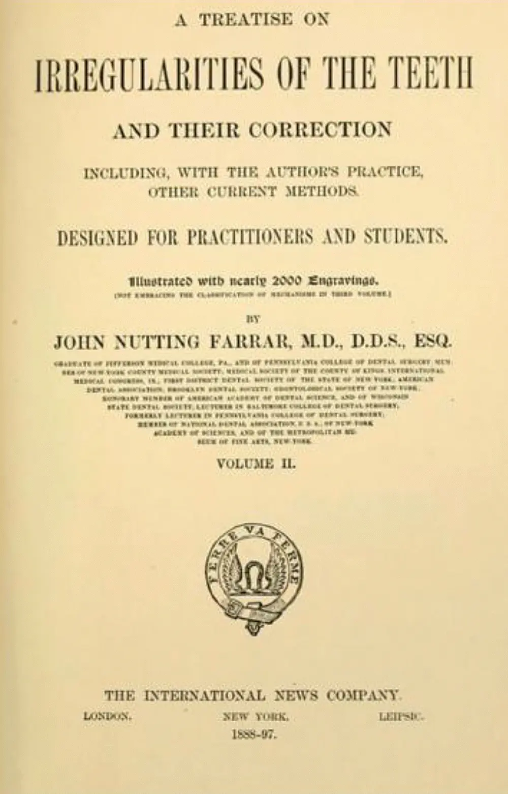
Figure 1.7 The front page of the book A Treatise on the Irregularities of the Teeth and their Correction by John Nutting Farrar.
(Source: Picture courtesy: https://openlibrary.org.)
Eugene Talbot, in his book titled Irregularities of Teeth and their Treatment (1888) rightly mentioned that “without the knowledge of etiology, no one can successfully correct the deformities as is evident in the many failures by men who profess to make this a specialty.” He argued that every case of malocclusion is different, making it difficult to classify, and proposed customizing appliances suited for each patient. He was the first to use X rays as a diagnostic aid in orthodontics, to identify abnormal and broken roots, locate third molars, and expose absorption of roots and alveolar process due to OTM.
Orthodontic tooth movement in the twentieth and twenty‐first centuries: From light microscopy to tissue engineering and stem cells
Histological studies of paradental tissues during tooth movement
Chappin Harris, in 1839, published a book titled The Dental Art, which stated that OTM in the socket depends on resorption and deposition of bone, but it took more than 60 years to have the first histological picture of this phenomenon, which was provided by Carl Sandstedt ( Figure 1.8). Sandstedt’s experimental studies of tooth movements in dogs were first published in German in 1901, and later in English (Sandstedt, 1904, 1905). His systematic way of conducting experiments was evident from the incorporation of a control group from the same litter as his two experimental dogs. A sectional fixed appliance was inserted in the upper jaw, which was subjected to repeated activations for palatal tipping of the upper incisors over a three‐week period. Histological sections of the incisor areas were prepared to assess tissue changes. In order to document positional changes of the teeth, plaster casts and radiographs were obtained. With these experiments, he could observe stretching of the periodontal ligament (PDL) in tension sites and narrowing of this tissue in pressure sites. He demonstrated new bone formation in areas of tension, while resorption was observed in areas of compression. In the compressed periodontium, he initially saw signs of necrosis (hyalinization), and described it as “an obviously degenerated product, a hyaline transformation of the connective tissue, in which regenerative processes take place … the old mortified tissue is resorbed and substituted by granulation tissue.” He further noted that “at the limit of the hyaline zone, the alveolar wall presents a deep, undermining notch filled by proliferating cells as in resorptive areas.” Furthermore, “the intensive resorptive process even attacked the incisor itself deeply into the dentine,” and he assumed that this process is a common secondary effect of OTM. Figure 1.9is a photograph of a cross section of a premolar root, showing areas of necrosis in the PDL, as well as multiple osteoclasts in Howship’s lacunae at the PDL–alveolar bone interface. These cells were, in Sandsted’s opinion, the main cells responsible for force‐induced tooth movement (Persson, 2005; Bister and Meikle, 2013).
He ended his landmark article by proposing a role for bone bending in the tooth movement process in line with the thinking provided by Kingsley and Farrar.
In 1911/1912, Oppenheim reported that tooth‐moving forces caused complete transformation (remodeling) of the entire alveolar process, indicating that orthodontic force effects spread beyond the limits of the PDL. E.H. Angle, the father of modern orthodontics, invited Oppenheim to lecture to his students, who accepted Oppenheim’s hypothesis enthusiastically. Oppenheim, the proponent of “the law of bone transformation,” rejected both the pressure/tension hypothesis supported by the histological evidence of Sandstedt, and the theory of bone bending hypothesis advanced by Kingsley and Farrar, based on the elastic properties of bone. Oppenheim’s experiments were conducted on mandibular deciduous incisors of baboons (the number of animals he used and the appliances he used remain ambiguous) and suggested that only very light forces evoke the required tissue responses. He stated that an increase in the force levels will produce occlusion of the vascular supply, as well as damage to the PDL and the other supporting tissues, and that the tooth will act as a one‐armed lever when light forces were applied, and like a two‐armed lever during the application of heavy forces. He also demonstrated how alveolar bone is restored structurally and functionally during the retention period (Noyes, 1945). As a proponent of bone transformation and Wolff ’s law, Oppenheim received acceptance from Angle, as it supported his thoughts in the matter. Oppenheim was also supported by Noyes, one of Angle’s followers and an established histologist.
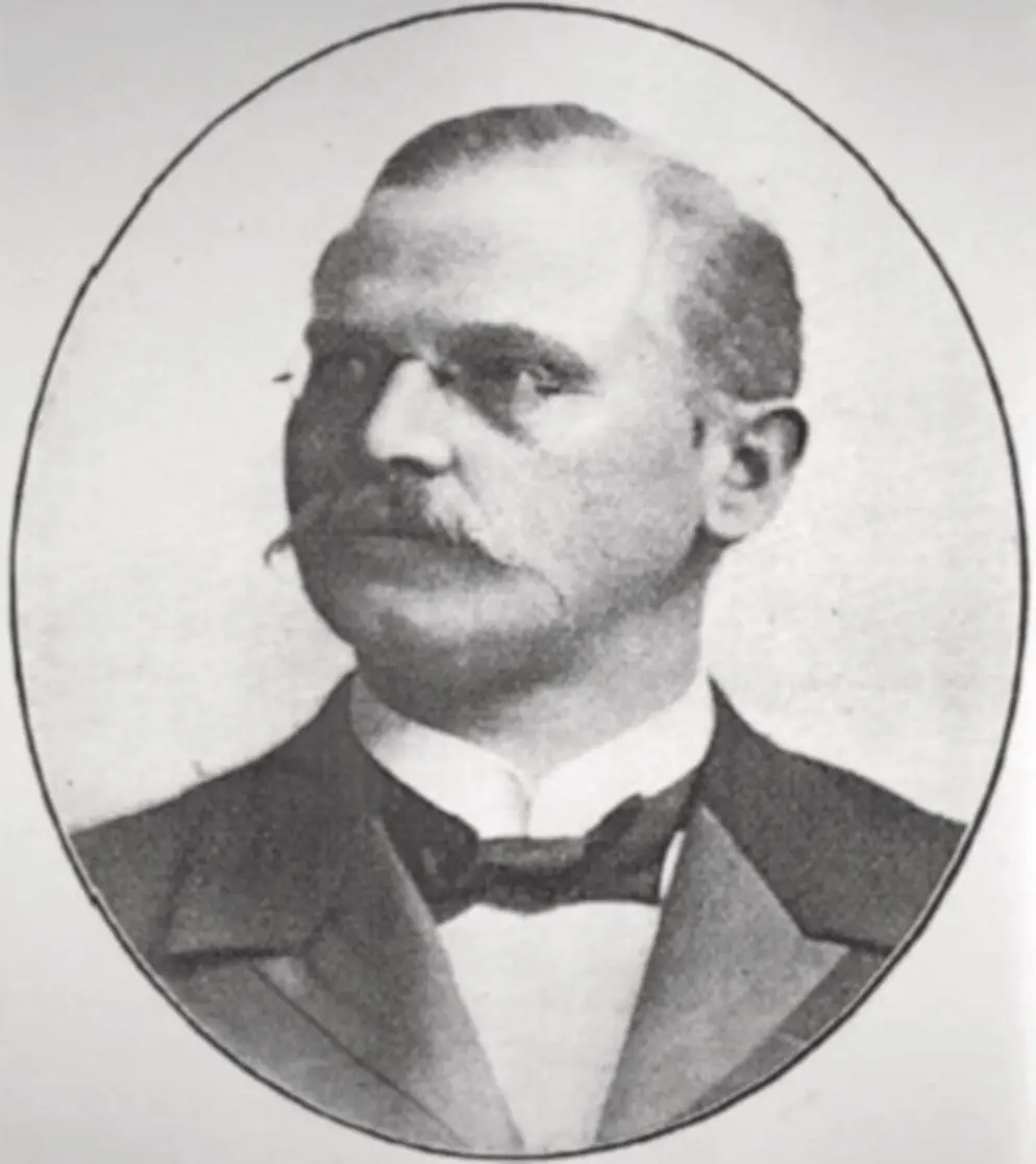
Figure 1.8 Carl Sandstedt (1860–1904), the father of biology of orthodontic tooth movement.
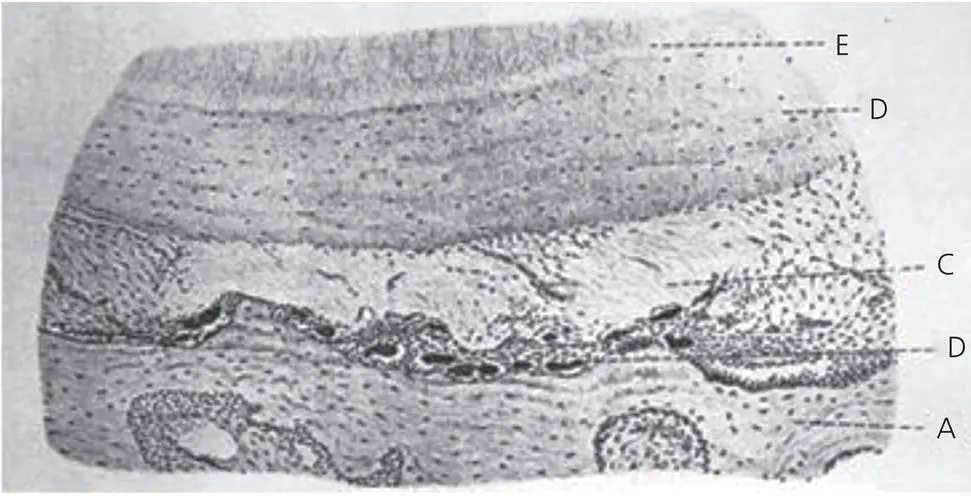
Figure 1.9 A figure from Carl Sandstedt’s historical article in 1904, presenting a histological picture of a dog premolar in cross section, showing the site of PDL compression, including an osteoclastic front and necrotic (hyalinized) areas.
Oppenheim’s research highlighted common concepts, shared by orthodontists and orthopedists, who were convinced that both specialties should be based upon a thorough knowledge of bone biology, particularly in relation to mechanical forces and their cellular reactions. However, it became evident that in orthodontics the PDL, in addition to bone, is a key tissue with regards to OTM.
Working on Macacus rhesus monkeys in 1926, Johnson, Appleton, and Rittershofer reported the first experiment where they recorded the relationship between the magnitude of the applied force and the distance in which it was active. In 1930, Grubrich reported surface resorptions in teeth subjected to orthodontic forces, a finding confirmed by Gruber in 1931. Even before these histological observations of surface changes were reported, Ketcham ( Figure 1.10) (1927, 1929) presented, radiographic evidence that root resorption may result from the application of faulty mechanics and the existence of some unknown systemic factors. Schwarz (1932) conducted extensive experiments on premolars in dogs, using known force levels for each tooth. The effects of orthodontic force magnitude on the dog’s paradental tissue responses were examined with light microscopy. Schwarz classified orthodontic forces into four degrees of biological efficiency:
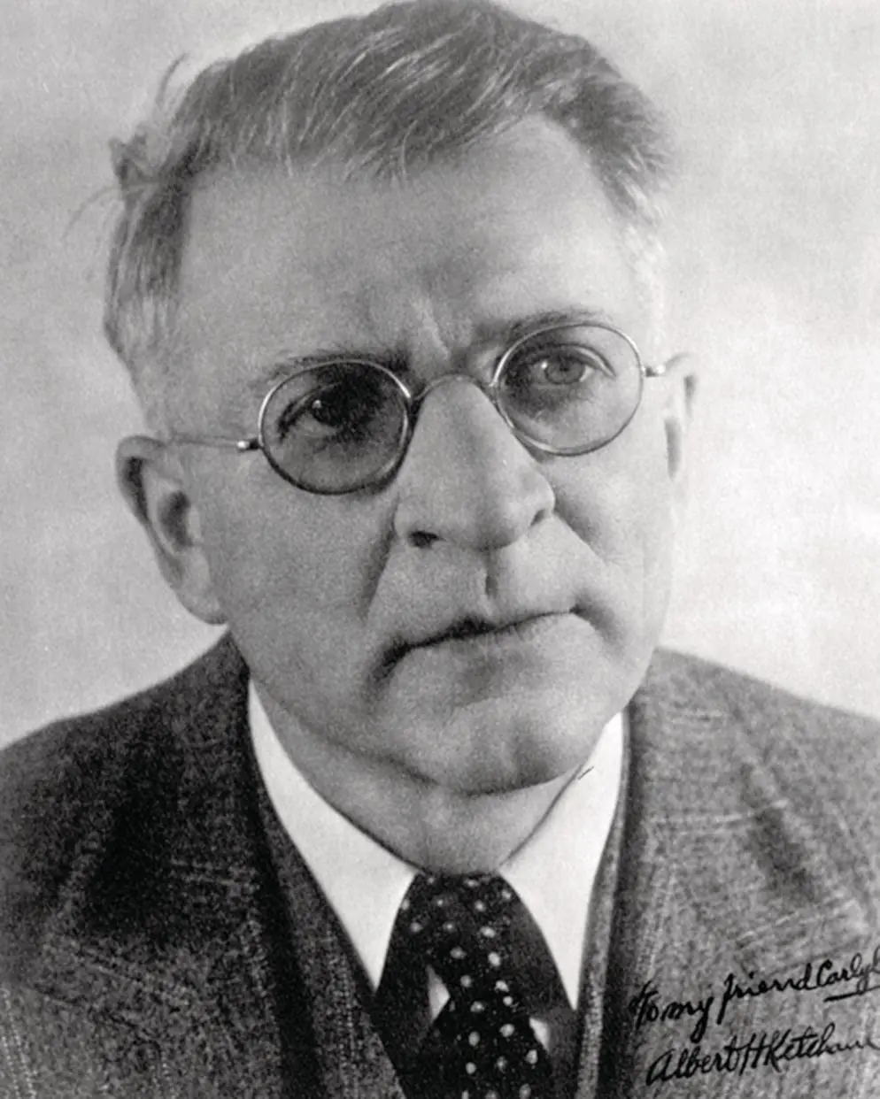
Figure 1.10 Albert Ketcham (1870–1935), who presented the first radiographic evidence of root resorption. He was also instrumental in forming the American Board of Orthodontics.
(Source: Siersma, 2015. Reproduced with permission of Elsevier.)
below threshold stimulus;
most favorable – about 20 g/cm2 of root surface, where no injury to the PDL is observed;
medium strength, which stops the PDL blood flow, but with no crushing of tissues;
Читать дальшеИнтервал:
Закладка:
Похожие книги на «Biological Mechanisms of Tooth Movement»
Представляем Вашему вниманию похожие книги на «Biological Mechanisms of Tooth Movement» списком для выбора. Мы отобрали схожую по названию и смыслу литературу в надежде предоставить читателям больше вариантов отыскать новые, интересные, ещё непрочитанные произведения.


Обсуждение, отзывы о книге «Biological Mechanisms of Tooth Movement» и просто собственные мнения читателей. Оставьте ваши комментарии, напишите, что Вы думаете о произведении, его смысле или главных героях. Укажите что конкретно понравилось, а что нет, и почему Вы так считаете.