Clinical Pancreatology for Practising Gastroenterologists and Surgeons
Здесь есть возможность читать онлайн «Clinical Pancreatology for Practising Gastroenterologists and Surgeons» — ознакомительный отрывок электронной книги совершенно бесплатно, а после прочтения отрывка купить полную версию. В некоторых случаях можно слушать аудио, скачать через торрент в формате fb2 и присутствует краткое содержание. Жанр: unrecognised, на английском языке. Описание произведения, (предисловие) а так же отзывы посетителей доступны на портале библиотеки ЛибКат.
- Название:Clinical Pancreatology for Practising Gastroenterologists and Surgeons
- Автор:
- Жанр:
- Год:неизвестен
- ISBN:нет данных
- Рейтинг книги:4 / 5. Голосов: 1
-
Избранное:Добавить в избранное
- Отзывы:
-
Ваша оценка:
Clinical Pancreatology for Practising Gastroenterologists and Surgeons: краткое содержание, описание и аннотация
Предлагаем к чтению аннотацию, описание, краткое содержание или предисловие (зависит от того, что написал сам автор книги «Clinical Pancreatology for Practising Gastroenterologists and Surgeons»). Если вы не нашли необходимую информацию о книге — напишите в комментариях, мы постараемся отыскать её.
was first published sixteen years ago, the knowledge and clinical management of pancreatic diseases have developed markedly. Thanks to the development of translational research and the “from bench to bedside” concept, much progress from the lab has been applied to clinical practice. Additionally, several highly relevant clinical trials published over the last decade have resulted in the updating and optimisation of clinical guidelines.
A new and validated classification of the severity and complications of acute pancreatitis that is firmly rooted in clinical practice has become the basis for the development of minimally invasive approaches to pancreatic necrosis. The etiopathogenic knowledge of chronic pancreatitis and other pancreatopaties, like that associated with diabetes mellitus, has developed significantly. Increased study of cystic pancreatic tumours, which has been reflected in the publication of several guidelines and consensus reports over the last few years, is especially important. Most research efforts have focused on pancreatic cancer, which have led and will further lead to a significant increase in the therapeutic armamentarium against this devastating disease. Finally, many newly published studies have changed the concept, causes, clinical relevance, diagnosis and treatment of exocrine pancreatic insufficiency. Updates based on these developments and more are included in the new edition of
.
This new edition of
is a result of the collaboration between the world's leading experts in each area of clinical pancreatology, with the aim of facilitating gastroenterologists, surgeons, oncologists, internists, nutritionists, diabetologists, paediatricians, radiologists, pathologists and other specialists in their daily clinical practice. This book is an indispensable update providing leading knowledge in clinical pancreatology.
Clinical Pancreatology for Practising Gastroenterologists and Surgeons — читать онлайн ознакомительный отрывок
Ниже представлен текст книги, разбитый по страницам. Система сохранения места последней прочитанной страницы, позволяет с удобством читать онлайн бесплатно книгу «Clinical Pancreatology for Practising Gastroenterologists and Surgeons», без необходимости каждый раз заново искать на чём Вы остановились. Поставьте закладку, и сможете в любой момент перейти на страницу, на которой закончили чтение.
Интервал:
Закладка:
27 27 Papachristou GI, Takahashi N, Chahal P, et al. Peroral endoscopic drainage/debridement of walled‐off pancreatic necrosis. Ann Surg 2007; 245(6):943–951.
28 28 Freeman ML, Werner J, van Santvoort HC, et al. Interventions for necrotizing pancreatitis: summary of a multidisciplinary consensus conference. Pancreas 2012; 41(8):1176–1194.
29 29 van Santvoort HC, Besselink MG, Bakker OJ, et al. A step‐up approach or open necrosectomy for necrotizing pancreatitis. N Engl J Med 2010; 362(16):1491–1502.
30 30 Hollemans RA, Bakker OJ, Boermeester MA, et al. Superiority of step‐up approach vs open necrosectomy in long‐term follow‐up of patients with necrotizing pancreatitis. Gastroenterology 2019; 156(4):1016–1026.
31 31 Van Brunschot S, van Grinsven J, van Santvoort HC, et al. Endoscopic or surgical step‐up approach for infected necrotising pancreatitis: a multicentre randomised trial. Lancet 2018; 391(10115):51–58.
32 32 Bang JY, Arnoletti JP, Holt BA, et al. An endoscopic transluminal approach, compared with minimally invasive surgery, reduces complications and costs for patients with necrotizing pancreatitis. Gastroenterology 2019; 156(4):1027–1040.e3.
33 33 Khan MA, Kahaleh M, Khan Z, et al. Time for a changing of guard: from minimally invasive surgery to endoscopic drainage for management of pancreatic walled‐off necrosis. J Clin Gastroenterol 2019; 53(2):81–88.
34 34 Adler DG, Shah J, Nieto J, et al. Placement of lumen‐apposing metal stents to drain pseudocysts and walled‐off pancreatic necrosis can be safely performed on an outpatient basis: a multicenter study. Endosc Ultrasound 2019; 8(1):36–42.
35 35 Abu Dayyeh BK, Mukewar S, Majumder S, et al. Large‐caliber metal stents versus plastic stents for the management of pancreatic walled‐off necrosis. Gastrointest Endosc 2018; 87(1):141–149.
36 36 Bang JY, Navaneethan U, Hasan MK, et al. Non‐superiority of lumen‐apposing metal stents over plastic stents for drainage of walled‐off necrosis in a randomised trial. Gut 2019; 68(7):1200–1209.
37 37 Mohan BP, Jayaraj M, Asokkumar R, et al. Lumen apposing metal stents in drainage of pancreatic walled‐off necrosis, are they any better than plastic stents? A systematic review and meta‐analysis of studies published since the revised Atlanta classification of pancreatic fluid collections. Endosc Ultrasound 2019; 8(2):82–90.
38 38 Chen YI, Yang J, Friedland S, et al. Lumen apposing metal stents are superior to plastic stents in pancreatic walled‐off necrosis: a large international multicenter study. Endosc Int Open 2019; 7(3):E347–E354.
39 39 Chen YI, Barkun AN, Adam V, et al. Cost‐effectiveness analysis comparing lumen‐apposing metal stents with plastic stents in the management of pancreatic walled‐off necrosis. Gastrointest Endosc 2018; 88(2):267–276.e1.
17 Management of Acute Pancreatic Pseudocyst : When to Observe, When and How to Drain?
Muhammad F. Dawwas1 and Kofi W. Oppong2
1 Jewish Hospital and St. Mary’s HealthCare, Louisville, KY, USA
2 Freeman Hospital, and Institute of Cellular Medicine, Newcastle University, Newcastle upon Tyne, UK
Introduction
The development of pancreatic fluid collections may complicate all forms of pancreatic disease, including acute pancreatitis, chronic pancreatitis, pancreatic duct injury by abdominal surgery or blunt trauma, and even pancreatic malignancy. The Revised Atlanta Classification ( Table 17.1) has categorized pancreatic fluid collections into four subgroups according to the length of time since onset of pancreatic injury, presence of a mature encapsulating wall, and magnitude of solid necrotic component [1]. Pancreatic pseudocysts are walled‐off fluid collections with no or minimal solid component usually developing in the setting of chronic pancreatitis, or at least four weeks after the onset of acute pancreatitis or other pancreatic duct injury. Pathologically, pancreatic pseudocysts are surrounded by a wall of fibrous or inflammatory tissue and lack an epithelial lining. The contents are dependent on communication or otherwise with the pancreatic duct. If there is communication, the cyst will contain pancreatic juice rich in amylase, lipase, and zymogens. If there is no communication, the fluid is serous and protease‐free. Pancreatic pseudocysts are more common in chronic pancreatitis than acute pancreatitis, complicating 20–40% of cases of chronic pancreatitis compared with 5–16% of episodes of acute pancreatitis. This chapter focuses on the management of pancreatic pseudocysts; the management of other types of pancreatic fluid collections is discussed in Chapter 16.
Evaluation
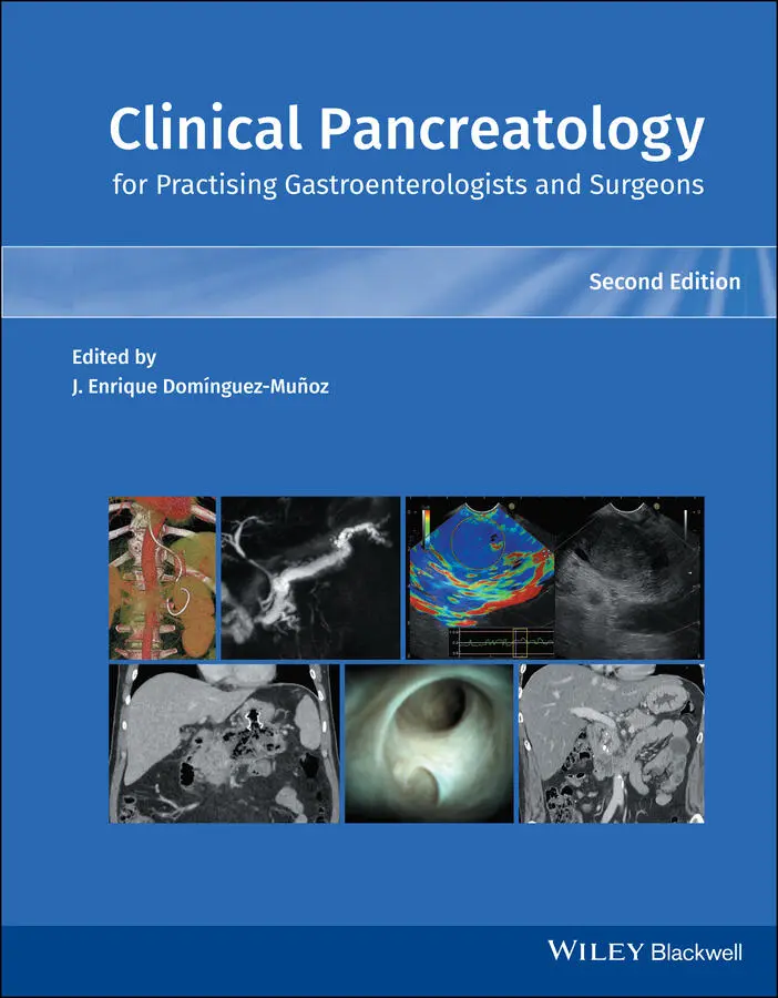
Critical appraisal of the patient’s history, physical examination findings, laboratory profile, and imaging abnormalities is a fundamental prerequisite for appropriate management of pancreatic pseudocysts and exclusion of their mimics. A multidisciplinary approach, enlisting the expertise of interventional endoscopists, interventional radiologists, and pancreatico‐biliary surgeons in the evaluation of this complex patient population is highly desirable. Because an interventional approach to management is not risk‐free and a significant proportion of pseudocysts can regress or even resolve spontaneously, only patients with sizeable (>6 cm diameter) pseudocysts causing significant symptoms or complications, such as intractable abdominal pain, nausea, vomiting, malnutrition, other evidence of gastric outlet obstruction, sepsis, gastrointestinal bleeding, biliary obstruction, compression of major peripancreatic vasculature and/or features of abdominal compartment syndrome, require drainage therapy [2–7] ( Figure 17.1). In contrast, it is reasonable to withhold or delay intervention in the setting of asymptomatic or minimally symptomatic pancreatic pseudocysts, following them up with serial cross‐sectional imaging instead as there is no immediate clinical benefit to be gained by the patient and the risk of intervention may outweigh the risk of developing a complication due to the pseudocyst. Patient comorbidity is particularly relevant in this context. In cases of diagnostic uncertainty as to whether a small pseudocyst is symptomatic, it is possible to simply needle‐aspirate the pseudocyst to dryness under endoscopic ultrasound (EUS) guidance and evaluate the subsequent clinical course. In general, the larger the pseudocyst, the greater the incidence of compressive symptoms, and the lower the probability of not only spontaneous resolution but, in our experience, also that of intervention‐related complications. Conversely, transluminal stent‐assisted drainage of collections smaller than 6 cm in diameter potentially increases the procedural risk of perforation and pseudocyst wall dehiscence and is therefore discouraged. Spontaneous resolution is more common in pseudocysts secondary to acute pancreatitis. Factors associated with a reduced likelihood of spontaneous resolution include communication with the main pancreatic duct, presence of multiple cysts, increase in size during follow‐up, and presence of a pancreatic duct stricture. Additionally, pseudocysts occurring in the setting of chronic pancreatitis with imaging evidence of calcification are unlikely to spontaneously resolve.
Table 17.1 Revised definitions of morphological features of acute pancreatitis.
| Interstitial edematous pancreatitisAcute inflammation of the pancreatic parenchyma and peripancreatic tissues, but without recognizable tissue necrosis:Pancreatic parenchyma enhancement by intravenous contrast agentNo findings of peripancreatic necrosis Necrotizing pancreatitisInflammation associated with pancreatic parenchymal necrosis and/or peripancreatic necrosis:Lack of pancreatic parenchymal enhancement by intravenous contrast agent and/orPresence of findings of peripancreatic necrosis Acute peripancreatic fluid collectionPeripancreatic fluid associated with interstitial edematous pancreatitis with no associated peripancreatic necrosis. This term applies only to areas of peripancreatic fluid seen within the first four weeks after onset of interstitial edematous pancreatitis and without the features of a pseudocyst:Occurs in the setting of interstitial edematous pancreatitisHomogeneous collection with fluid densityConfined by normal peripancreatic fascial planesNo definable wall encapsulating the collectionAdjacent to pancreas (no intrapancreatic extension) Pancreatic pseudocystAn encapsulated collection of fluid with a well‐defined inflammatory wall usually outside the pancreas with minimal or no necrosis. This entity usually occurs more than four weeks after onset of interstitial edematous pancreatitis to mature:Well circumscribed, usually round or ovalHomogeneous fluid densityNo nonliquid componentWell‐defined wall, i.e. completely encapsulatedMaturation usually requires more than four weeks after onset of acute pancreatitis; occurs after interstitial edematous pancreatitis Acute necrotic collectionA collection containing variable amounts of both fluid and necrosis associated with necrotizing pancreatitis; the necrosis can involve the pancreatic parenchyma and/or the peripancreatic tissues:Occurs only in the setting of acute necrotizing pancreatitisHeterogeneous and nonliquid density of varying degrees in different locations (some appear homogeneous early in their course)No definable wall encapsulating the collectionLocation: intrapancreatic and/or extrapancreatic Walled‐off necrosisA mature encapsulated collection of pancreatic and/or peripancreatic necrosis that has developed a well‐defined inflammatory wall. This usually occurs more than four weeks after onset of necrotizing pancreatitis:Heterogeneous with liquid and nonliquid density with varying degrees of loculations (some may appear homogeneous)Well‐defined wall, i.e. completely encapsulatedLocation: intrapancreatic and/or extrapancreaticMaturation usually requires four weeks after onset of acute necrotizing pancreatitis |

Figure 17.1 CT scan of 12‐cm collection causing a degree of gastric outflow obstruction, associated portal vein occlusion, and varices in stomach wall.
Читать дальшеИнтервал:
Закладка:
Похожие книги на «Clinical Pancreatology for Practising Gastroenterologists and Surgeons»
Представляем Вашему вниманию похожие книги на «Clinical Pancreatology for Practising Gastroenterologists and Surgeons» списком для выбора. Мы отобрали схожую по названию и смыслу литературу в надежде предоставить читателям больше вариантов отыскать новые, интересные, ещё непрочитанные произведения.


Обсуждение, отзывы о книге «Clinical Pancreatology for Practising Gastroenterologists and Surgeons» и просто собственные мнения читателей. Оставьте ваши комментарии, напишите, что Вы думаете о произведении, его смысле или главных героях. Укажите что конкретно понравилось, а что нет, и почему Вы так считаете.