Fernando Suarez - Periodontics
Здесь есть возможность читать онлайн «Fernando Suarez - Periodontics» — ознакомительный отрывок электронной книги совершенно бесплатно, а после прочтения отрывка купить полную версию. В некоторых случаях можно слушать аудио, скачать через торрент в формате fb2 и присутствует краткое содержание. Жанр: unrecognised, на английском языке. Описание произведения, (предисловие) а так же отзывы посетителей доступны на портале библиотеки ЛибКат.
- Название:Periodontics
- Автор:
- Жанр:
- Год:неизвестен
- ISBN:нет данных
- Рейтинг книги:5 / 5. Голосов: 1
-
Избранное:Добавить в избранное
- Отзывы:
-
Ваша оценка:
Periodontics: краткое содержание, описание и аннотация
Предлагаем к чтению аннотацию, описание, краткое содержание или предисловие (зависит от того, что написал сам автор книги «Periodontics»). Если вы не нашли необходимую информацию о книге — напишите в комментариях, мы постараемся отыскать её.
Periodontics — читать онлайн ознакомительный отрывок
Ниже представлен текст книги, разбитый по страницам. Система сохранения места последней прочитанной страницы, позволяет с удобством читать онлайн бесплатно книгу «Periodontics», без необходимости каждый раз заново искать на чём Вы остановились. Поставьте закладку, и сможете в любой момент перейти на страницу, на которой закончили чтение.
Интервал:
Закладка:
ENAMEL PEARLS
An enamel pearlis defined as a focal mass of enamel that has formed apical to the CEJ and is typically located in the area between the roots of molars. 1Historically, enamel pearls have also been referred to as droplets , nodules , globules , knots , and exostoses , based on their surface morphology. 46,56Interestingly, some authors referred to them as enamelomas as an early inaccurate delineation of an odontogenic tumor. 57,58These enamel anomalies are described as spheroid masses with a mean diameter of 0.96 mm and a prevalence of 4.6%. They are generally located at the coronal third of the root with a mean distance of 2.8 mm from the CEJ. 59Moskow and Canut performed a literature review and reported an incidence rate ranging from 1.1% to 9.7% (mean: 2.69%) and a predilection for maxillary third and second molars. 60Furthermore, Cavanha introduced a classification based on macroscopic and microscopic observations based on the number, location, and composition of the enamel pearls ( Table 5-3). 61
TABLE 5-3 Classification of enamel pearls 61
| Enamel pearls | Extradental | Solitary | Enamel nodules in periodontiumEnamel-cementum nodules in periodontium |
| Adherent | True enamel pearlEnamel-dentine pearlEnamel-dentine-pulp pearl | ||
| Intradental | CoronalCervicalRadicular |
The formation of extradental enamel pearls is attributed to activity of the Hertwig epithelial root sheath (HERS) that failed to detach from the dental surface after root formation. 62Interestingly, Slavkin et al developed a model to explore the HERS differentiation as well as root, cementum, and bone formation. 63They noted deposits that resembled enamel pearls during the differentiation, disintegration, and detachment of the HERS. Conversely, others speculated that enamel pearl formation occurs at the early stages of tooth development because they have been associated with dentin and pulp. 64In fact, this theory could explain the formation of intradental enamel pearls resulting from focal ameloblastic activity of invaginated cells or entrapped enamel epithelium from the cervical loop during root formation within newly forming dentin. 65Nevertheless, the process of formation of enamel pearls at some locations remains to be further elucidated. 60
Furcations
FURCATION MORPHOLOGY
The furcation morphology has been a subject of interest for multitude of investigations, including research evaluating the effectiveness of mechanical instrumentation. 66,67Bower noted that 81% of the furcations had a diameter less than 1 mm, while 58% were less than 0.75 mm in diameter. 66At the time, such findings were clinically relevant because the diameter of tested curettes (ie, Gracey, Columbia, and McCall) had a blade face width ranging between 0.75 and 1.1 mm, resulting in limitations for mechanical instrumentation. Similarly, Chiu et al reported that 49% of furcation entrance dimensions were equal to or less than 0.75 mm, which reinforced the need to use narrow instruments and ultrasonic devices with narrow tips for the management of furcation defects. 67
LOCATION OF FURCATION ENTRANCE
Gher and Dunlap performed a series of studies to determine variations in the root anatomy. 13,68,69One study reported that the mean distance from the CEJ to the furcation entrance for maxillary first molars was 3.6 mm, 4.2 mm, and 4.8 mm for mesial, buccal, and distal furcations, respectively 68(Fig 5-2). In a similar study using mandibular first molars, the mean distance was 4 mm for both buccal and lingual furcation entrances. 69Ultimately, the furcation anatomy of maxillary first premolars was explored, and authors reported a mean distance of 6 mm from the CEJ 70( Table 5-4). 68–75
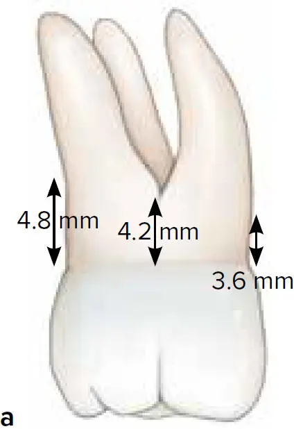
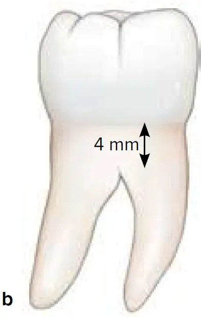
Fig 5-2 Location of furcation entrance of maxillary (a) and mandibular (b) first molars.
TABLE 5-4 Location of furcation entrance with respect to the CEJ
| Tooth type | Authors | Distance from CEJ to furcation entrance (mm) |
| Maxillary first molars | Gher and Dunlap 68 | Mesial: 3.6Buccal: 4.2Distal: 4.8 |
| Kerns et al 71 | Mesial: 4.73Buccal: 4.11Distal: 4.66 | |
| Maxillary second molars | Kerns et al 71 | Mesial: 6.40Buccal: 4.29Distal: 4.83 |
| Mandibular first molars | Dunlap and Gher 69 | Buccal/lingual: 4 |
| Mandelaris et al 72 | Buccal: 3.19Lingual: 4.08 | |
| Kerns et al 71 | Buccal: 3.27Lingual: 4.28 | |
| Wheeler 73 | Buccal: 3Lingual: 4 | |
| Mandibular second molars | Mandelaris et al 72 | Buccal: 3.09Lingual: 4.27 |
| Kerns 71 | Buccal: 3.28Lingual: 3.83 | |
| Maxillary first premolars | Gher and Vernino 70 | 6 |
| Booker and Loughlin 74 | 7.9 | |
| Joseph et al 75 | Range of 7.6–7.9 |
CLASSIFICATION OF FURCATION DEFECTS
Classification systems for furcations were developed to help determine the extension of the defect, tooth prognosis, and treatment approaches. These classifications were mostly developed with the use of a Nabers probe. Both horizontal and vertical components represent the primary endpoints of these classification systems. 1,76–94 Table 5-5summarizes some of the most commonly employed classifications. 1,76,83,84,87,90
TABLE 5-5 Classification systems for furcation defects
| Authors | Criteria |
| Glickman 76 | Pattern of destruction: Horizontal and vertical component.Grade I: Furcation area without gross or radiographic evidence of loss of alveolar bone loss ( incipient defect ).Grade II: Bone loss in one or more aspects of the furcation area, but a portion of the alveolar bone and periodontal membrane remains intact (also known as cul-de-sac lesion ).Grade III: Alveolar bone destruction permits the complete passage of a probe through the furcation. Entrance might be occluded by gingival tissues ( through-and-through defect ).Grade IV: Alveolar bone destruction creates an open area through which a probe can be passed without difficulty. The entrance is exposed and clearly visible to clinical examination. |
| Hamp et al, 83Lindhe and Nyman 87 | Pattern of destruction: Horizontal component.Degree I: Horizontal loss of periodontal support less than 3 mm.Degree II: Horizontal loss of periodontal support exceeding 3 mm, but not encompassing the total width of the furcation.Degree III: Horizontal “through-and-through” destruction of the periodontal tissue in the furcation. |
| Nyman and Lindhe 84 | Pattern of destruction: Horizontal component.Class I: Horizontal loss of periodontal support not exceeding one-third of the width of the tooth.Class II: Horizontal loss of periodontal support exceeding one-third of the width of the tooth, but not encompassing the total width of the furcation.Class III: Horizontal “through-and-through” destruction of the periodontal tissue in the furcation. |
| Tarnow and Fletcher 90 | Pattern of destruction: Vertical component.Subclass of Lindhe and Nyman 87classification.Subclass A: 0–3 mm probable depth from the roof of the furcation.Subclass B: 4–6 mm probable depth from the roof of the furcation.Subclass C: 7 mm or greater probable depth from the roof of the furcation. |
| American Academy of Periodontology Glossary of Periodontal Terms 1 | Pattern of destruction: Horizontal component.Class I: Incipient loss of bone limited to the furcation flute that does not extend horizontally.Class II: A variable degree of bone loss in a furcation, but not extending completely through the furcation.Class III: Bone loss extending completely through the furcation. |
FURCATION ARROW
Интервал:
Закладка:
Похожие книги на «Periodontics»
Представляем Вашему вниманию похожие книги на «Periodontics» списком для выбора. Мы отобрали схожую по названию и смыслу литературу в надежде предоставить читателям больше вариантов отыскать новые, интересные, ещё непрочитанные произведения.


Обсуждение, отзывы о книге «Periodontics» и просто собственные мнения читателей. Оставьте ваши комментарии, напишите, что Вы думаете о произведении, его смысле или главных героях. Укажите что конкретно понравилось, а что нет, и почему Вы так считаете.