Fernando Suarez - Periodontics
Здесь есть возможность читать онлайн «Fernando Suarez - Periodontics» — ознакомительный отрывок электронной книги совершенно бесплатно, а после прочтения отрывка купить полную версию. В некоторых случаях можно слушать аудио, скачать через торрент в формате fb2 и присутствует краткое содержание. Жанр: unrecognised, на английском языке. Описание произведения, (предисловие) а так же отзывы посетителей доступны на портале библиотеки ЛибКат.
- Название:Periodontics
- Автор:
- Жанр:
- Год:неизвестен
- ISBN:нет данных
- Рейтинг книги:5 / 5. Голосов: 1
-
Избранное:Добавить в избранное
- Отзывы:
-
Ваша оценка:
Periodontics: краткое содержание, описание и аннотация
Предлагаем к чтению аннотацию, описание, краткое содержание или предисловие (зависит от того, что написал сам автор книги «Periodontics»). Если вы не нашли необходимую информацию о книге — напишите в комментариях, мы постараемся отыскать её.
Periodontics — читать онлайн ознакомительный отрывок
Ниже представлен текст книги, разбитый по страницам. Система сохранения места последней прочитанной страницы, позволяет с удобством читать онлайн бесплатно книгу «Periodontics», без необходимости каждый раз заново искать на чём Вы остановились. Поставьте закладку, и сможете в любой момент перейти на страницу, на которой закончили чтение.
Интервал:
Закладка:
Biofilm and Calculus
Biofilmformation is a progressive and dynamic process that facilitates bacterial attachment to tooth structures through van der Waals forces, glycocalyx, and lectin-like receptors within a salivary pellicle and negative surface charges mediated by teichoic acid of gram-positive bacteria. 14–16The shift from gram-positive to gram-negative bacteria occurs as subgingival plaquedevelops and is influenced by biofilm thickness and gingival crevicular fluid. The process of mineralization of biofilm into dental calculushas been suggested by Genco et al 16to undergo four theoretical processes: booster mechanism, epitaxic concept, inhibition theory, and transformation theory.
The inorganic and organic composition of dental calculus have been extensively explored. 17–28Major inorganic components of dental calculus are calcium, phosphorus, carbonate, sodium, magnesium, potassium, and trace elements such as fluoride and zinc. 16Moreover, four major crystalline forms can be noted within mature calculus, including hydroxyapatite, octacalcium phosphate (OCP), whitlockite (WHT), and brushite. 18,20,22On the other hand, proteins (50%–60%), carbohydrates (12%–20%), and lipids (10%–15%) represent about 15% to 20% of the dry weight of mature supragingival plaque. 29Major differences in the characteristics of dental calculus exist depending on the location. Table 5-1summarizes the characteristics of both supragingival and subgingival calculus. 30
TABLE 5-1 Characteristics of supragingival and subgingival calculus *
| Supragingival | Subgingival | |
| Location | Coronal to gingival margin | Apical to gingival margin |
| Color | Yellow/white | Brown/black |
| Distribution | Adjacent to salivary duct openings | Randomly around the oral cavity |
| Composition | Low concentration of Ca, Mg, F, Sr, and ZnHigh concentration of carbonate and Mn | High concentration of Ca, Mg, and FLower concentration of carbonateMore irregular distribution of F |
| Mineral content and source | Average of 37% from saliva by volume | Average of 58% from gingival crevicular fluid by volume |
| Crystal type | Mostly OCP and hydroxyapatite | Mostly WHT |
| Formation | Heterogenous nucleation and crystal growthMore variable calcification | Heterogenous nucleation and crystal growthMore homogenous calcification |
| Microorganisms | More filamentous, faster growing | Less filamentous, slower growing |
| Morphology | Heterogenous with small needle-shaped (100 nm), large-ribbon-like and bundle/rosettes (1–50 nm) crystals | Several crystal types (< 50 nm): spiny, crusty, nodular, ledge/ring, individual islands, smooth veneers and finger/fern-like |
| Pathogenic potential | Little evidence | Associated with periodontal disease |
*Adapted and modified from Roberts-Harry and Clerehugh. 30
Ca, calcium; Mg, magnesium; F, fluoride; Sr, strontium; Zn, zinc; Mn, manganese; Na, sodium.
MECHANISM OF CALCULUS ATTACHMENT
In 1953, Zander explored the mechanisms of calculus attachment upon 50 freshly extracted teeth. 31Four main types of attachment were identified: (1) secondary cuticle, (2) direct attachment into irregularities of cementum, (3) penetration into cementum, and (4) mechanical retention in areas of resorption. In addition, various forms of combinations were also described ( Box 5-1). 31Types II (20%) and III (10%) were noted as the most frequent modalities of calculus attachment, and the cementoenamel junction (CEJ) was the favored site for calculus formation. 31
BOX 5-1 Mode of calculus attachment to cementum 31
| Type IType IIType IIIType IVType VType VIType VIIType VIIIType IXType X | Secondary cuticleDirect attachment into irregularities of cementumPenetration into cementumMechanical retention in areas of resorptionCombination of Types III and IVCombination of Types II, III, and IVCombination of Types I, II, III, and IVCombination of Types I and IICombination of Types II and IIICombination of Types II and IV |
Following Zander’s findings, multiple authors questioned specific types of calculus attachment and attempted to employ more sophisticated technology to test his conclusions. 19,32–40Notably, Canis et al rejected the possibility that microorganisms can penetrate the cementum surface and considered this phenomenon as an artifact due to superimposition of a detached cementum onto the tooth structure during sample preparation. 41These findings were confirmed using light microscopy, scanning electron microscopy (SEM), and transmission electron microscopy (TEM).
Developmental Deformities
ENAMEL PROJECTIONS
Defined as an apical extension of enamel usually toward a furcation, 1 enamel projections(also known as cervical enamel projections [CEPs]) are common anatomical variations where a definitive projection of enamel extends into the furcation area, preventing true attachment of periodontal ligament (PDL) fibers upon the root surface 7(Fig 5-1).
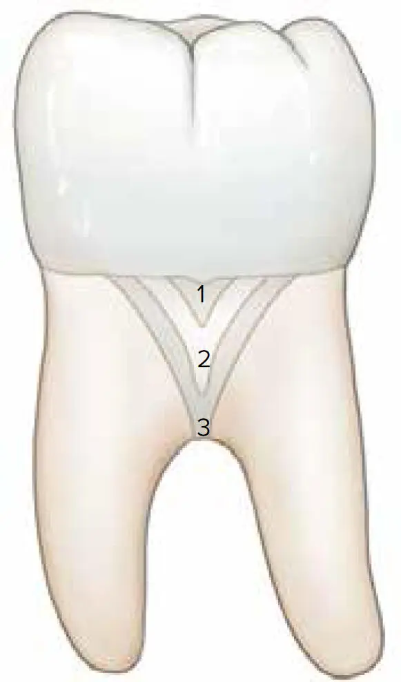
Fig 5-1 Classification of cervical enamel projections.
Early observations in dental anatomy described how the enamel dips into the furcation area of multirooted teeth in a tongue-like fashion. 42–48A landmark article by Masters and Hoskins examined extracted teeth with CEPs and suggested a grading system to determine the severity of these projections ( Box 5-2). 7The authors reported the prevalence of CEPs in mandibular and maxillary molars as 28.6% and 17%, respectively. In addition, clinical observations revealed that 90% of isolated furcation involvementswere associated with CEPs and mainly affected buccal furcation entrances. 7
BOX 5-2 Grading system for cervical enamel projections by Masters and Hoskins 7
| Grade IGrade IIGrade III | A distinct change in CEJ attitude with enamel projecting toward the bifurcation.Enamel projection approaching the furcation but not actually making contact with it.Enamel projection extending into the furcation proper. |
A myriad of studies continued exploring the prevalence, incidence, and association between CEPs and furcation involvement ( Table 5-2). 7,49–55Variations within these investigations might arise from differences in tooth type, reason for tooth extraction (eg, caries, severe periodontitis, endodontic failure), and patient population. With the exception of Leib et al, 50most studies confirmed a positive correlation between CEPs and furcation defects. Notably, Hou and Tsai reported a high prevalence (63.2%) of furcation defects associated with CEPs and intermediate bifurcational ridges (IBRs) affecting primarily mandibular molars. 55
TABLE 5-2 Prevalence of enamel projections
| Authors | Material and methods | Main findings |
| Masters and Hoskins 7 | Extracted teethPopulation not specified | Prevalence:– Mandibular molars: 28.6%– Maxillary molars: 17%90% of isolated furcation involvements were associated with CEP |
| Grewe et al 49 | Extracted teethPopulation not specified | Prevalence:– Mandibular: 25.2%– Maxillary: 15.8%Frequency (in order):– Mandibular second molars– Maxillary second molars– Mandibular first molars– Mandibular third molars– Maxillary third molars– Maxillary first molars |
| Leib et al 50 | Extracted teethPopulation not specified | Prevalence:– Mandibular molars: 25.4%– Maxillary molars: 21.9%Not associated with furcation defects. |
| Bissada and Abdelmalek 51 | Egyptian skulls | Incidence:– Overall: 8.6%Frequency (in order):– Mandibular second molars– Maxillary second molars– Mandibular first molars– Mandibular third molars– Maxillary third molars– Maxillary first molars |
| Tsatsas et al 52 | Extracted teethPopulation not specified | Prevalence:– Overall: 29.9% |
| Swan and Hurt 53 | East Indian skulls | Prevalence:– Overall: 32.6%– Mandibular molars: 33.7%– Maxillary molars: 31.4%Frequency (in order):– Mandibular second molars– Maxillary second molars– Maxillary third molars– Mandibular first molars– Mandibular third molars– Maxillary first molars |
| Hou and Tsai 54 | Surgical accessPopulation: Taiwanese | Prevalence:– Overall: 45.2%Frequency (in order):– Mandibular first molars– Maxillary first molars– Mandibular second molars– Maxillary second molars |
| Hou and Tsai 55 | Hopeless teeth with Class III FIPopulation: Taiwanese | Prevalence:– FI with CEPs and IBR: 63.2%– FI with CEPs alone: 21.8%– FI with IBR alone: 2.3% |
FI, furcation involvement; IBR, intermediate bifurcational ridge.
Читать дальшеИнтервал:
Закладка:
Похожие книги на «Periodontics»
Представляем Вашему вниманию похожие книги на «Periodontics» списком для выбора. Мы отобрали схожую по названию и смыслу литературу в надежде предоставить читателям больше вариантов отыскать новые, интересные, ещё непрочитанные произведения.


Обсуждение, отзывы о книге «Periodontics» и просто собственные мнения читателей. Оставьте ваши комментарии, напишите, что Вы думаете о произведении, его смысле или главных героях. Укажите что конкретно понравилось, а что нет, и почему Вы так считаете.