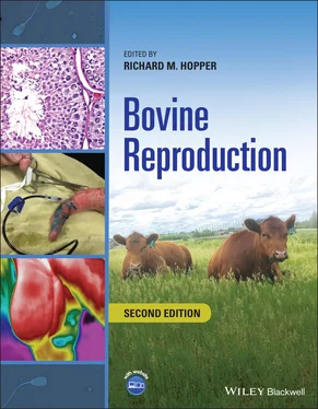
Bovine Reproduction
Здесь есть возможность читать онлайн «Bovine Reproduction» — ознакомительный отрывок электронной книги совершенно бесплатно, а после прочтения отрывка купить полную версию. В некоторых случаях можно слушать аудио, скачать через торрент в формате fb2 и присутствует краткое содержание. Жанр: unrecognised, на английском языке. Описание произведения, (предисловие) а так же отзывы посетителей доступны на портале библиотеки ЛибКат.
- Название:Bovine Reproduction
- Автор:
- Жанр:
- Год:неизвестен
- ISBN:нет данных
- Рейтинг книги:4 / 5. Голосов: 1
-
Избранное:Добавить в избранное
- Отзывы:
-
Ваша оценка:
- 80
- 1
- 2
- 3
- 4
- 5
Bovine Reproduction: краткое содержание, описание и аннотация
Предлагаем к чтению аннотацию, описание, краткое содержание или предисловие (зависит от того, что написал сам автор книги «Bovine Reproduction»). Если вы не нашли необходимую информацию о книге — напишите в комментариях, мы постараемся отыскать её.
A complete resource for practical, authoritative information on all aspects of bovine theriogenology Bovine Reproduction
Bovine Reproduction
Bovine Reproduction
Bovine Reproduction — читать онлайн ознакомительный отрывок
Ниже представлен текст книги, разбитый по страницам. Система сохранения места последней прочитанной страницы, позволяет с удобством читать онлайн бесплатно книгу «Bovine Reproduction», без необходимости каждый раз заново искать на чём Вы остановились. Поставьте закладку, и сможете в любой момент перейти на страницу, на которой закончили чтение.
Интервал:
Закладка:
16 Chapter 17 Figure 17.1 Inverted L. Ventral branches of labeled T13 and L1 to L4 are des... Figure 17.2 Proximal paravertebral. Figure 17.3 Distal paravertebral. Figure 17.4 (a) Needle placement for caudal epidural. Source: Image courtesy... Figure 17.5 Dorsal view (a) and lateral view (b) of S3, S4, and S5 foramina ... Figure 17.6 Sacral paravertebral. Figure 17.7 Injection of 2 ml local anesthetic in the skin at the ischiorect... Figure 17.8 Needle placement for pudendal nerve block. Figure 17.9 Pudendal nerve block.
17 Chapter 18 Figure 18.1 Ultrasound of normal testis in transverse plane. Mediastinum tes... Figure 18.2 Sagittal ultrasound of normal bovine testicle. Mediastinum testi... Figure 18.3 Normal thermographic pattern of bull scrotum. Figure 18.4 Thermograph of scrotum of bull with unilateral pathology. Note d... Figure 18.5 Ultrasound of scrotum of a bull with a hydrocele. Note anechoic ... Figure 18.6 Bull with acute bilateral scrotal enlargement secondary to traum... Figure 18.7 Inguinal hernia in a bull. Note characteristic swelling confined... Figure 18.8 Generalized edema of scrotal wall due to Mycoplasma weyenoii . Figure 18.9 Elliptical skin incision on lateral aspect of scrotum. Figure 18.10 Testicle within parietal vaginal tunic bluntly dissected from s... Figure 18.11 Parietal vaginal tunic incised and testicle exposed. Figure 18.12 Schematic of double ligation and transection of spermatic cord ... Figure 18.13 Transected stump of vaginal tunic following inverting suture cl... Figure 18.14 Schematic of inverting closure of vaginal tunic. Figure 18.15 Ligation of spermatic cord for a closed castration. Figure 18.16 Scrotal skin incision closed with continuous interlocking patte... Figure 18.17 Postoperative bandaging of scrotum to minimize swelling. Figure 18.18 Resection and anastomosis of herniated bowel removed from ingui... Figure 18.19 Incision over inguinal ring. Figure 18.20 Herniated bowel exposed after incising parietal vaginal tunic.... Figure 18.21 Exposure of inguinal ring. MB, Medial border of inguinal ring; ... Figure 18.22 Caudal retraction of internal abdominal oblique (IAO) muscle. M... Figure 18.23 Sutures preplaced through medial and lateral borders of inguina... Figure 18.24 Completed closure of inguinal ring.
18 Chapter 19 Figure 19.1 Penile fibropapilloma identified during routine BBSE. Figure 19.2 Proper placement of towel clamp under dorsal apical ligament to ... Figure 19.3 Subcutaneous injection of local anesthetic over dorsum of penis.... Figure 19.4 Catheterization of urethra. Figure 19.5 Penile hair ring. Figure 19.6 Urethral fistula that is of a size and location to require repai... Figure 19.7 Persistent frenulum preventing complete straightening of erect p... Figure 19.8 Persistent frenulum with two branches attached from prepuce to p... Figure 19.9 Very short persistent frenulum preventing separation of penis an... Figure 19.10 Transfixation ligatures on each end of persistent frenulum. Figure 19.11 Excision of frenulum adjacent to transfixation ligatures. Figure 19.12 Completed excision of frenulum. Figure 19.13 Severe preputial stenosis due to frostbite. Figure 19.14 Paraphimosis secondary to preputial laceration. Figure 19.15 Phimosis secondary to preputial laceration. Figure 19.16 Preputial prolapse on presentation. Note “elephant trunk” appea... Figure 19.17 Moderately severe preputial prolapse with tear on ventral aspec... Figure 19.18 Fresh preputial prolapse with edema and minimal necrosis. Figure 19.19 Preputial prolapse with severe laceration and minimal edema. Figure 19.20 Preputial prolapse with laceration and severe edema. Figure 19.21 Soaking injured prepuce. Figure 19.22 Orthopedic stockinette applied over prolapsed prepuce. Figure 19.23 Latex tube inserted into preputial lumen for urine drainage. Figure 19.24 Prepuce bandage with Elasticon and with tube held in place. Figure 19.25 Support sling fashioned from burlap material and bungee cords.... Figure 19.26 Reusable bull diaper canvass construction with eyelets for atta... Figure 19.27 PVC pipe prepared for use in “ring technique.” Figure 19.28 Ring sutured in place, inside prepuce. Figure 19.29 Penis extended in preparation for circumcision. Note loose exce... Figure 19.30 Determination of length of free portion of penis. Figure 19.31 Proximal end of free portion indicated by surgeon’s left forefi... Figure 19.32 Length of excess prepuce measured from end of sheath (surgeon's... Figure 19.33 (a) Circumferential and longitudinal skin incisions in preputia... Figure 19.34 Dissection and removal of preputial skin and scar tissue. Figure 19.35 Subcutaneous closure with continuous pattern. Note this closure... Figure 19.36 Preputial epithelium suture closure. Figure 19.37 Preputial epithelium closed with staples. Figure 19.38 Penrose drain sutured over free portion of penis for urine drai... Figure 19.39 Prepuce bandaged with Penrose drain within rigid tube. Figure 19.40 Preputial scar. This scar did not restrict extension of penis, ... Figure 19.41a Scar dissected in transverse plane. Figure 19.41b Tissue stretched and incised area takes on oval appearance. Cl... Figure 19.42 Extensive fibrosis. Penis is observed following manual extensio... Figure 19.43 Scar dissected. Figure 19.44 Closure with bootlace pattern in longitudinal orientation. Figure 19.45 Schematic of closing incision with bootlace suture pattern when... Figure 19.46 Triangular flap of skin removed from sheath (apex of triangle o... Figure 19.47 Prepuce elevated through incision in sheath. Figure 19.48 Incision into preputial cavity to create stoma. Figure 19.49 Free portion of penis exteriorized through preputial stoma. Figure 19.50 Preputial epithelium apposed to skin of sheath with interrupted... Figure 19.51 Penrose drain sutured over free portion of penis. Figure 19.52 Penrose drain over penis through preputial stoma for urine drai... Figure 19.53 Postoperative image of bull with preputial stoma. (Note Penrose... Figure 19.54 Completely healed preputial stoma ready for attempted semen col... Figure 19.55 Hematoma location. Figure 19.56 Rent in tunica albuginea. Figure 19.57 Spiral deviation. Figure 19.58 Ventral deviation. Figure 19.59 Harvest of fascia. Figure 19.60 Prepared fascia graft. Figure 19.61 Placement of fascia graft. Figure 19.62 Suturing graft to tunic. Figure 19.63 Closure of apical ligament. Figure 19.64 Closure of penile and preputial epithelium.
19 Chapter 20 Figure 20.1 Urethral calculi visible on preputial hairs. Figure 20.2 Subcutaneous peripenile swelling typically extends from the base... Figure 20.3 Severe hydronephrosis in a mature bull. Figure 20.4 Placement of a Foley catheter in the tube cystotomy procedure.... Figure 20.5 Placement of a Foley catheter in the flank tube cystotomy proced... Figure 20.6 Utilization of stay sutures to assist with bladder manipulation ... Figure 20.7 Placement of a plastic or rubber (source‐ automobile inner tube)... Figure 20.8 Perineal urethrostomy showing spatulation of the urethra and use...
20 Chapter 21 Figure 21.1 Procedure for vasectomy. Figure 21.2 Procedure for epididymectomy. Figure 21.3 Circumferential incision 4 cm from the preputial orifice is perf... Figure 21.4 Ventral midline incision extending caudally with circumferential... Figure 21.5 Use of a cold sterilized PVC pipe to facilitate tunneling of pen... Figure 21.6 Closure of new preputial orifice with interrupted sutures and ve... Figure 21.7 Exteriorization of the penis through the incision and identifica... Figure 21.8 Preplacement of sutures through the dorsal third of the penis an... Figure 21.9 Securing the stay sutures for penopexy. Figure 21.10 Site for incision for ventral fistula. Figure 21.11 (a and b) Suturing of preputial mucosa to the sheath skin. Figure 21.12 Excision of 5 mm of the preputial epithelium and sheath skin ju...
21 Chapter 22 Figure 22.1 (a) Ov, ovary, Uh, uterine horn, F, follicle, 22.1 (b) Corpus lu... Figure 22.2 The internal reproductive tract from oblique angle. V, Vagina; C... Figure 22.3 The internal reproductive tract from dorsal view. V, Vagina; C, ... Figure 22.4 Structures of the ovary. Ov, Ovary; Inf, infundibulum; Ut, uteri... Figure 22.5 Dorsal view of reproductive tract. U, Uterus body; Ov, ovary; Uh... Figure 22.6 Uterine horns positioned to better view the intercornual ligamen... Figure 22.7 The uterine lumen. Pm, Perimetrium; Mm, myometrium; Em, endometr... Figure 22.8 Vaginal and cervical lumen. V, Vagina; C, cervix; U, body uterus... Figure 22.9 Close‐up of vaginal and cervical lumen allowing visualization of... Figure 22.10 External view of perineum. A, Anus; Vs, vestibule; Uo, urethral... Figure 22.11 Arterial blood supply to the reproductive tract. Aa, Abdominal ...
Читать дальшеИнтервал:
Закладка:
Похожие книги на «Bovine Reproduction»
Представляем Вашему вниманию похожие книги на «Bovine Reproduction» списком для выбора. Мы отобрали схожую по названию и смыслу литературу в надежде предоставить читателям больше вариантов отыскать новые, интересные, ещё непрочитанные произведения.
Обсуждение, отзывы о книге «Bovine Reproduction» и просто собственные мнения читателей. Оставьте ваши комментарии, напишите, что Вы думаете о произведении, его смысле или главных героях. Укажите что конкретно понравилось, а что нет, и почему Вы так считаете.


