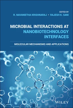
Microbial Interactions at Nanobiotechnology Interfaces
Здесь есть возможность читать онлайн «Microbial Interactions at Nanobiotechnology Interfaces» — ознакомительный отрывок электронной книги совершенно бесплатно, а после прочтения отрывка купить полную версию. В некоторых случаях можно слушать аудио, скачать через торрент в формате fb2 и присутствует краткое содержание. Жанр: unrecognised, на английском языке. Описание произведения, (предисловие) а так же отзывы посетителей доступны на портале библиотеки ЛибКат.
- Название:Microbial Interactions at Nanobiotechnology Interfaces
- Автор:
- Жанр:
- Год:неизвестен
- ISBN:нет данных
- Рейтинг книги:4 / 5. Голосов: 1
-
Избранное:Добавить в избранное
- Отзывы:
-
Ваша оценка:
- 80
- 1
- 2
- 3
- 4
- 5
Microbial Interactions at Nanobiotechnology Interfaces: краткое содержание, описание и аннотация
Предлагаем к чтению аннотацию, описание, краткое содержание или предисловие (зависит от того, что написал сам автор книги «Microbial Interactions at Nanobiotechnology Interfaces»). Если вы не нашли необходимую информацию о книге — напишите в комментариях, мы постараемся отыскать её.
Microbial Interactions at Nanobiotechnology Interfaces — читать онлайн ознакомительный отрывок
Ниже представлен текст книги, разбитый по страницам. Система сохранения места последней прочитанной страницы, позволяет с удобством читать онлайн бесплатно книгу «Microbial Interactions at Nanobiotechnology Interfaces», без необходимости каждый раз заново искать на чём Вы остановились. Поставьте закладку, и сможете в любой момент перейти на страницу, на которой закончили чтение.
Интервал:
Закладка:
Table of Contents
1 Cover
2 Title Page
3 Copyright Page
4 Preface
5 List of Contributors
6 1 Shape‐ and Size‐Dependent Antibacterial Activity of Nanomaterials Objectives 1.1 Introduction 1.2 Synthesis of Nanomaterials 1.3 Classification of NMs 1.4 Application of NMs 1.5 Bacterial Resistance to Antibiotics 1.6 Microbial Resistance: Role of NMs 1.7 Antibacterial Application of NMs 1.8 Interaction of NMs with Bacteria 1.9 Antibacterial Mechanism of NMs 1.10 Factors Affecting the Antibacterial Activity of NMs 1.11 Influence of Size on the Antibacterial Activity and Mechanism of Action of Nanomaterials 1.12 Influence of Shape on the Antibacterial Activity and Mechanism of Action of Nanomaterials 1.13 Effects of Functionalization on the Antimicrobial Property of Nanomaterials 1.14 Conclusion and Future Perspectives Questions and Answers References Keywords
7 2 Size‐ and Shape‐Selective Synthesis of DNA‐Based Nanomaterials and Their Application in Surface‐Enhanced Raman Scattering Objectives 2.1 Introduction 2.2 Mechanism of Surface‐Enhanced Raman Scattering (SERS) 2.3 Size‐ and Shape‐Selective Synthesis of Metal NPs with DNA for SERS Studies Questions and Answers Acknowledgements References Academic Profile
8 3 Surface Modification Strategies to Control the Nanomaterial–Microbe Interplay Objectives 3.1 Introduction 3.2 Factors Influencing NM–Microbe Cross talk 3.3 Surface Functionalization 3.4 Characterization of NM–Microbe Interactions 3.5 Toxicity of the Surface‐Modified NMs 3.6 Challenges and Future Perspectives Questions and Answers References
9 4 Surface Functionalization of Nanoparticles for Stability in Biological Systems Objectives 4.1 Introduction 4.2 Major Processes Affecting NP Stability in Biological Media 4.3 Measures to Enhance NP Stability in Biological Systems 4.4 Conclusion and Future Perspectives 4.5 Summary Questions and Answers References
10 5 Molecular Mechanisms Behind Nano‐Cancer Therapeutics Objectives 5.1 Nanotechnology at Nano–Bio Interfaces 5.2 Armory of Nanomedicine at Nano–Bio Interfaces 5.3 Nanoparticle Edge in Modulating Biological Process 5.4 Intracellular Uptake and Trafficking of Nanoparticle 5.5 Challenges in Clinical Applications 5.6 Conclusion Questions and Answers Acknowledgements References
11 6 Protein Nanoparticle Interactions and Factors Influencing These Interactions Objectives 6.1 Introduction 6.2 Types and Biomedical Application of Nanoparticles 6.3 Methods and Mechanisms of Nanomaterials Synthesis 6.4 Routes of Entry of Nanoparticles into Biological System 6.5 Rationale for Studying Nanoparticles–Protein Interactions 6.6 Formation of Protein Corona 6.7 Nanoparticles‐Induced Structural Changes in Proteins 6.8 Factors Influencing Corona Formation 6.9 Interaction of Nanoparticles with Cells and Their Uptake 6.10 Pleiotrophic Effect of Nanoparticles 6.11 Analytical Methods to Study Nanoparticles–Protein Interaction Questions and Answers References
12 7 Interaction Effects of Nanoparticles with Microorganisms Employed in the Remediation of Nitrogen‐Rich Wastewater Objectives 7.1 Introduction 7.2 Bacterial Nitrification Process 7.3 Effect of NPs on Denitrifying Bacteria 7.4 Impact of Nanoparticles on Nitrogen Removal 7.5 Conclusion Take Home Message Questions and Answers Acknowledgements References
13 8 Silver‐Based Nanoparticles for Antibacterial Activity Objectives 8.1 Introduction 8.2 Historical Background of Silver 8.3 Synthesis Procedures of Silver Nanoparticles 8.4 Biological Application of Silver Nanoparticles 8.5 Bacterial Infection and Antibiotic Resistance 8.6 Nanosilver for Antibacterial Therapy 8.7 Influence of Size and Shape of Silver Nanoparticles as Antibacterial Agents 8.8 Nanosilver and Its Mechanism of Action for Antibacterial Therapy 8.9 Application of Silver Nanoparticle in Commercial Products 8.10 Toxicity of Silver Nanoparticles 8.11 Future Prospective and Challenges 8.12 Conclusion Take Home Message Questions and Answers Acknowledgments References
14 9 Microbial Gold Nanoparticles and Their Biomedical Applications Objectives 9.1 Introduction 9.2 Microbial Gold Nanoparticles Synthesis 9.3 Applications of Microbial Gold Nanoparticles 9.4 Conclusion Acknowledgements Take Home Message Questions and Answers References
15 10 Nano‐Bio Interactions and Their Practical Implications in Agriculture 10.1 Introduction 10.2 Engineered Nanomaterials and Agriculture 10.3 Summary References
16 11 Biogeochemical Interactions of Bioreduced Uranium Nanoparticles 11.1 Introduction 11.2 Coupled Biogeochemical Mechanisms and Interactions of U in the Subsurface 11.3 Biogenic Uraninite Precipitation and Its Nanoparticulate Forms 11.4 Re‐oxidation and Stability of Bioreduced Uranium 11.5 Summary and Conclusions Questions and Answers References
17 12 Characterization and Quantification of Mobile Bioreduced Uranium Phases 12.1 Introduction 12.2 Characterization of Biogenic U(IV) 12.3 Quantification of Mobile Bioreduced U(IV) Nanoparticles 12.4 Summary and Conclusions Questions and Answers References
18 Index
19 End User License Agreement
List of Tables
1 Chapter 1 Table 1.1 Various approaches for NM synthesis (Ealias & Saravanakumar, 2017... Table 1.2 Bandgap energy and activation wavelength for various metal oxide ...
2 Chapter 2 Table 2.1 Comparison of metal–DNA assemblies and their corresponding SERS a...
3 Chapter 7Table 7.1 The effect of various NPs to the expression of nitrifying and den...
4 Chapter 8Table 8.1 Various biological application of silver nanoparticles.
5 Chapter 9Table 9.1 Synthesis of gold nanoparticles by bacteria.Table 9.2 Synthesis of gold nanoparticles by algae.Table 9.3 Synthesis of gold nanoparticles by fungi.
List of Illustrations
1 Chapter 1 Figure 1.1 Schematics of typical methods for synthesis of NMs. Figure 1.2 Schematic representation of photoactivated ROS generation and ant... Figure 1.3 Bacterial cell wall structure of (a) Gram‐positive bacteria (b) G... Figure 1.4 Schematic of the antibacterial mechanism of NMs. Figure 1.5 Schematic of the factors influencing the antimicrobial activity o... Figure 1.6 Schematic of the key factors that contribute to the antimicrobial...
2 Chapter 2 Figure 2.1 Schematization of DNA with Ag for the formation of Ag–DNA. Lower ... Figure 2.2 (a–d) TEM images of Ag–DNA taken at different magnifications show... Figure 2.3 (a) Model taken for calculating induced magnetic field. (b) SPR b... Figure 2.4 (a) EM enhancement profile for N = 2. (b) N = 14. (c) 2D array us... Figure 2.5 (a) Upper panel shows the overall depiction of Ag–DNA for SERS an... Figure 2.6 FT‐IR spectral characteristics of Au–DNA nanowires. Figure 2.7 Upper panel shows the TEM images of Au–DNA and the lower panel sh... Figure 2.8 Electronic spectral features of DNA aided Rh NPs. Figure 2.9 Schematic illustration of small‐, medium‐, and large‐sized Rh NPs... Figure 2.10 Schematic illustration of Pd NPs over DNA, CTAB and PVA scaffold... Figure 2.11 Upper panel shows TEM images of Pd–DNA nanowires and lower panel... Figure 2.12 (a–d) HR‐TEM images of Pt–DNA with chain‐like structure. (e, f) ... Figure 2.13 Representation of 532 nm laser with Pt–DNA and MB probe for SERS... Figure 2.14 Schematization of Rh–DNA shown on the left‐hand side. The visual... Figure 2.15 Upper panel shows the morphology of Rh–DNA from TEM. Lower panel... Figure 2.16 Schematization of Re–DNA by a chemical reduction method. Figure 2.17 (a) Upper panel shows the morphologies of Re–DNA and (b) lower p... Figure 2.18 Schematization of Re–DNA organosol by a chemical reduction metho... Figure 2.19 SERS application of Re–DNA organosol by a chemical reduction met... Figure 2.20 Schematic of Os–DNA organosol for SERS application. Figure 2.21 Upper panel shows the morphologies of Os–DNA organosol and lower... Figure 2.22 Upper panel shows the SPR band of Os–DNA prepared by microwave c... Figure 2.23 Upper panel shows wire‐like morphologies of Os–DNA and the lower...
Читать дальшеИнтервал:
Закладка:
Похожие книги на «Microbial Interactions at Nanobiotechnology Interfaces»
Представляем Вашему вниманию похожие книги на «Microbial Interactions at Nanobiotechnology Interfaces» списком для выбора. Мы отобрали схожую по названию и смыслу литературу в надежде предоставить читателям больше вариантов отыскать новые, интересные, ещё непрочитанные произведения.
Обсуждение, отзывы о книге «Microbial Interactions at Nanobiotechnology Interfaces» и просто собственные мнения читателей. Оставьте ваши комментарии, напишите, что Вы думаете о произведении, его смысле или главных героях. Укажите что конкретно понравилось, а что нет, и почему Вы так считаете.
