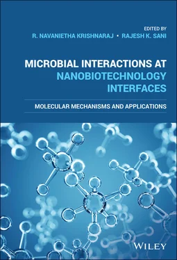
Microbial Interactions at Nanobiotechnology Interfaces
Здесь есть возможность читать онлайн «Microbial Interactions at Nanobiotechnology Interfaces» — ознакомительный отрывок электронной книги совершенно бесплатно, а после прочтения отрывка купить полную версию. В некоторых случаях можно слушать аудио, скачать через торрент в формате fb2 и присутствует краткое содержание. Жанр: unrecognised, на английском языке. Описание произведения, (предисловие) а так же отзывы посетителей доступны на портале библиотеки ЛибКат.
- Название:Microbial Interactions at Nanobiotechnology Interfaces
- Автор:
- Жанр:
- Год:неизвестен
- ISBN:нет данных
- Рейтинг книги:4 / 5. Голосов: 1
-
Избранное:Добавить в избранное
- Отзывы:
-
Ваша оценка:
- 80
- 1
- 2
- 3
- 4
- 5
Microbial Interactions at Nanobiotechnology Interfaces: краткое содержание, описание и аннотация
Предлагаем к чтению аннотацию, описание, краткое содержание или предисловие (зависит от того, что написал сам автор книги «Microbial Interactions at Nanobiotechnology Interfaces»). Если вы не нашли необходимую информацию о книге — напишите в комментариях, мы постараемся отыскать её.
Microbial Interactions at Nanobiotechnology Interfaces — читать онлайн ознакомительный отрывок
Ниже представлен текст книги, разбитый по страницам. Система сохранения места последней прочитанной страницы, позволяет с удобством читать онлайн бесплатно книгу «Microbial Interactions at Nanobiotechnology Interfaces», без необходимости каждый раз заново искать на чём Вы остановились. Поставьте закладку, и сможете в любой момент перейти на страницу, на которой закончили чтение.
Интервал:
Закладка:
W. Aadinath Department of Biotechnology Bhupat and Jyoti Mehta School of Biosciences Indian Institute of Technology Madras Chennai India
Srishti Agarwal Bio‐Nano Electronics Research Center Graduate School of Interdisciplinary New Science Toyo University Kawagoe Saitama Japan
R. Akhil Department of Biotechnology Bhupat and Jyoti Mehta School of Biosciences Indian Institute of Technology Madras Chennai India
Papia Basuthakur Department of Applied Biology CSIR‐Indian Institute of Chemical Technology Hyderabad Telangana State India Academy of Scientific and Innovative Research (AcSIR) Ghaziabad Uttar Pradesh India
Achintya N. Bezbaruah Department of Civil and Environmental Engineering North Dakota State University Fargo ND USA
Parmita Chawley Department of Environmental Science and Engineering Indian Institute of Technology (Indian School of Mines) Dhanbad Jharkhand India
Ann‐Marie Fortuna United States Department of Agriculture Grazingland Research Laboratory Agricultural Research Service El Reno OK USA
Shagufta Haque Department of Applied Biology CSIR‐Indian Institute of Chemical Technology Hyderabad Telangana State India Academy of Scientific and Innovative Research (AcSIR) Ghaziabad Uttar Pradesh India
Sheeja Jagadevan Department of Environmental Science and Engineering Indian Institute of Technology (Indian School of Mines) Dhanbad Jharkhand India
K. Karthick Electrochemical Process Engineering (EPE) Division CSIR‐Central Electrochemical Research Institute (CECRI) Karaikudi Tamil Nadu India
Senthilguru Kulanthaivel Department of Biochemical Engineering and Biotechnology Indian Institute of Technology Delhi New Delhi India
Subrata Kundu Electrochemical Process Engineering (EPE) Division CSIR‐Central Electrochemical Research Institute (CECRI) Karaikudi Tamil Nadu India
Uday Suryakant Maharana School of Biotechnology KIIT University Bhubaneswar Odisha India
R. Mala Department of Biotechnology Mepco Schlenk Engineering College Sivakasi Tamil Nadu India
Dindyal Mandal School of Biotechnology KIIT University Bhubaneswar Odisha India
Prashant Mishra Department of Biochemical Engineering and Biotechnology Indian Institute of Technology Delhi New Delhi India
Sourav Mishra School of Biotechnology KIIT University Bhubaneswar Odisha India
Vignesh Muthuvijayan Department of Biotechnology Bhupat and Jyoti Mehta School of Biosciences Indian Institute of Technology Madras Chennai India
Bijayananda Panigrahi School of Biotechnology KIIT University Bhubaneswar Odisha India
Chitta Ranjan Patra Department of Applied Biology CSIR‐Indian Institute of Chemical Technology Hyderabad Telangana State India Academy of Scientific and Innovative Research (AcSIR) Ghaziabad Uttar Pradesh India
Aravind Kumar Rengan Department of Biomedical Engineering Indian Institute of Technology Kandi, Sangareddy Hyderabad India
Arpita Roy Department of Applied Biology CSIR‐Indian Institute of Chemical Technology Hyderabad Telangana State India Academy of Scientific and Innovative Research (AcSIR) Ghaziabad Uttar Pradesh India
D. Sakthi Kumar Bio‐Nano Electronics Research Center Graduate School of Interdisciplinary New Science Toyo University Kawagoe Saitama Japan
Rajesh K. Sani Department of Chemical and Biological Engineering South Dakota School of Mines and Technology Rapid City SD USA
S. Sevinç Şengör Bobby B. Lyle School of Engineering Southern Methodist University Dallas TXUSADepartment of Environmental Engineering Middle East Technical University Ankara, Turkey
Rohit Kumar Singh School of Biotechnology KIIT University Bhubaneswar Odisha India
Surya Prakash Singh Department of Biomedical Engineering Indian Institute of Technology Kandi, Sangareddy Hyderabad India Academy of Scientific and Innovative Research (AcSIR) Ghaziabad Uttar Pradesh India
T. K. Vasudha Department of Biotechnology Bhupat and Jyoti Mehta School of Biosciences Indian Institute of Technology Madras Chennai India
1 Shape‐ and Size‐Dependent Antibacterial Activity of Nanomaterials
Senthilguru Kulanthaivel and Prashant Mishra
Department of Biochemical Engineering and Biotechnology, Indian Institute of Technology Delhi, New Delhi, India
Objectives
To understand the importance of studying nanomaterials‐bio interface and the factors that dictate it.
To understand the current situation of antibiotic resistance of microbes and their effect on worldwide healthcare.
To know the strategies to overcome the antibiotic resistance mechanism and role of nanomaterials in combatting it.
To understand the mechanism of action of antimicrobial nanomaterials and the factors that influence their antimicrobial properties.
To understand the key role of size and shape of the nanomaterials on the antimicrobial properties of nanomaterials.
1.1 Introduction
Over the past three decades “Nanotechnology” has emerged as a promising strategy to overcome impasses that have accumulated in various fields of science and technology (Albanese, Tang, & Chan, 2012). Nanomaterials (NMs) are defined as minuscule structures having at least one of their dimensions equal to or between 1 and 100 nm. Since there is no single universally accepted definition, so far various organizations have given their own definition to the term “NMs” (Boverhof et al., 2015). US Food and Drug Administration (USFDA) defines nanomaterial as “materials that have at least one dimension in the range of approximately 1–100 nm and exhibit dimension dependent phenomena” (Bleeker et al., 2012). As per Environmental Protection Agency (EPA) definition, “NMs can exhibit unique properties dissimilar than the equivalent chemical compound in a larger dimension.” Similarly, the International Organization for Standardization (ISO) defines it as “material with any external nanoscale dimension or having internal nanoscale surface structure.” EU Commission has described it as “a manufactured or natural material that possesses unbound, aggregated or agglomerated particles where external dimensions are between 1–100 nm size range” (Jeevanandam et al., 2018).
NMs generally exist in the shape of spheres, cubes, rods, tubes, flowers, and platelets (Machado et al., 2015). NMs in the nanoscale dimensions possess a high surface area‐to‐volume ratio and also a high number of atoms/molecules present on the surface rather than the interior of the materials. These are the properties that majorly contribute to the unique functionality of the nanoscale materials, which vary from the bulk of the same material. Therefore, modulation in their structural properties such as change of size or shape will significantly affect their optical, electrical, magnetic, and biological activity (He et al., 2010; Machado et al., 2015). This is one of the distinct advantages of nanotechnology where by engineering the design or production parameters we can modulate the functionality of the NMs specific to particular application (Machado et al., 2015). Hence, the recent studies in nanotechnology are majorly focused on understanding the effect of physicochemical properties such as size, shape, and surface chemistry of the material on the optical, electrical, magnetic, and biological activities.
Considering the aforementioned advantages, NMs have found enormous applications in various fields and products such as cosmetics, catalysts, fillers, biomedical devices, and semiconductors. As per data report from 2015, approximately more than 1800 products from 622 companies in 32 countries contain engineered NMs. In summary, 762 (i.e. around 42%) of the total products are used in the health and fitness category where silver is the most predominantly used NM, in almost 435 products, which are around 24% of the total. Further, about 528 products (i.e. 29% of total) contain NMs as liquid suspension where dermal contact is highly possible. Hence, the abundant application of these materials is leading towards a long‐term co‐existence of such NMs with living systems which may result in adverse toxicological effects to the living bodies. In this context, it is necessary to study the effect of these materials on biological entities such as proteins, DNA, RNA, cell membranes, cell organelles, cells, tissues, and organs. The interactions between the biological system and NMs strongly depend upon the environment and the biophysico‐chemical property of the nano‐bio interface (Nel et al., 2009). As discussed, size, shape, and surface chemistry are the most important factors that govern the physicochemical properties of the NM that in turn throws light at the nano‐bio interface. Understanding the effect of these physicochemical factors and extrapolating them toward the interactions at the nano‐bio interface would help us to design or engineer NMs for specific applications with an added advantage of minimal toxicity to living bodies. There are a number of analytical tools to study the interaction of nano‐biomolecules/proteins. Among them the most employed are mass spectroscopy, Fourier transform infrared spectroscopy, circular dichroism spectroscopy, Raman spectroscopy, nuclear magnetic resonance, UV–vis spectroscopy, surface plasma resonance, quartz crystal balance, atomic force microscopy, fluorescence correlation spectroscopy, fluorescence spectroscopy, and isothermal calorimetry (Saptarshi, Duschl, & Lopata, 2013). Among the different strategies, mass spectroscopy‐based proteomics is the most preferred. Even though the technique is a qualitative measure of proteins bound to NMs, such as nanoparticles (NPs), it can be applied over a wide range of NMs. UV–vis spectroscopy is employed to measure changes observed in the adsorption spectrum caused by NM–protein interaction. Similar to UV–vis spectroscopy, fluorescence spectroscopy is employed to measure changes in the fluorescence spectra caused by the binding of protein on to NMs. Surface plasma resonance is used to study changes in electrons' oscillation on the surface of metal NMs as a result of protein interaction with NM. Isothermal calorimetry analysis is employed to determine the binding constant and other thermodynamic parameters of the nano/bio interface (Saptarshi et al., 2013). Quartz crystal balance is used to measure changes in mass on the surface of oscillating quartz caused by the NM–protein interaction. In a study, adsorption of proteins myoglobin, bovine serum albumin, and cytochrome over the surface of gold NPs was studied using quartz crystal balance (Kaufman et al., 2007). Confocal Raman spectroscopy and confocal spectroscopy can be employed to study and visualize NM–protein interaction and intake of NMs into cells by fluorescent labeling of NPs. In recent times, a combination of these techniques has been strategically employed to study the different aspects of NM–protein/biomolecule interaction. NMs can be synthesized through different routes such as chemical, physical, and green methods. Changes in the synthesis methods, concentration of reactants, and conditions can definitely modulate the morphological parameters (size and shape) of NMs. Taking this into account, the selection of synthesis route also plays an important role in governing NMs' morphological features and their functions. Keeping the aforementioned perspectives in mind, this chapter describes the effects of size and shape of NMs on their biological activity.
Читать дальшеИнтервал:
Закладка:
Похожие книги на «Microbial Interactions at Nanobiotechnology Interfaces»
Представляем Вашему вниманию похожие книги на «Microbial Interactions at Nanobiotechnology Interfaces» списком для выбора. Мы отобрали схожую по названию и смыслу литературу в надежде предоставить читателям больше вариантов отыскать новые, интересные, ещё непрочитанные произведения.
Обсуждение, отзывы о книге «Microbial Interactions at Nanobiotechnology Interfaces» и просто собственные мнения читателей. Оставьте ваши комментарии, напишите, что Вы думаете о произведении, его смысле или главных героях. Укажите что конкретно понравилось, а что нет, и почему Вы так считаете.


