Microbial Interactions at Nanobiotechnology Interfaces
Здесь есть возможность читать онлайн «Microbial Interactions at Nanobiotechnology Interfaces» — ознакомительный отрывок электронной книги совершенно бесплатно, а после прочтения отрывка купить полную версию. В некоторых случаях можно слушать аудио, скачать через торрент в формате fb2 и присутствует краткое содержание. Жанр: unrecognised, на английском языке. Описание произведения, (предисловие) а так же отзывы посетителей доступны на портале библиотеки ЛибКат.
- Название:Microbial Interactions at Nanobiotechnology Interfaces
- Автор:
- Жанр:
- Год:неизвестен
- ISBN:нет данных
- Рейтинг книги:4 / 5. Голосов: 1
-
Избранное:Добавить в избранное
- Отзывы:
-
Ваша оценка:
Microbial Interactions at Nanobiotechnology Interfaces: краткое содержание, описание и аннотация
Предлагаем к чтению аннотацию, описание, краткое содержание или предисловие (зависит от того, что написал сам автор книги «Microbial Interactions at Nanobiotechnology Interfaces»). Если вы не нашли необходимую информацию о книге — напишите в комментариях, мы постараемся отыскать её.
Microbial Interactions at Nanobiotechnology Interfaces — читать онлайн ознакомительный отрывок
Ниже представлен текст книги, разбитый по страницам. Система сохранения места последней прочитанной страницы, позволяет с удобством читать онлайн бесплатно книгу «Microbial Interactions at Nanobiotechnology Interfaces», без необходимости каждый раз заново искать на чём Вы остановились. Поставьте закладку, и сможете в любой момент перейти на страницу, на которой закончили чтение.
Интервал:
Закладка:
1.8 Interaction of NMs with Bacteria
Based on the cell wall structure, bacteria are divided into two categories as Gram‐positive bacteria and Gram‐negative bacteria. Gram‐positive bacteria have a thick peptidoglycan layer ranging from 15 to 100 nm. Further, they contain a phosphate‐containing polymeric chain of teichoic acid that is responsible for the negative charge of the bacteria (Neu, 1992). In the case of Gram‐negative bacteria, an extra hydrophobic lipid bilayer is present over a thin peptidoglycan layer (20–50 nm). The presence of an extra lipid layer limits the permeability of several hydrophilic antimicrobial agents, which is the reason for the high resistant nature of Gram‐negative bacteria (Gupta, Landis, & Rotello, 2016). The negative surface charge of bacteria is due to the lipids and carbohydrates of the lipopolysaccharide layer. Hence, the structure of the bacterial cell wall determines the interaction of bacteria with NMs. Schematics of cell wall structure of Gram‐positive and Gram‐negative bacteria are given in Figure 1.3as detailed by Hajipour et al. (2012)
In an earlier study, a homogenous distribution of cetyltrimethylammonium bromide (CTAB) coated gold NPs was observed over the Bacillus cereus . This was explained on the basis of electrostatic interaction between negatively charged teichoic acid moieties on the bacteria and positively charged gold NPs (Berry et al., 2005). In another study, it was reported that mannose functionalized gold NPs bound to the pili of Gram‐negative E. coli . It was attributed to the receptor–ligand interaction between mannose and lectin containing pili (Lin et al., 2002). Later, Hayden et al. (2012) suggested that the positively charged NMs exhibited high toxicity over bacteria. This electrostatic interaction might be a plausible reason for the spatialized aggregation of cationic and hydrophobic gold NPs on the negatively charged bacterial membrane (Hayden et al., 2012). The interaction of NPs with the membrane generally effects in membrane blebbing, tubule formation, and other membrane damage.
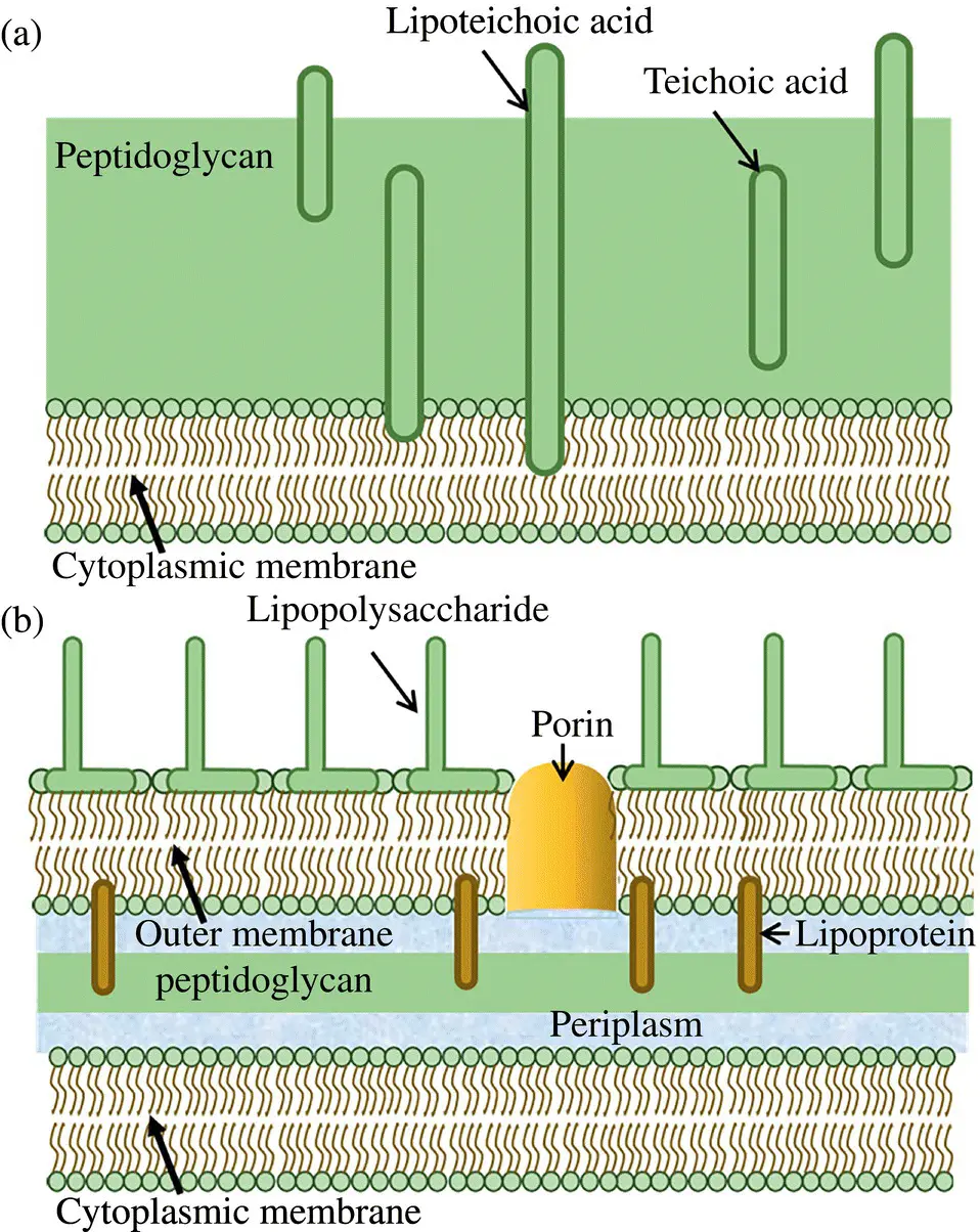
Figure 1.3 Bacterial cell wall structure of (a) Gram‐positive bacteria (b) Gram‐negative bacteria.
1.9 Antibacterial Mechanism of NMs
NMs exert different mechanisms of action against microbes as represented in Figure 1.4. Most of the time, the antimicrobial systems employ multiple mechanisms to take over the multiple resistance mechanisms developed by microorganisms, thereby increasing the antimicrobial efficiency. In brief, different mechanisms of action of NMs are given below:
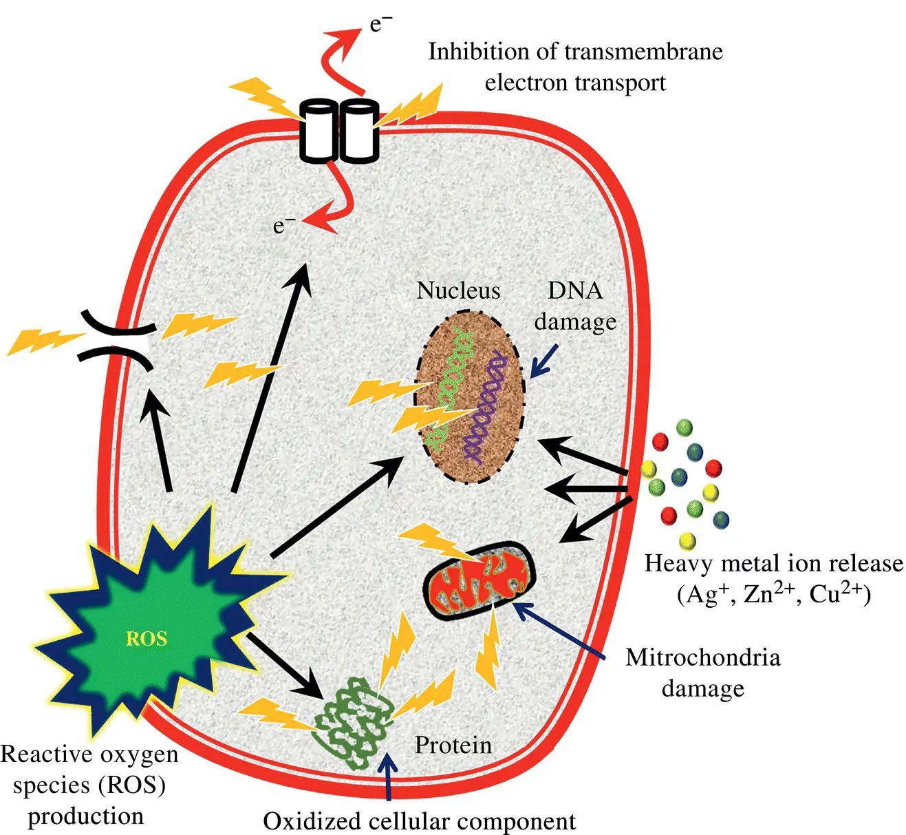
Figure 1.4 Schematic of the antibacterial mechanism of NMs.
Cell membrane disruption: Interaction of the NMs with the surface of the microbes causes mechanical damage to cell wall, which in turn leads to cell wall disruption followed by leakage of the cytoplasmic content and subsequent cell death (Pal, Tak, & Song, 2007).
DNA damage: NMs or metal ions from NMs diffuse through the cell wall and effectively interact with DNA and affect or modify the morphology of the DNA. This, in turn, interrupts the duplication or replication of DNA, leading to cell death (Feng et al., 2000).
Release of metal ions: Metal ions released from the NM diffuse into the cells and bind to thiol‐containing proteins including enzymes and compromise their function. In addition to that, metal ions generally function as cofactors for a number of enzymes. Hence homeostasis of metal ions is very important for the survival of the microorganism. Metal ions released from the NM diffuse in excess into bacterial cells, affecting the homeostasis leading to dysfunction of proteins and enzymes (Feng et al., 2000).
Interrupted transmembrane electron transport: Often the interaction of NM with cell wall damages the cell wall and hampers the electron transport chain leading to the disruption of cellular respiration and eventual cell death (Lemire, Harrison, & Turner, 2013).
Oxidative stress: Few of the NMs such as metals and metal oxides prominently induce production of ROS, which includes hydroxyl free radicals, superoxides, and hydrogen peroxide. The produced free radicals further oxidize cell wall components causing their disruption and act on the internal components of cells. In few cases, the oxidation continues till the entire cell is oxidized into CO2 and water. In some specific NMs such as carbon NMs, oxidative stress is induced without the involvement of ROS, which acts by electron transfer mechanism (Jacoby et al., 1998). Schematic of the antibacterial mechanism of NMs is given in the Figure 1.4as described by Hajipour et al. (2012).
1.10 Factors Affecting the Antibacterial Activity of NMs
As discussed earlier, the activity of any NM system depends on its physicochemical properties such as size, shape, zeta potential, crystal structure, charge, and other factors. The physicochemical factors' influence on the surface area, surface energy, and atomic ligand deficiency dictates the behavior of NMs, which in turn affects the activity of NMs. Hence it is very important to study the effect of these factors on the activity of the NMs. Schematic of the factors influencing the antimicrobial activity of the NMs is given the Figure 1.5as detailed by Daima and Bansal (2015).
1.10.1 Size
A key advantage of any NM system is its higher surface area‐to‐volume ratio in comparison to the micro and macro structures. Practically it is possible to contain a significantly high number of smaller NMs in comparison to bigger particles in the same volume. High surface area‐to‐volume ratio owing to an increased number of particles results in exposure of greater numbers of atoms, which could increase the activity. Considering the case of antibacterial system, the formation of biofilm is a key event in the development of resistant bacteria. Exposure of a greater number of atoms on the surface results in increased interaction of NM surface with bacteria, increasing the number of reactive oxygen species at a faster rate followed by inhibition or elimination of the bacteria. Supporting this fact, several recent studies have shown that the size of NMs plays a critical role in dictating the antimicrobial property of the NM.
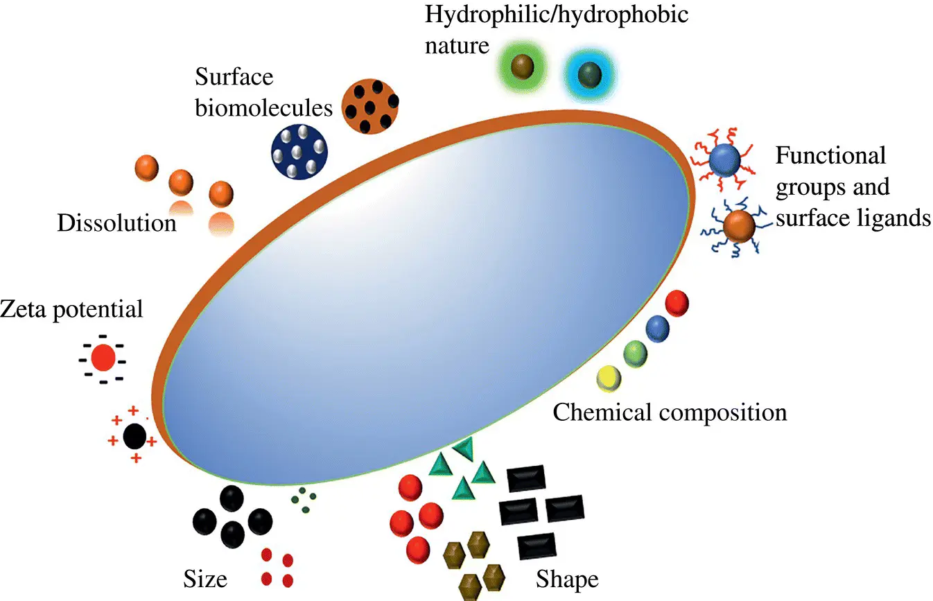
Figure 1.5 Schematic of the factors influencing the antimicrobial activity of the NMs.
Generally, the smaller the size of NM the higher will be the surface area with a high chance of prolonged interaction with microbial system and high diffusion through the cell membrane in comparison to bigger NMs with smaller surface area (Gurunathan et al., 2014). In the case of silver NPs, the size of the NPs clearly influences the surface area exposure, release rate of silver ions, and the antimicrobial efficacy of the particles. Similarly, ZnO NPs of smaller size (12 nm) had better antimicrobial activity in comparison to larger particles of size 45 nm due to its high cell permeability (Padmavathy & Vijayaraghavan, 2008). In another study involving the TiO 2and silica NPs, the antimicrobial property and the mechanism of action of the system were influenced by the size of titanium nanotubes (Çalışkan et al., 2014). In contrast, a study involving three different sizes of Mg(OH) 2, the smallest NPs had the least antibacterial effect (Pan et al., 2013). Thus, it is necessary to consider the effect of other factors also with size in determining the mechanism action of NMs.
Читать дальшеИнтервал:
Закладка:
Похожие книги на «Microbial Interactions at Nanobiotechnology Interfaces»
Представляем Вашему вниманию похожие книги на «Microbial Interactions at Nanobiotechnology Interfaces» списком для выбора. Мы отобрали схожую по названию и смыслу литературу в надежде предоставить читателям больше вариантов отыскать новые, интересные, ещё непрочитанные произведения.


Обсуждение, отзывы о книге «Microbial Interactions at Nanobiotechnology Interfaces» и просто собственные мнения читателей. Оставьте ваши комментарии, напишите, что Вы думаете о произведении, его смысле или главных героях. Укажите что конкретно понравилось, а что нет, и почему Вы так считаете.