Daniel Buser - 30 Years of Guided Bone Regeneration
Здесь есть возможность читать онлайн «Daniel Buser - 30 Years of Guided Bone Regeneration» — ознакомительный отрывок электронной книги совершенно бесплатно, а после прочтения отрывка купить полную версию. В некоторых случаях можно слушать аудио, скачать через торрент в формате fb2 и присутствует краткое содержание. Жанр: unrecognised, на английском языке. Описание произведения, (предисловие) а так же отзывы посетителей доступны на портале библиотеки ЛибКат.
- Название:30 Years of Guided Bone Regeneration
- Автор:
- Жанр:
- Год:неизвестен
- ISBN:нет данных
- Рейтинг книги:3 / 5. Голосов: 1
-
Избранное:Добавить в избранное
- Отзывы:
-
Ваша оценка:
30 Years of Guided Bone Regeneration: краткое содержание, описание и аннотация
Предлагаем к чтению аннотацию, описание, краткое содержание или предисловие (зависит от того, что написал сам автор книги «30 Years of Guided Bone Regeneration»). Если вы не нашли необходимую информацию о книге — напишите в комментариях, мы постараемся отыскать её.
30 Years of Guided Bone Regeneration — читать онлайн ознакомительный отрывок
Ниже представлен текст книги, разбитый по страницам. Система сохранения места последней прочитанной страницы, позволяет с удобством читать онлайн бесплатно книгу «30 Years of Guided Bone Regeneration», без необходимости каждый раз заново искать на чём Вы остановились. Поставьте закладку, и сможете в любой момент перейти на страницу, на которой закончили чтение.
Интервал:
Закладка:
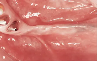
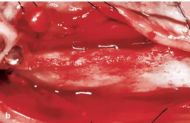
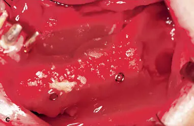
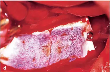
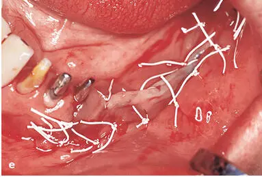
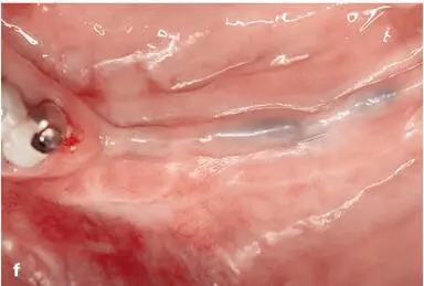
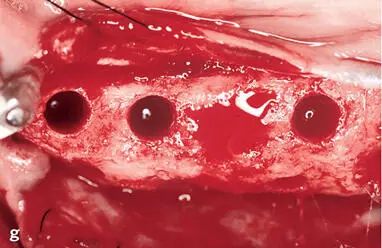
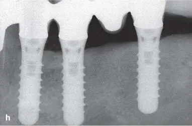
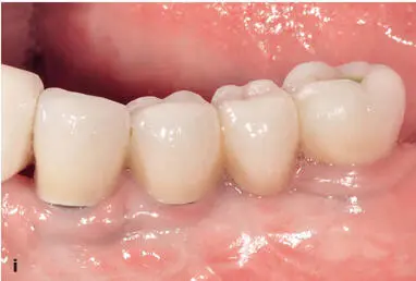
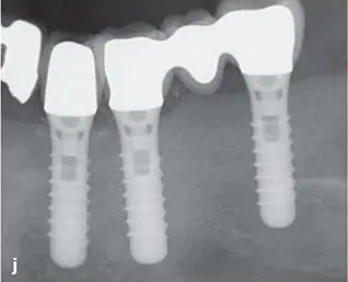
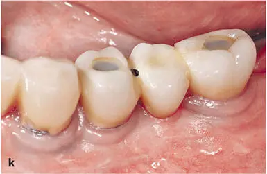
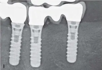
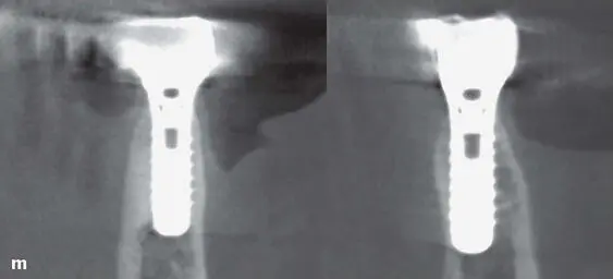
Fig 1-2Case 1. (a) Preoperative status (1991). Distal extension situation in the right maxilla of a man with a healed ridge. Two titanium implants were planned to allow a fixed prosthesis. (b) Both implants were placed, resulting in a crestal dehiscence defect at the mesial implant. The cortical bone surface was perforated with a small round bur to open the marrow cavity and stimulate bleeding in the defect area. (c) Locally harvested bone chips were applied to support the ePTFE membrane and to stimulate new bone formation in the defect area. (d) A bioinert ePTFE membrane was applied to function as a physical barrier. The punched membrane was stabilized around the necks of both implants. (e) Following incision of the periosteum, the surgery was completed with a tension-free primary wound closure. (f) Clinical status 4 months after implant surgery. The wound healing was uneventful. (g) Reopening after 4 months of healing. A second surgery was necessary to remove the nonresorbable membrane. (h) The clinical status following membrane removal showed successful bone regeneration in the defect area at both implants. (i) Longer healing caps were applied, and the soft tissue margins were adapted and secured in place with interrupted sutures. (j) Two weeks later, the soft tissues had healed, and both implants could be restored with a single crown. (k) The clinical status at the 15-year follow-up examination (2006) showed a satisfactory treatment outcome with stable peri-implant soft tissues. (l) Radiographic follow-up at 15 years: The bone crest levels were stable around both implants, which are splinted. (m) In 2010 (19 years after the initial surgery), an additional implant was placed in the canine site as late implant placement with a flapless approach. The clinical view during surgery showed stable peri-implant soft tissue at both implants in the premolar sites. (n) During perioperative examination of the canine implant site, a CBCT scan was taken. The orofacial cuts showed a thick facial bone wall for both premolar implants, which had been in function for 19 years at the time. (o) Clinical status after completion of the new single crown at the canine site. The treatment outcome was very satisfactory considering when the GBR procedure was done (1991). (p) Periapical radiograph after completion of therapy. The two tissue-level implants in the premolar sites had been in function for 19 years, and both showed stable peri-implant bone crest levels. This was the final follow-up examination, as the patient sadly developed dementia and passed away a few years later.
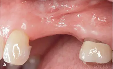
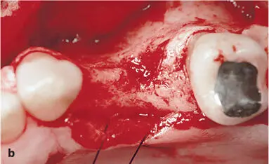
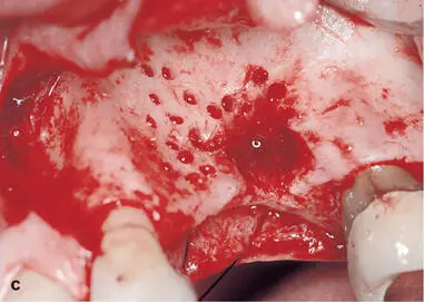
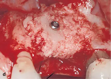
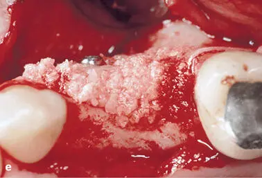
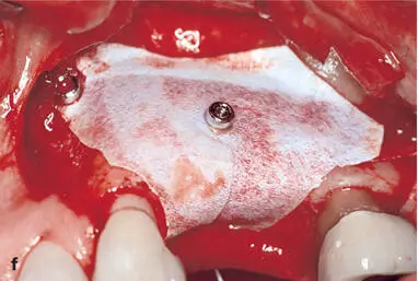
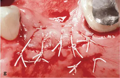
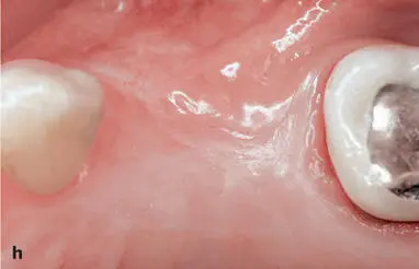
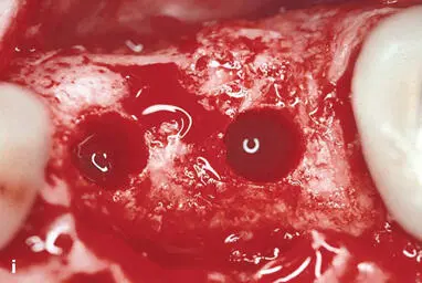
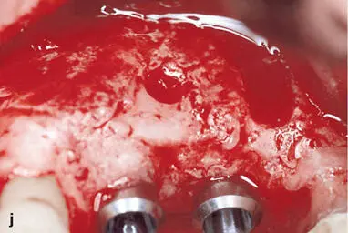
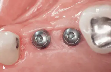
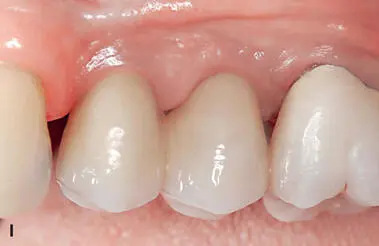
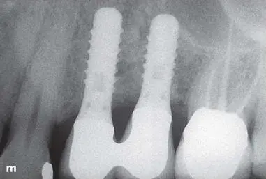
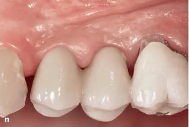
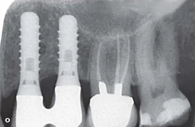
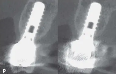
Fig 1-3Case 2. (a) Preoperative view (1994). The buccal view of this woman’s left maxilla shows two missing premolars. The buccal aspect is flattened. (b) The occlusal view during surgery shows a significant buccal flattening and a buccal bone defect in the area of the second premolar. (c) Prior to block application, the entire buccal bone surface was perforated to open the marrow cavity. The bone defect was debrided from scar tissues. (d) An autogenous block graft harvested from the chin was applied and fixed with a fixation screw. Bone chips were used to augment the entire surrounding area. (e) The occlusal view shows the volume of the augmented ridge. (f) Buccal view of the applied ePTFE membrane to cover the augmented ridge as a bioinert barrier membrane. (g) Primary wound closure was achieved with several mattress and interrupted single sutures using 4-0 and 5-0 ePTFE sutures. (h) Six months after ridge augmentation, the clinical status shows healthy soft tissues following a healing period free from complications. (i) Following flap elevation and membrane removal, the occlusal view demonstrates an excellent ridge volume and thick buccal bone wall following implant bed preparation. (j) The buccal view confirms successful ridge augmentation. The block graft can still be recognized, and it is covered in some areas with newly formed bone. (k) Clinical status following 3 months of nonsubmerged healing for both implants. The peri-implant mucosa was healthy and included a nice band of keratinized mucosa. (l) Clinical status at the 10-year examination (2005) shows the two splinted implant crowns. The peri-implant mucosa was stable with no signs of a peri-implant pathology. (m) The periapical radiograph at the 10-year examination confirms stable bone crest levels around the two tissue-level implants with a hybrid design. (n) The 25-year follow-up examination (2019) shows the clinical status with quite healthy peri-implant mucosa, although the plaque control is no longer perfect in this elderly patient (age 86). (o) The periapical radiograph confirms stable bone crest levels at both tissue-level implants. (p) The CBCT scan shows fully intact, thick buccal bone walls for the implants in the first premolar (left) and second premolar (right) sites.
Интервал:
Закладка:
Похожие книги на «30 Years of Guided Bone Regeneration»
Представляем Вашему вниманию похожие книги на «30 Years of Guided Bone Regeneration» списком для выбора. Мы отобрали схожую по названию и смыслу литературу в надежде предоставить читателям больше вариантов отыскать новые, интересные, ещё непрочитанные произведения.


Обсуждение, отзывы о книге «30 Years of Guided Bone Regeneration» и просто собственные мнения читателей. Оставьте ваши комментарии, напишите, что Вы думаете о произведении, его смысле или главных героях. Укажите что конкретно понравилось, а что нет, и почему Вы так считаете.