Adrian Becker - Orthodontic Treatment of Impacted Teeth
Здесь есть возможность читать онлайн «Adrian Becker - Orthodontic Treatment of Impacted Teeth» — ознакомительный отрывок электронной книги совершенно бесплатно, а после прочтения отрывка купить полную версию. В некоторых случаях можно слушать аудио, скачать через торрент в формате fb2 и присутствует краткое содержание. Жанр: unrecognised, на английском языке. Описание произведения, (предисловие) а так же отзывы посетителей доступны на портале библиотеки ЛибКат.
- Название:Orthodontic Treatment of Impacted Teeth
- Автор:
- Жанр:
- Год:неизвестен
- ISBN:нет данных
- Рейтинг книги:4 / 5. Голосов: 1
-
Избранное:Добавить в избранное
- Отзывы:
-
Ваша оценка:
Orthodontic Treatment of Impacted Teeth: краткое содержание, описание и аннотация
Предлагаем к чтению аннотацию, описание, краткое содержание или предисловие (зависит от того, что написал сам автор книги «Orthodontic Treatment of Impacted Teeth»). Если вы не нашли необходимую информацию о книге — напишите в комментариях, мы постараемся отыскать её.
Provides protocols for common cases as well as complex and rare presentations Contains individual chapters on the specific aspects of the diagnosis and treatment of impaction in each of the different types of teeth Covers prevalence, etiology, diagnosis, attitudes to treatment, treatment timing, treatment methods, and prognosis Features more than 1,000 high-quality color images and illustrations
remains essential reading for all specialist orthodontists, academic researchers and instructors, oral and maxillofacial surgeons, and advanced students in orthodontics.
Orthodontic Treatment of Impacted Teeth — читать онлайн ознакомительный отрывок
Ниже представлен текст книги, разбитый по страницам. Система сохранения места последней прочитанной страницы, позволяет с удобством читать онлайн бесплатно книгу «Orthodontic Treatment of Impacted Teeth», без необходимости каждый раз заново искать на чём Вы остановились. Поставьте закладку, и сможете в любой момент перейти на страницу, на которой закончили чтение.
Интервал:
Закладка:
There should be no problem identifying which of the two radiographs is the ortho‐radial view and which was taken from the distally deviated aspect, simply by comparing the relative distortion of the erupted teeth on the two radiographs. However, by radiographically ‘labelling’ the deviated receptor with the placement of a paper clip in one corner or by using a different receptor size for the deviated view, such as an occlusal‐sized receptor, this distinction will be simplified.
Let us assume that a right unerupted canine is palatally placed ( Figure 4.5), so that this tooth will be close to the middle of the picture obtained in both radiographs. In the first picture (direction B), where the tube is directed over the designated canine area of the ridge, the lateral incisor root will be on the right of the picture. If the canine is also mesially displaced, there will be some overlap of the canine crown and the lateral incisor root. On the second picture, taken from the front (direction A), the right lateral incisor root and the crown of the palatal canine will be in the middle of the picture, superimposed on one another, to a much greater degree.
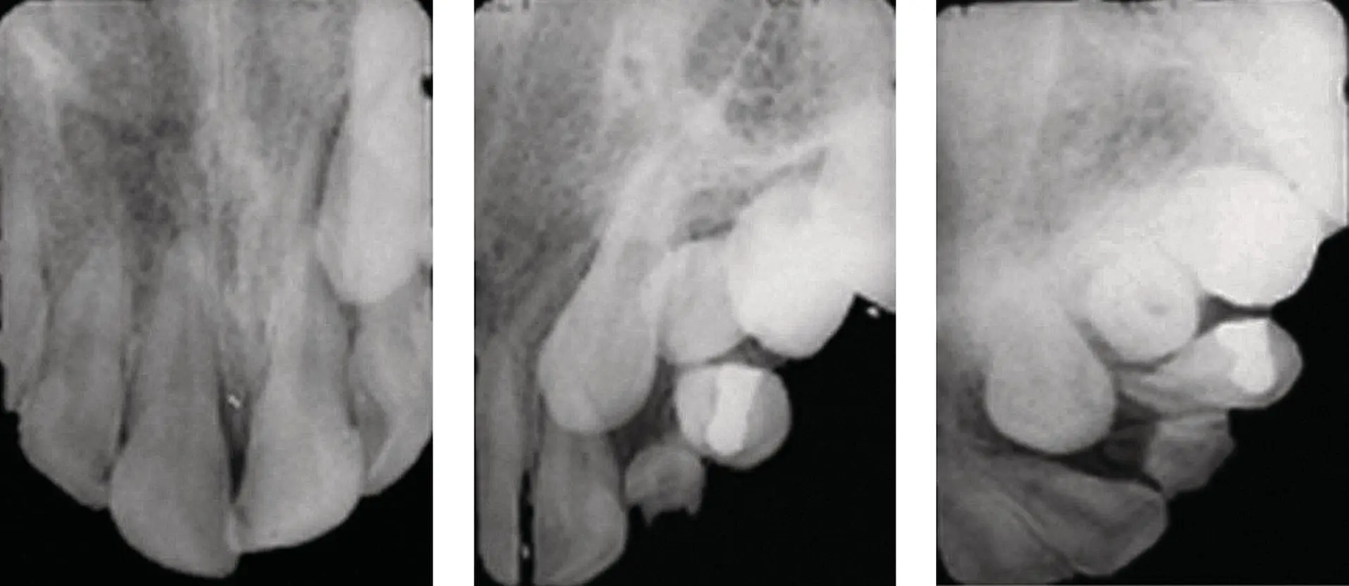
Fig. 4.4 The left periapical view, oriented for the central incisors, shows the crown of the canine superimposed on the distal half of the central incisor root. The middle radiograph, rotated 30° to the left, shows the canine overlapping only the lateral incisor root. By rotating the central beam a further 30°, superimposition of the canine over the lateral incisor root has been eliminated. The canine is palatally displaced.
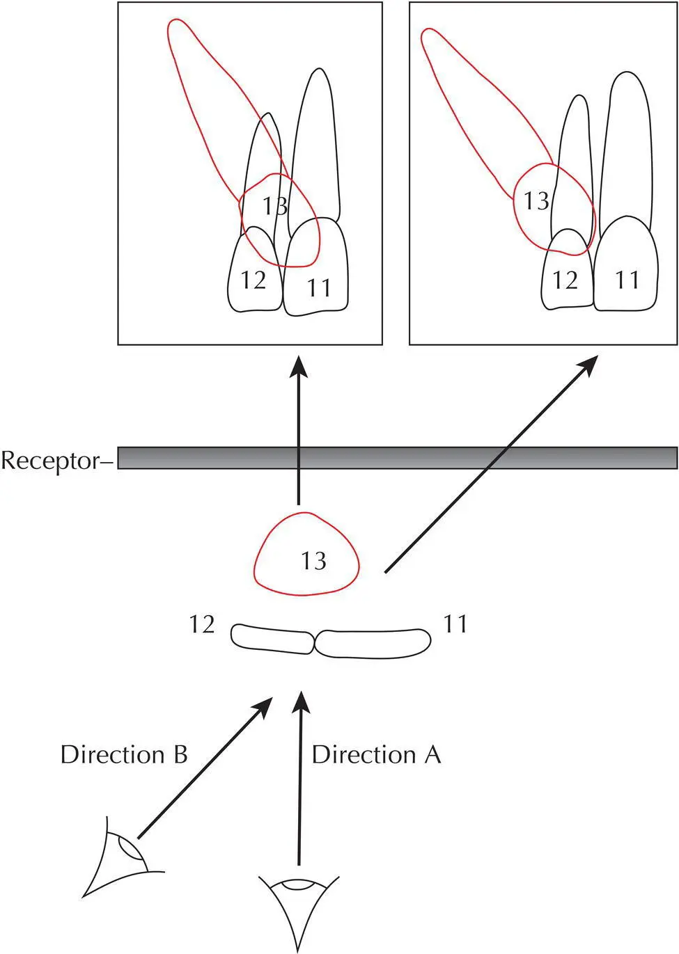
Fig. 4.5 A diagrammatic representation of the parallax method. If the observer’s eye peers along the axis of the X‐ray beam in each case, the image on the radiograph will be easy to reconstruct.
Source: Reproduced from previous edition. Adrian Becker, The Orthodontic Treatment of Impacted Teeth , 2nd ed., 2007 with the kind permission of Informa Healthcare – Books.
Jacobs [7, 8] enjoins the observer to demonstrate this principle by the simple act of using the right eye, purporting to be the X‐ray tube, and holding up two fingers vertically, at eye level, with one finger obscuring the other. If the observer now closes this eye and opens the other, without moving the head and fingers, the new vantage point for inspection will have resulted in a visual separation of the two fingers. Through the left eye, the obscured finger will have ‘moved’ to the left of the forward finger, to become partially visible. Transferring this to the radiographic context, in the second picture, the tooth furthest from the tube (i.e. the palatal tooth) will ‘move’ in the same direction that the X‐ray tube has travelled from the first exposure.
This parallax method has its limitations, although it is very useful in cases where there is a minimal height discrepancy between the erupted and unerupted adjacent teeth. However, when the canine is high and the periapical view shows no superimposition of the canine over the roots of the erupted teeth, or where the superimposition is only in the apical area, then the overall picture may be very misleading. Accordingly, a different method of localization should be used, as follows: the periapical view is directed from above the occlusal plane and in an oblique downward and medial direction, which distances the palatal canine from the roots of the other teeth and makes it appear higher than the anatomy of the maxilla would allow. While it may prove useful in locating the position of the crown of the impacted tooth, it is not adequate to the task of accurately placing the root apex and thereby defining the orientation of its long axis. These are important parameters when assessing treatment difficulty and prognosis during the treatment planning stage and are absolutely critical for the successful resolution of an impacted tooth, as we shall see in the following chapters.
A useful variant of the same technique is the vertical parallax, in which two radiographs are taken of the area, with the central ray of one periapical radiograph being more steeply angled in the vertical plane than the other. In this manner, the separation of the images in the more steeply angled (above the occlusal plane) radiograph will result in a palatal tooth being more superiorly related vis‐à‐vis the target tooth than in the regular radiograph.
Unfortunately, the parallax method in general offers a relatively low degree of reliability. In a study to evaluate the usefulness of its two variants [6], six experienced orthodontists were given the case records of 39 patients with ectopic canines. The cases were evaluated twice, once using radiographs that showed vertical parallax and once with radiographs that featured horizontal parallax, although the parallax pairs were not revealed to the examiners as being of the same individual. In 83% of cases the correct positional diagnosis was made with the horizontal method, while only 68% of cases were correctly diagnosed with the vertical method. These results expose the method as being too crude, or the experts insufficiently discerning, for it to be relied upon with any degree of confidence. Thus, while often useful to obtain an initial overall impression, the method should certainly be backed up by more reliable diagnostic radiographic methods before a final treatment plan is presented to the patient.
In the incisor region, an unerupted permanent incisor may be associated with one or two supernumerary teeth (mesiodentes). The parallax method is insufficiently clear in these cases due to the presence of two or three hard tissue entities in the bone, superimposed on the outline of the roots of the deciduous teeth and at varying heights in the alveolus.
The question arises as to whether the parallax principles may be applied to other types of receptor combinations, possibly with a greater degree of reliability. A vertical imaging discrepancy between teeth in the line of the arch and those that are buccally or palatally displaced can be created between the panoramic view and the periapical/anterior occlusal views ( Figure 4.6). The panoramic view is a rotational tomograph, with the cone of the machine pointing upwards with a very small 7° tilt from below the occlusal plane, as it circles around the head of the patient. Because this view is recorded when the receptor is on the buccal side of the teeth and the cone directed from the palatal aspect, this is equivalent to a 7° tilt of the X‐ray cone, when translated into a buccal‐to‐palatal direction.
By contrast, the direction of the central ray in an anterior occlusal view (60–65°) or periapical view (50–55°) is angulated much more steeply to the receptor. These will both show superimposition of an ectopic tooth over the tooth in the line of the arch, but it is the degree of vertical discrepancy between these radiographs and the panoramic view that will reveal the position of the displaced tooth.
The same panoramic view, with the X‐ray beam hitting this area when the cone is at the back of the patient’s head, will project the anterior midline area in its postero‐anterior aspect. The canine and premolar regions will be projected from an increasingly angulated viewpoint, as the X‐ray cone moves from the back to the side of the head. The molar and retromolar areas will be projected from the side, on the same revolving receptor, as a consequence of the further rotation of the X‐ray beam.
Читать дальшеИнтервал:
Закладка:
Похожие книги на «Orthodontic Treatment of Impacted Teeth»
Представляем Вашему вниманию похожие книги на «Orthodontic Treatment of Impacted Teeth» списком для выбора. Мы отобрали схожую по названию и смыслу литературу в надежде предоставить читателям больше вариантов отыскать новые, интересные, ещё непрочитанные произведения.


Обсуждение, отзывы о книге «Orthodontic Treatment of Impacted Teeth» и просто собственные мнения читателей. Оставьте ваши комментарии, напишите, что Вы думаете о произведении, его смысле или главных героях. Укажите что конкретно понравилось, а что нет, и почему Вы так считаете.