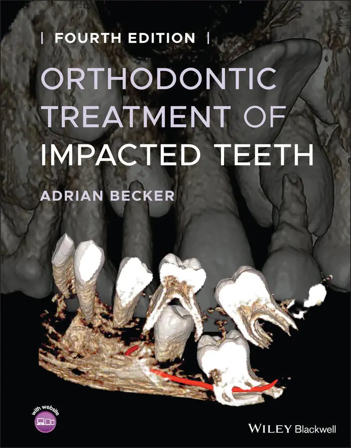
Adrian Becker - Orthodontic Treatment of Impacted Teeth
Здесь есть возможность читать онлайн «Adrian Becker - Orthodontic Treatment of Impacted Teeth» — ознакомительный отрывок электронной книги совершенно бесплатно, а после прочтения отрывка купить полную версию. В некоторых случаях можно слушать аудио, скачать через торрент в формате fb2 и присутствует краткое содержание. Жанр: unrecognised, на английском языке. Описание произведения, (предисловие) а так же отзывы посетителей доступны на портале библиотеки ЛибКат.
- Название:Orthodontic Treatment of Impacted Teeth
- Автор:
- Жанр:
- Год:неизвестен
- ISBN:нет данных
- Рейтинг книги:4 / 5. Голосов: 1
-
Избранное:Добавить в избранное
- Отзывы:
-
Ваша оценка:
Orthodontic Treatment of Impacted Teeth: краткое содержание, описание и аннотация
Предлагаем к чтению аннотацию, описание, краткое содержание или предисловие (зависит от того, что написал сам автор книги «Orthodontic Treatment of Impacted Teeth»). Если вы не нашли необходимую информацию о книге — напишите в комментариях, мы постараемся отыскать её.
Provides protocols for common cases as well as complex and rare presentations Contains individual chapters on the specific aspects of the diagnosis and treatment of impaction in each of the different types of teeth Covers prevalence, etiology, diagnosis, attitudes to treatment, treatment timing, treatment methods, and prognosis Features more than 1,000 high-quality color images and illustrations
remains essential reading for all specialist orthodontists, academic researchers and instructors, oral and maxillofacial surgeons, and advanced students in orthodontics.
Orthodontic Treatment of Impacted Teeth — читать онлайн ознакомительный отрывок
Ниже представлен текст книги, разбитый по страницам. Система сохранения места последней прочитанной страницы, позволяет с удобством читать онлайн бесплатно книгу «Orthodontic Treatment of Impacted Teeth», без необходимости каждый раз заново искать на чём Вы остановились. Поставьте закладку, и сможете в любой момент перейти на страницу, на которой закончили чтение.
Интервал:
Закладка:
8 Chapter 8Fig. 8.1 The canine in the line of the arch. (a) The palpable bulge in the s...Fig. 8.2 (a–c) Extreme mesial inclination of a line‐of‐the‐arch impacted can...Fig. 8.3 A maxillary canine has erupted in an abnormal location. Is this pri...Fig. 8.4 Cone beam computed tomography transparency presentation of 3D scree...Fig. 8.5 (a) A mesio‐angular, labially impacted maxillary canine (#23) is hi...Fig. 8.6 (a, b) Clinical views showing an over‐retained deciduous right maxi...Fig. 8.7 3D screenshots of the high left maxillary canine, which is labial t...Fig. 8.8 The ‘window of opportunity’. (a) The anterior section of a panorami...Fig. 8.9 (a, b) A general panoramic view and a 3D cone beam computed tomogra...
9 Chapter 9Fig. 9.1 (a) Right‐side molar region of a panoramic radiograph of a 6.10‐yea...Fig. 9.2 An extreme example of resorption of the entire root of the central ...Fig. 9.3 (a) A section of the panoramic view of a female patient aged 12 yea...Fig. 9.4 (a) The panoramic view of the anterior maxilla in this 13‐year‐old ...Fig. 9.5 Root resorption, space opening and spontaneous eruption. (a) The le...Fig. 9.6 (a) A poorly executed panoramic radiograph of a female patient aged...Fig. 9.7 (a) Initial clinical intra‐oral views of the dentition. (b, c) Sect...Fig. 9.8 The anterior portion of a panoramic view. (a) Resorption of the lat...Fig. 9.9 Enlarged dental follicle and no apparent incisor root resorption. (...Fig. 9.10 (a) Intra‐oral view of the teeth in occlusion before treatment, in...
10 Chapter 10Fig. 10.1 An impacted canine had resisted attempts to mechanically erupt it....Fig. 10.2 (a) Periapical radiograph showing the central incisors at approxim...Fig. 10.3 The ‘red herring’ case. (a) An apparently simple class I malocclus...Fig. 10.4 An advanced invasive cervical root resorption lesion in an impacte...Fig. 10.5 (a) The maxillary right first premolar is impacted and is apparent...Fig. 10.6 (a) A longitudinal slice of an infra‐occluded right mandibular mol...Fig. 10.7 (a) The practitioner’s intra‐oral photographs taken approximately ...Fig. 10.8 (a) From the pre‐treatment records of the patient. The blue dotted...Fig. 10.9 A typical ‘pinhole’ pre‐eruptive intra‐coronal resorption lesion i...Fig. 10.10 (a) A panoramic view of the mixed dentition, with a lingual holdi...Fig. 10.11 Unerupted second mandibular molar with large semi‐lunar pre‐erupt...Fig. 10.12 (a) A dilacerate central incisor with a ‘small’ pre‐eruptive intr...Fig. 10.13 (a) The initial photographic intra‐oral records. (b) Pre‐treatmen...Fig. 10.14 Periapical radiograph of a 63‐year‐old patient with bilaterally i...
11 Chapter 11Fig. 11.1 A bilateral case of impacted first permanent molars, with complete...Fig. 11.2 Although this appears to be a unilateral case of left molar impact...Fig. 11.3 (a) Incomplete eruption of the maxillary first permanent molar, du...Fig. 11.4 A series of six panoramic views covering a nine‐year follow‐up of ...Fig. 11.5 (a) Panoramic view of a 4‐year‐old boy with an impacted first mola...Fig. 11.6 Four different cases of impaction of second molars with different ...Fig. 11.7 (a, b) A coil spring is threaded onto a sectional archwire, which ...Fig. 11.8 (a) Button attachments bonded buccally and lingually to an impacte...Fig. 11.9 Initial panoramic and periapical radiographs. (a) A second permane...Fig. 11.10 (a) The pre‐treatment intra‐oral views of the teeth in occlusion....Fig. 11.11 (a) Pre‐treatment panoramic view at age 13.2 years showing second...Fig. 11.12 The ‘banana’ maxillary third molar. (a) In the panoramic view of ...Fig. 11.13 The dental age of this patient is 14–15 years, given that the roo...Fig. 11.14 In this 18‐year‐old male patient, the banana third molars have er...Fig. 11.15 (a) Occlusal view of the mandibular dentition to show the severel...Fig. 11.16 (a) A normal occlusion of the posterior teeth is present on the r...
12 Chapter 12Fig. 12.1 The crown of the horizontally impacted right mandibular canine is ...Fig. 12.2 (a) Panoramic view of the left mandibular canine, which has been g...Fig. 12.3 (a) Lateral cephalometric radiograph shows the canine to be comple...Fig. 12.4 (a) A transmigrated mandibular left canine had traversed the midli...Fig. 12.5 (a) A late‐developing left second premolar, horizontally oriented....Fig. 12.6 (a–c) Serial periapical views of a failed attempt to bond an edgew...Fig. 12.7 (a) The mandibular second premolar is very late developing both in...Fig. 12.8 (a) Intra‐oral views of the completed case of a 12‐year‐old child ...Fig. 12.9 (a) The pre‐treatment panoramic view of a 17‐year‐old female with ...Fig. 12.10 (a) Characteristic extreme tipping of the right permanent first m...Fig. 12.11 (a) The complex interrelations between the first permanent molar ...Fig. 12.12 Panoramic radiographic follow‐up over a 10‐year period. (a) At ag...
13 Chapter 13Fig. 13.1 A vertically impacted second mandibular molar, prevented from erup...Fig. 13.2 A very large composite odontoma has limited the space in which the...Fig. 13.3 (a) Bilaterally impacted mandibular third molars associated with r...Fig. 13.4 (a) A section of a panoramic film showing a first permanent molar ...Fig. 13.5 (a) An 11‐year‐old boy with infra‐occluded maxillary left first pe...Fig. 13.6 (a) A section of the low‐quality panoramic film of a male 18‐year‐...Fig. 13.7 From the case illustrated in Figure 6.17. (a) Inset, early develop...Fig. 13.8 (a) Panoramic radiograph at age 10 years. The right mandibular qua...Fig. 13.9 (a) The initial panoramic radiograph of a female patient at age 16...Fig. 13.10 (a) A 3D screenshot taken from the cone beam computed tomography ...
14 Chapter 14Fig. 14.1 (a) A dentigerous cyst surrounding the crown of the mandibular rig...Fig. 14.2 A large cyst occupies much of the right side of the maxilla. (a) T...Fig. 14.3 (a) An incomplete root canal treatment has been performed in the m...Fig. 14.4 The anterior portion of a panoramic view showing cystic enlargemen...Fig. 14.5 Two similar situations arising from different causes. (a) A radicu...Fig. 14.6 (a) Pre surgery. (b) Immediately post surgery. (c) The same view s...Fig. 14.7 Marsupializing a cyst of dental origin. The yellow dotted line rep...Fig. 14.8 (a) Dentigerous cyst radiography – the initial panoramic view show...Fig. 14.9 (a) The initial film taken in January 2007, shortly before surgica...Fig. 14.10 (a) Buccal and palatal swelling indicating a cyst (arrows), due t...
15 Chapter 15Fig. 15.1 (a) Anterior and (b) occlusal views of a non‐vital and discoloured...Fig. 15.2 (a) Impacted right maxillary central incisor, replaced (b) by poor...Fig. 15.3 The location of the dilacerate central incisor in a 24‐year‐old pa...Fig. 15.4 Impacted maxillary right third molar, following loss of second mol...Fig. 15.5 Radiographic views of the anterior maxilla (a) in the panoramic vi...Fig. 15.6 (a) Left side of panoramic film, showing an unerupted second molar...
16 Chapter 16Fig. 16.1 Lingual appliance (Incognito) in case of palatally impacted #13.Fig. 16.2 Space maintenance with a closed‐coil spring.Fig. 16.3 Open surgical exposure of the canine.Fig. 16.4 Small distance between palatal canine and lingual archwire.Fig. 16.5 (a) Elastic thread tied between the buccal eyelet of the impacted ...Fig. 16.6 Canine auxiliary ligated under main lingual arch and to canine eye...Fig. 16.7 Nickel–titanium archwire inserted through the palatal eyelet.Fig. 16.8 Loss of anchorage in treatment of a palatally impacted canine in a...Fig. 16.9 Elastic traction from the eyelet bonded on the palatal aspect of t...Fig. 16.10 Ballista spring tied into mini screw. (a) Passive state. (b) Acti...Fig. 16.11 Elastic traction from a secondary labial eyelet to a labially sit...Fig. 16.12 Canine moved buccally underneath the lingual archwire.Fig. 16.13 (a) Micro implant inserted at the appointment for the canine expo...Fig. 16.14 (a, b) Beta‐titanium spring in its passive mode, bonded to the im...
Читать дальшеИнтервал:
Закладка:
Похожие книги на «Orthodontic Treatment of Impacted Teeth»
Представляем Вашему вниманию похожие книги на «Orthodontic Treatment of Impacted Teeth» списком для выбора. Мы отобрали схожую по названию и смыслу литературу в надежде предоставить читателям больше вариантов отыскать новые, интересные, ещё непрочитанные произведения.


Обсуждение, отзывы о книге «Orthodontic Treatment of Impacted Teeth» и просто собственные мнения читателей. Оставьте ваши комментарии, напишите, что Вы думаете о произведении, его смысле или главных героях. Укажите что конкретно понравилось, а что нет, и почему Вы так считаете.