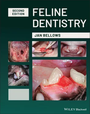
Jan Bellows - Feline Dentistry

Здесь есть возможность читать онлайн «Jan Bellows - Feline Dentistry» — ознакомительный отрывок электронной книги совершенно бесплатно, а после прочтения отрывка купить полную версию. В некоторых случаях можно слушать аудио, скачать через торрент в формате fb2 и присутствует краткое содержание. Жанр: unrecognised, на английском языке. Описание произведения, (предисловие) а так же отзывы посетителей доступны на портале библиотеки ЛибКат.

Feline Dentistry: краткое содержание, описание и аннотация

Предлагаем к чтению аннотацию, описание, краткое содержание или предисловие (зависит от того, что написал сам автор книги «Feline Dentistry»). Если вы не нашли необходимую информацию о книге — напишите в комментариях, мы постараемся отыскать её.

The most up-to-date version of the leading resource on veterinary dentistry in cats The Second Edition of
delivers a comprehensive exploration of the specific considerations required to provide dental care to cats that emphasizes their unique needs.
The updated Second Edition includes brand-new material and approximately 300 new images illustrating diseases, conditions, and procedures discussed within the book. The new edition combines the pathology and treatment information to provide additional context which helps make it more clinically relevant. The book also offers:
A thorough introduction to feline oral assessment, including anatomy, oral examinations, radiology, and charting Comprehensive explorations of dental pathology and treatment in cats, including necessary equipment and materials and anesthesia and pain control Practical discussions of dental pathology prevention in felines, including plaque and tartar control Perfect for veterinary general practitioners and veterinary students,
will also be useful to veterinary technicians seeking a one-stop, visual resource on feline-specific dentistry.
Feline Dentistry — читать онлайн ознакомительный отрывок

Ниже представлен текст книги, разбитый по страницам. Система сохранения места последней прочитанной страницы, позволяет с удобством читать онлайн бесплатно книгу «Feline Dentistry», без необходимости каждый раз заново искать на чём Вы остановились. Поставьте закладку, и сможете в любой момент перейти на страницу, на которой закончили чтение.

Тёмная тема
Сбросить

Интервал:

Закладка:

Сделать

4 Chapter 4Figure 4.1 (a) Feline Grimace Scale parameters and point values. (b) Patient...Figure 4.2 (a) General physical examination while cat resides in bottom half...Figure 4.3 (a) Veterinarian and assistant evaluating a cat's mouth in the ex...Figure 4.4 (a) Findings of saliva stained muzzle and right forelimb secondar...Figure 4.5 (a) Left facial swelling secondary to osteomyelitis treated with ...Figure 4.6 Left‐sided decreased palpebral size secondary to caudal oral cavi...Figure 4.7 (a) sliding the lip caudally reveals supernumerary left mandibula...Figure 4.8 Normal position of the maxillary/mandibular incisors and canines....Figure 4.9 Marked mandibular distoclusion and asymmetry.Figure 4.10 (a) Dorsal tongue surface ulceration consistent with calicivirus...Figure 4.11 (a) Normal teeth and gingiva. (b) Tooth resorption, gingivitis a...Figure 4.12 (a) Catheter placed. (b) Ultrasonic dental scaling.Figure 4.13 Dental assistant obtaining full‐mouth intraoral radiographs.Figure 4.14 Visual tooth‐by‐tooth examination and probing.Figure 4.15 Dental assistant arranges radiographs for doctor to view while c...Figure 4.16 (a) Tooth resorption affecting the right maxillary canine root a...Figure 4.17 (a) Clinically evident tooth resorption of the left maxillary th...Figure 4.18 (a) Application of SANOS ®, a dental sealant. (b) Applicatio...Figure 4.19 Right maxillary canine root canal therapy.Figure 4.20 (a) Painful type 2 tooth resorption exposed to the oral cavity. ...Figure 4.21 (a) Right maxillary fourth premolar penetrating the gingiva bucc...Figure 4.22 (a) Marked malocclusion. (b) Malocclusion corrected by tooth mov...Figure 4.23 (a) Sutured surgical site. (b) Post‐extraction radiograph confir...Figure. 4.24 Patient in sternal recumbency in the recovery cage.Figure 4.25 Client report is prepared.Figures 4.26 (a) Review of case with client at release. (b) Demonstration ap...Figure 4.27 Oral assessment treatment and prevention visit workflow.Figure 4.28 (a) Feline deciduous tooth chart. (b) Radiograph findings.Figure 4.29 (a) Feline adult chart and (b) Radiograph findings.Figure 4.30 Completed feline dental chart (VIN).Figure 4.31 American Animal Hospital Association Feline dental charts.Figure 4.32 (a) Electronic feline deciduous tooth pathology chart. (b) Elect...Figure 4.33 (a) Electronic feline patient pathology dental chart. (b) Electr...Figure 4.34 (a) Two‐handed electronic charting (veterinarian only). (b) Four...Figure 4.35 Normal rostral occlusion,Figure 4.36 Marked mandibular mesioclusion in a “normal” Persian cat.Figure 4.37 Marked mandibular distoclusion – note the palatal penetration fr...Figure 4.38 Normal incisor, canine, premolar relationships.Figure 4.39 Tongue ulceration secondary to calici virus.Figure 4.40 Feline eosinophilic granuloma affecting the hard palate. Feline Dentistry - изображение 2Figure 4.41 Ranula in a cat.Figure 4.42 Caudal stomatitis.Figure 4.43 (a) Clinically missing right mandibular third premolar (note too...Figure 4.44 Persistent left maxillary canine tooth, note the buccal swelling...Figure 4.45 (a) Supernumerary left maxillary second premolars. (b) Supernume...Figure 4.46 Williams probe – with markings at – 1, 2, 3, 5, 7, 8, 9, 10 mm....Figure 4.47 (a) Right maxillary canine palatal defect. (b) Abnormal 3 mm pro...Figure 4.48 7mm palatal attachment loss.Figure 4.49 (a) Shepherd's hook explorer. (b) Orban explorer, and (c) ODU 11...Figure 4.50 (a) Suspected tooth resorption in a cat's left mandibular canine...Figure 4.51 (a) Gingival recession revealing the furcation of the right mand...Figure 4.52 bleeding on probing.Figure 4.53 Normal gingiva with no evidence of inflammation.Figure 4.54 (a) Marginal gingivitis on a cat's right maxillary fourth premol...Figures 4.55 (a–d) Examples of calculus indices (Table 4.6).Figure 4.56 Gingival enlargement around the right mandibular first molar.Figure 4.57 (a) Gingival recession of the left maxillary fourth premolar wit...Figure 4.58 Left maxillary canine complicated and right maxillary canine unc...Figure 4.59 Uncomplicated crown fractured left mandibular molar.Figure 4.60 (a) Acute complicated fractured left maxillary canine. (b) Chron...Figure 4.61 (a) Complicated crown root fracture of the left maxillary fourth...Figure 4.62 (a) Left maxillary canine crown wear due to malpositioned left m...Figure 4.63 Intrinsic staining.Figure 4.64 Intraoral radiograph of stage 2 tooth resorption affecting the l...Figure 4.65 (a) Stage 3 tooth resorption affecting a cat's left mandibular m...Figure 4.66 (a) Tooth resorption of a cat's right mandibular third premolar ...Figure 4.67 (a) Stage 4b tooth resorption of the right mandibular molar. (b)...Figure 4.68 (a) Cat's left mandibular canine with clinical tooth resorption ...


5 Chapter 5Figure 5.1 (a) Dental hard tissue superimposition in skull film resulting in...Figure 5.2 (a) Wall‐mounted X‐ray generator.(b) Handheld X‐ray generator...Figure 5.3 Sensor bite damage.Figure 5.4 Size 1 and 2 DR sensors.Figure 5.5 CR film sizes.Figure 5.6 Full mouth intraoral radiograph series.Figure 5.7 (a–q) Examples of intraoral digital software screens.Figure 5.8 (a) Foreshortened image. (b) Elongated image.Figure 5.9 (a) Parallel technique to expose the left cheek teeth in a cat. (...Figure 5.10 (a) Bisecting angle used to image the right maxillary cheek teet...Figure 5.11 (a) Sensor placed in the cat's mouth for exposure of maxillary i...Illustration 5.1 Parallel technique and bisecting angle technique. Illustrat...Figure 5.12 (a) Lateral oblique positioning of the PID and sensor for a cat'...Figure 5.13 (a) PID and sensor positioned to obtain image of the cat's right...Figure 5.14 (a) Zygomatic arch superimposed upon the left maxillary fourth p...Figure 5.15 (a) Position of PID and sensor to expose the mandibular incisors...Figure 5.16 (a) Sensor parallel to the mandibular cheek teeth with PID posit...Figure 5.17 (a) Right TMJ arrowed on a cat's skull. (b) Tube head and patien...Figure 5.18 (a) Clinical appearance of cat with a right rostrodorsal TMJ lux...Figure 5.19 (a) Radiograph of right caudoventral TMJ luxation. (b) Mandibula...Figure 5.20 Radiograph of cat's incisive bone.Figure 5.21 (a) Normal left lateral maxilla. (b) Sagittal section maxilla.Figure 5.22 Various radiograph appearances of the mental foramina: (a) Rostr...Figure 5.23 (a–d) radiograph appearances of normal mandibular symphyses.Figure 5.24 Right mandibular canal.Figure 5.25 Lamina dura and periodontal ligament space surrounding the right...Figure 5.26 Enlarged periodontal ligament space with advanced periodontal di...Figure 5.27 (a) Mandibular incisors (b) Normal level of bone support support...Figure 5.28 Stage 2 periodontal disease affecting the right mandibular fourt...Figure 5.29 Horizontal bone loss consistent with stage 3 moderate periodonta...Figure 5.30 (a) Vertical bone loss secondary to advanced (stage 4) periodont...Figure 5.31 (a) Stage 4 periodontal disease affecting the left mandibular mo...Figure 5.32 Stage 2 furcation involvement,Figure 5.33 (a) Area of inflammation affecting the left mandibular molar. (b...Figure 5.34 (a) Right and left maxillary canine chronic alveolar osteitis. (...Figure 5.35 (a) Super erupted left mandibular canine. (b) Radiograph reveali...Figure 5.36 (a) Apparent apical closure of a cat's fractured left maxillary ...Figure 5.37 (a) Left maxillary canine root canal space enlarged compared to ...Figure 5.38 Periapical (radicular) cyst associated with the right mandibular...Figure 5.39 (a) Discolored left mandibular canine crown. (b) Radiograph reve...Figure 5.40 (a) Stage 2 tooth resorption affecting the right mandibular cani...Illustration 5.2 Types of tooth resorption.Figure 5.41 (a) Left mandibular molar affected by Type 1 tooth resorption (b...Figure 5.42 (a) Loss of detail and increased bone production of the caudal o...Figure 5.43 (a) CT imaging of a cat with facial swelling.(b) Patient in ...Figure 5.44 Evaluation of a TMJ in a multi‐planar reconstruction view. (a) N...Figure 5.45 (a) Clinical image of caudal maxillary lesion. (b) CT image of l...Figure 5.46 (a–c) Patient presented with history of pain on tooth brushing. ...Figure 5.47 (a–e) Patient presented with bilateral non‐healing maxillary can...Figure 5.48 (a–d) Patient presented with increased plaque and calculus over ...Figure 5.49 (a–e) refractory stomatitis despite reported full mouth extracti...Figure 5.50 (a–d) Patient presented with barely visible tooth resorption of ...Figure 5.51 (a–c) Patient presented for anorexia and weight loss, oral exam ...Figure 5.52 The cat had fallen from a tree and presented with oral cavity bl...Figure 5.53 (a) Right facial swelling. (b) Two‐dimensional image of the embe...

Читать дальше
Тёмная тема
Сбросить

Интервал:

Закладка:

Сделать

Похожие книги на «Feline Dentistry»

Представляем Вашему вниманию похожие книги на «Feline Dentistry» списком для выбора. Мы отобрали схожую по названию и смыслу литературу в надежде предоставить читателям больше вариантов отыскать новые, интересные, ещё непрочитанные произведения.


Отзывы о книге «Feline Dentistry»

Обсуждение, отзывы о книге «Feline Dentistry» и просто собственные мнения читателей. Оставьте ваши комментарии, напишите, что Вы думаете о произведении, его смысле или главных героях. Укажите что конкретно понравилось, а что нет, и почему Вы так считаете.

x