Jan Bellows - Feline Dentistry
Здесь есть возможность читать онлайн «Jan Bellows - Feline Dentistry» — ознакомительный отрывок электронной книги совершенно бесплатно, а после прочтения отрывка купить полную версию. В некоторых случаях можно слушать аудио, скачать через торрент в формате fb2 и присутствует краткое содержание. Жанр: unrecognised, на английском языке. Описание произведения, (предисловие) а так же отзывы посетителей доступны на портале библиотеки ЛибКат.
- Название:Feline Dentistry
- Автор:
- Жанр:
- Год:неизвестен
- ISBN:нет данных
- Рейтинг книги:3 / 5. Голосов: 1
-
Избранное:Добавить в избранное
- Отзывы:
-
Ваша оценка:
Feline Dentistry: краткое содержание, описание и аннотация
Предлагаем к чтению аннотацию, описание, краткое содержание или предисловие (зависит от того, что написал сам автор книги «Feline Dentistry»). Если вы не нашли необходимую информацию о книге — напишите в комментариях, мы постараемся отыскать её.
delivers a comprehensive exploration of the specific considerations required to provide dental care to cats that emphasizes their unique needs.
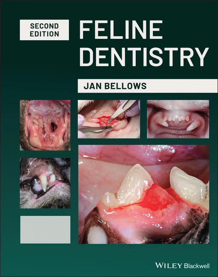
The updated Second Edition includes brand-new material and approximately 300 new images illustrating diseases, conditions, and procedures discussed within the book. The new edition combines the pathology and treatment information to provide additional context which helps make it more clinically relevant. The book also offers:
A thorough introduction to feline oral assessment, including anatomy, oral examinations, radiology, and charting Comprehensive explorations of dental pathology and treatment in cats, including necessary equipment and materials and anesthesia and pain control Practical discussions of dental pathology prevention in felines, including plaque and tartar control Perfect for veterinary general practitioners and veterinary students,
will also be useful to veterinary technicians seeking a one-stop, visual resource on feline-specific dentistry.
Feline Dentistry — читать онлайн ознакомительный отрывок
Ниже представлен текст книги, разбитый по страницам. Система сохранения места последней прочитанной страницы, позволяет с удобством читать онлайн бесплатно книгу «Feline Dentistry», без необходимости каждый раз заново искать на чём Вы остановились. Поставьте закладку, и сможете в любой момент перейти на страницу, на которой закончили чтение.
Интервал:
Закладка:
2 Chapter 2 Figure 2.1 Dental operatory. Figure 2.2 Dental operatory. Figure 2.3 Multiple table dental operatory allowing many procedures to be pe... Figure 2.4 (a) Four‐handed dentistry.(b) Six‐handed dentistry. Figure 2.5 (a) and (b) Dentistry suite.(c) Illustration of a one‐table d... Figure 2.6 (a) Head held at improper angle. (b) Proper head/neck angle. Figure 2.7 (a) Unencumbered area under the working end of the operatory tabl... Figure 2.8 Dental operatory where the shelves of dental materials are locate... Figure 2.9 Water‐cooled electric micromotor unit. Figure 2.10 Nitrogen‐powered delivery system. Figure 2.11 (a) Self‐contained dental delivery system with a large air compr... Figure 2.12 Dental air compressor unit positioned to power multiple delivery... Figure 2.13 Suction unit for multiple workstation suction. Figure 2.14 Chair‐side dental storage. Figure 2.15 Drawer containing sterilized instruments for performance of peri... Figure 2.16 Dental operatory with adequate storage. Figure 2.17 Dental operatory with mobile and stationary storage areas. Figure 2.18 (a) Cassette holding wing‐tipped elevators. (b) Sterilizable fel... Figure 2.19 Ceiling‐mounted spotlight. Figure 2.20 Proper head angulation (left) and improper head position causing... Figure 2.21 (a) Front lens‐mounted loupes and (b) through the lens‐mounted l... Figure 2.22 Properly fitted telescopic loupes resulting in functional head t... Figure 2.23 Fiber‐illuminated high‐speed handpiece. Figure 2.24 Floor stand X‐ray generator, sensor, and monitor. Figure 2.25 Wall‐mounted X‐ray generator to service two treatment tables.... Figure 2.26 Handheld X‐ray generator. Figure 2.27 Three digital sensors – from right to left sizes 0, 1, and 2. Figure 2.28 Portable x‐ray generator and CR processing system. Figure 2.29 Wall‐mounted anesthesia unit and easily accessible monitors and ... Figure 2.30 Patient monitoring unit. Figure 2.31 (a) Adult Feline dental chart.(b) Electronic dental chart (h... Figure 2.32 (a) Shepherd’s hook dental explorer – #23 dental explorer (Cisla... Figure 2.33 (a) UNC‐15 periodontal probe with markings at every 1 mm and bar... Figure 2.34 Dental mirror. Figure 2.35 (a) Various sized mouth props – Dentalaire. (b) Incorrect use of... Figure 2.36 Magnification and illumination and operator safety equipment use... Figure 2.37 Sickle scaler. Figure 2.38 (a) Columbia 13/14 universal curette. (b) Area‐specific curette ... Figure 2.39 (a) Ultrasonic scaler with illumination. (b) Stack insert. (c) T... Figure 2.40 IM3’s 42‐12 ferrite rod ultrasonic scaler operates at 43000Hz. Figure 2.41 Side of piezoelectric tip used to remove plaque and tartar. Figure 2.42 Tip wear and resulting efficiency. 2 mm loss in length can resul... Figure 2.43 (a–d) Piezoelectric scalers require a wrench to unscrew and repl... Figure 2.44 (a) Metallic prophy angle. (b) Disposable polishing angle and pa... Figure 2.45 Application of SANOS into the left maxillary canine sulcus. Figure 2.46 (a) OraVet® professional product gel applied to teeth and under ... Figure 2.47 (a) 2 mm bleeding periodontal pocket, (b) Application of Clindor... Figure 2.48 (a) #11 scalpel blade used to incise gingiva caudal to the right... Figure 2.49 Periotome. Figure 2.50 (a) Ex‐9 periosteal elevator. Figure 2.51 Freer periosteal elevator. Figure 2.52 (a) Dental luxating type elevator (Cislak). (b) Feline pen luxat... Figure 2.53 (a) Various‐sized wing‐tipped elevators in a cassette. (b) Winge... Figure 2.54 (a) Not recommended thick end of wing‐tipped elevator. (b) Recom... Figure 2.55 Root Forcep Z4658 (Cislak). Figure 2.56 (a) Curved extraction forceps (#301 Forceps). (b) Curved extract... Figure 2.57 (a) Micro‐Friedman rongeur.(b) Rongeur used to deliver a can... Figure 2.58 WA‐1 Root tip pick. Figure 2.59 (a) Sharpening a wing‐tipped elevator. (b) Mechanical sharpening... Figure 2.60 (a) Diplomate extraction kit in instrument cassette.(b) Dr. ... Figure 2.61 (a) High‐ (left) and low‐speed (right) handpieces loaded on a de... Figure 2.62 (a) iM3 Advantage 4–1 nose cone. (b) iM3 LS Advantage motor. Figure 2.63 Contra‐angle attachment. Figure 2.64 High‐speed handpiece. Figure 2.65 Fiber‐optic illumination. Figure 2.66 (a) Lever control bur changer. (b) Push control. (c) Bur inserti... Figure 2.67 Bur design. Figure 2.68 Round bur on a LA shank. Figure 2.69 Round bur on a friction grip shank. Figure 2.70 Bur block with various friction grip burs. Figure 2.71 Storage container for multiple bur types and sizes. Figure 2.72 Friction grip, surgical and long burs. Figure 2.73 (a) Common types of burs used in operative dentistry. Figure 2.73 (b) Round bur used to remove a section of buccal alveolar bone ...Figure 2.74 Diamond bur.Figure 2.75 Football‐shaped diamond bur used to smooth the alveolar crest af...Figure 2.76 White stone used to complete a composite restoration.Figure 2.77 (a) Canister removal tool. (b) Canister turbine. (c) Turbine wit...Figure 2.78 VOHC seal of acceptance.Figure 2.79 VOHC accepted products to decrease the accumulation of plaque an...Figure 2.80 Cotton applicator used to wipe the daily accumulation of plaque ...Figure 2.81 Cat Bites ® to help decrease the accumulation of plaque.Figure 2.82 Barbed broach used to remove the pulp from a fractured cat's can...Figure 2.83 K‐file removing clean dentinal shavings.Figure 2.84 Various‐sized paper points 30 mm lengths.Figure 2.85 Various‐sized gutta percha points.Figure 2.86 GuttaFlow.Figure 2.87 Endodontic locking pliers.Figure 2.88 Retrograde amalgam carrier used to deliver MTA or calcium hydrox...Figure 2.89 Glass ionomer.Figure 2.90 Bottle and “lollipop” self‐etch (one‐step) adhesive.Figure 2.91 (a) Capsules and syringe flowable composite. (b) Light‐cured hyb...Figure 2.92 Curing light.Figure 2.93 (a) CO 2laser. (b) Diode laser, and (c) Therapy laser.Figure 2.94 (a) Protemp®. (b) Splint material used to stabilize a cat's symp...
3 Chapter 3Figure 3.1 Patient anesthetized, temperature control assist, vital parameter...Figure 3.2 Patient preanesthetic evaluation.Figure 3.3 (a) An anesthesia checkoff list. (b) Soda lime replacement remind...Figure 3.4 Pneumothorax, pneumomediastinum, pneumoperitoneum secondary to en...Figure 3.5 (a) Hypothermia prevented by using a radiant heat blanket HotDog®...Figure 3.6 (a) Stationary multiparameter monitor.(b) Portable multiparam...Figure 3.7 (a–e) Capnogram illustrations.Figure 3.8 Radiant heat system – HotDog.Figure 3.9 In the circuit CO 2monitor.Figure 3.10 Tail base pulse oximeter probe.Figure 3.11 (a–c) Infraorbital nerve block.Figure 3.12 Maxillary nerve block illustration.Figure 3.13 (a and b) Middle mental nerve block.Figure 3.14 (a) A median view of the point of entry of the inferior alveolar...Figure 3.15 Nerve block summary illustration.Figure 3.16 Fentanyl transdermal patch.
Читать дальшеИнтервал:
Закладка:
Похожие книги на «Feline Dentistry»
Представляем Вашему вниманию похожие книги на «Feline Dentistry» списком для выбора. Мы отобрали схожую по названию и смыслу литературу в надежде предоставить читателям больше вариантов отыскать новые, интересные, ещё непрочитанные произведения.


Обсуждение, отзывы о книге «Feline Dentistry» и просто собственные мнения читателей. Оставьте ваши комментарии, напишите, что Вы думаете о произведении, его смысле или главных героях. Укажите что конкретно понравилось, а что нет, и почему Вы так считаете.