Stephen R. Bolsover - Cell Biology
Здесь есть возможность читать онлайн «Stephen R. Bolsover - Cell Biology» — ознакомительный отрывок электронной книги совершенно бесплатно, а после прочтения отрывка купить полную версию. В некоторых случаях можно слушать аудио, скачать через торрент в формате fb2 и присутствует краткое содержание. Жанр: unrecognised, на английском языке. Описание произведения, (предисловие) а так же отзывы посетителей доступны на портале библиотеки ЛибКат.
- Название:Cell Biology
- Автор:
- Жанр:
- Год:неизвестен
- ISBN:нет данных
- Рейтинг книги:3 / 5. Голосов: 1
-
Избранное:Добавить в избранное
- Отзывы:
-
Ваша оценка:
Cell Biology: краткое содержание, описание и аннотация
Предлагаем к чтению аннотацию, описание, краткое содержание или предисловие (зависит от того, что написал сам автор книги «Cell Biology»). Если вы не нашли необходимую информацию о книге — напишите в комментариях, мы постараемся отыскать её.
Cell Biology: A Short Course
Cell Biology: A Short Course
Cell Biology: A Short Course
Cell Biology — читать онлайн ознакомительный отрывок
Ниже представлен текст книги, разбитый по страницам. Система сохранения места последней прочитанной страницы, позволяет с удобством читать онлайн бесплатно книгу «Cell Biology», без необходимости каждый раз заново искать на чём Вы остановились. Поставьте закладку, и сможете в любой момент перейти на страницу, на которой закончили чтение.
Интервал:
Закладка:
 ORGANELLES BOUNDED BY DOUBLE‐MEMBRANE ENVELOPES
The nucleus, mitochondrion, and chloroplast (in plants) are enclosed within an envelope consisting of two parallel membranes. These major cell organelles all contain the genetic material, DNA.
The Nucleus
The nucleus is often the most prominent cell organelle. It contains the genome,the cell's database, which is encoded in molecules of the nucleic acid,DNA. The nucleus is bounded by a nuclear envelopecomposed of two membranes separated by an intermembrane space ( Figure 2.3). The inner membrane of the nuclear envelope is lined by the nuclear lamina,a meshwork of laminproteins that provide rigidity to the nucleus and anchorage for the DNA. A two‐way traffic of proteins and nucleic acids between the nucleus and the cytoplasm passes through holes in the nuclear envelope called nuclear pores.The nucleus of a cell that is synthesizing proteins at a low level will have few nuclear pores. In cells that are undergoing active protein synthesis, however, virtually the whole nuclear surface is perforated.
IN DEPTH 2.1WATER, WATER (AND AQUAPORINS) EVERYWHERE
Our bodies are ~70% water. Water can readily diffuse into and out of cells through the process of osmosis. It is thus essential that we maintain correct water balance otherwise our cells would distort or even lyse. Osmosis can be defined as the movement of water across a membrane down its concentration gradient from a solution of low osmolarityto a solution of high osmolarity. Osmolarity is calculated by summing the molar concentration of all the solutes in a particular solution. The more concentrated a solution is, the lower its water concentration and the higher its osmolarity.
For most mammalian fluids, the osmolarity is approximately 300 mOsm/l. Hypertonic solutions have an osmolarity that is higher, whereas hypotonic solutions have an osmolarity that is lower. If cells were placed in a hypertonic solution, water would move out of the cell from the cytosol where the osmolarity is lower than the bathing medium. Cells shrink under these conditions. Conversely, water would move into the cell if placed in hypotonic solutions because the osmolarity of the cytosol is now higher relative to the bathing medium. This can cause lysis. Changes in osmolarity can occur in pathological situations.
Water is a hydrophilic molecule, obviously! Yet it can cross the hydrophobic membrane relatively quickly. Why? The answer is that membranes have water channels known as aquaporins. They facilitate the diffusion of water across the membrane. Aquaporins are expressed in all cells but are particularly abundant in red blood cells and kidney tubules. Consequently, the plasma membranes of these cells are highly permeable to water.
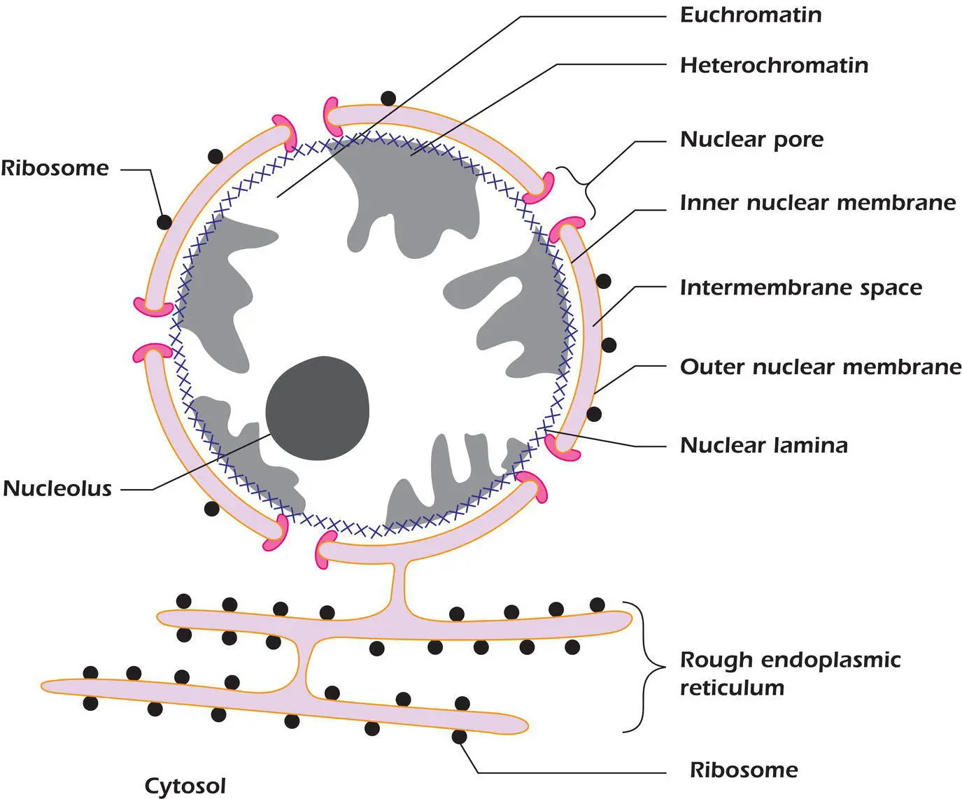
 Figure 2.3.The nucleus and the relationship of its membranes to those of the endoplasmic reticulum.
Within the nucleus, it is usually possible to recognize discrete areas. Much of it is occupied by chromatin, a complex of DNA and certain DNA‐binding proteins such as histones(page 39). In most cells, it is possible to discern two types of chromatin. A central region of lightly staining euchromatinis that portion of the cell's DNA database that is being actively read out by being transcribedinto RNA,another nucleic acid ( Chapter 5). In contrast the peripheral, darkly staining heterochromatinis the inactive portion of the genome where no RNA synthesis is occurring. The DNA in heterochromatin is densely packed, leading to its dark appearance.
Example 2.1 DNA Destruction in the Cytosol
An animal cell's own DNA should remain in the nucleus, except for the tiny amount that is within mitochondria. DNA in the cytosol will likely belong to a pathogen such as an invading virus. Cells therefore contain active DNAses in the cytosol that rapidly destroy DNA, while leaving RNA intact. It is to evade this defense mechanism that many viruses use RNA as their genetic material, even though RNA is a much less stable molecule than is DNA.
Unlike DNA, RNA is also found in the cytoplasm associated with particles called ribosomes whose function is to make proteins. Ribosomes are made in the nucleus, in specialized regions called nucleolithat form at specific nucleolar organizer regionsites on the DNA. These contain blocks of genes that code for the ribosomal RNA. Nuclear pores allow ribosomal subunits to exit the nucleus.
It should be stressed that the appearance of the nucleus we have described thus far relates to the cell in interphase,the period between successive rounds of cell division. As the cell enters mitosis ( Chapter 14) the organization of the nucleus changes dramatically. The DNA becomes more and more tightly packed and is revealed as a number of separate rods called chromosomes,of which there are usually 46 in human cells. The nucleolus disperses, and the nuclear envelope fragments. Upon completion of mitosis, these structural rearrangements are reversed and the nucleus resumes its typical interphase organization.
Mitochondria
Like nuclei, mitochondria are encapsulated by an outer and inner membrane ( Figure 2.4). Perhaps the most distinctive feature of mitochondria is that the inner membrane is markedly elaborated and folded to increase its surface area. These shelf‐like projections, named cristae,make mitochondria among the most easily recognizable organelles (e.g. Figure 1.4on page 8). The number of cristae, like the number of mitochondria themselves, depends upon the energy budget of the cell in which they are found. In muscle cells, which must contract and relax repeatedly over long periods of time, there are many mitochondria that contain numerous cristae; in fat cells, which generate little energy, there are few mitochondria and their cristae are less well developed. This gives a clue as to the function of mitochondria: they are the cell's power stations. Mitochondria produce the molecule adenosine triphosphate ( ATP )(page 35), the cell's main energy currency that provides the energy to drive a host of cellular reactions and mechanisms. Mitochondria make ATP through the process of oxidative phosphorylation whereby oxygen is used to pass electrons from energy intermediates to a series of protein complexes on the inner mitochondrial membrane known as the electron transport chain. This results in the transfer of H +out of the mitochondria and the generation of a concentration and voltage gradient. This gradient is subsequently tapped into by a protein known as ATP synthase which, as its name suggests, produces ATP. This process is essential for aerobic life and is the reason we breathe. We will return to ion gradients and the uses the cell puts them to in Chapter 9.

Figure 2.4.The mitochondrion.
Интервал:
Закладка:
Похожие книги на «Cell Biology»
Представляем Вашему вниманию похожие книги на «Cell Biology» списком для выбора. Мы отобрали схожую по названию и смыслу литературу в надежде предоставить читателям больше вариантов отыскать новые, интересные, ещё непрочитанные произведения.


Обсуждение, отзывы о книге «Cell Biology» и просто собственные мнения читателей. Оставьте ваши комментарии, напишите, что Вы думаете о произведении, его смысле или главных героях. Укажите что конкретно понравилось, а что нет, и почему Вы так считаете.