Stephen R. Bolsover - Cell Biology
Здесь есть возможность читать онлайн «Stephen R. Bolsover - Cell Biology» — ознакомительный отрывок электронной книги совершенно бесплатно, а после прочтения отрывка купить полную версию. В некоторых случаях можно слушать аудио, скачать через торрент в формате fb2 и присутствует краткое содержание. Жанр: unrecognised, на английском языке. Описание произведения, (предисловие) а так же отзывы посетителей доступны на портале библиотеки ЛибКат.
- Название:Cell Biology
- Автор:
- Жанр:
- Год:неизвестен
- ISBN:нет данных
- Рейтинг книги:3 / 5. Голосов: 1
-
Избранное:Добавить в избранное
- Отзывы:
-
Ваша оценка:
Cell Biology: краткое содержание, описание и аннотация
Предлагаем к чтению аннотацию, описание, краткое содержание или предисловие (зависит от того, что написал сам автор книги «Cell Biology»). Если вы не нашли необходимую информацию о книге — напишите в комментариях, мы постараемся отыскать её.
Cell Biology: A Short Course
Cell Biology: A Short Course
Cell Biology: A Short Course
Cell Biology — читать онлайн ознакомительный отрывок
Ниже представлен текст книги, разбитый по страницам. Система сохранения места последней прочитанной страницы, позволяет с удобством читать онлайн бесплатно книгу «Cell Biology», без необходимости каждый раз заново искать на чём Вы остановились. Поставьте закладку, и сможете в любой момент перейти на страницу, на которой закончили чтение.
Интервал:
Закладка:
Membrane contact sites serve a variety of functions. They are important for the trafficking of lipids around the cell and in the transfer of calcium between compartments, for example between the ER and mitochondria.
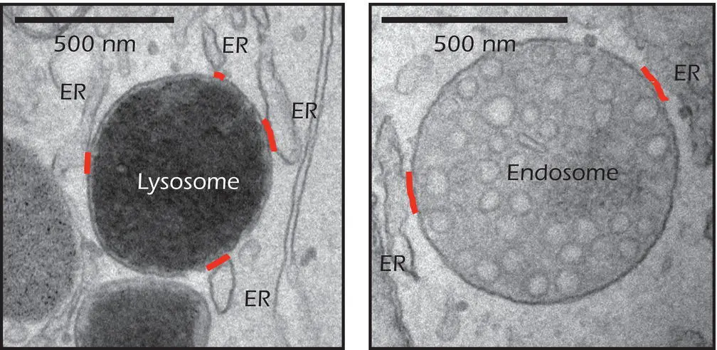
 Figure 2.5.Electron micrographs showing contact sites between the endoplasmic reticulum (ER) and lysosomes (left) and endosomes (right). Endosomes will be described in Chapter 12.
Source: Images by Bethan S. Kilpatrick, Clare E. Futter, and Sandip Patel, University College London. Reproduced by permission.
Cell Junctions
In multicellular organisms, and particularly in their epithelia, it is often necessary for neighboring cells within a tissue to be connected. This function is provided by cell junctions. In animal cells there are three types of junction. Those that form a tight seal between adjacent cells are known as tight junctions;those that allow communication between cells are known as gap junctions.A third class of cell junction anchors cells together, allowing the tissue to be stretched without tearing. These are called anchoring junctions.
Tight junctions are found wherever the flow of extracellular medium is to be restricted and are particularly common in epithelial cells, such as those lining the small intestine. The plasma membranes of adjacent cells are pressed together so tightly that no intercellular space exists between them ( Figure 1.6on page 9). Tight junctions between the epithelial cells of the intestine ensure that the only way that molecules can get from the lumen of the intestine to the blood supply that lies beneath is by passing through the cells, a route that can be selective.
Gap junctions are specialized structures that allow cell‐to‐cell communication in animals ( Figure 2.6). When two cells form a gap junction, ions and small molecules can pass directly from the cytosol of one cell to the cytosol of the other cell without going into the extracellular medium. Since ions can move through the junction, changes in electrical voltage are also rapidly transmitted from cell to cell by this route. In vertebrates, the structure that makes this possible is the connexon.When two compatible connexons meet, they form a tube, about 1.5 nm in diameter, that runs through the plasma membrane of the first cell, across the small gap between the cells, and through the plasma membrane of the second cell. This hole is large enough to allow through small organic molecules as well as ions, but it is too small for proteins or nucleic acids. The limit is a relative molecular mass (Mr) of about 1000. Gap junctions are especially important in the heart, where they allow an electrical signal to pass rapidly between all the cardiac muscle cells, ensuring that they all contract at the appropriate time. Each connexon is composed of six protein subunits called connexins that can twist against each other to open and close the central channel in a process called gating(page 147). This allows the cell to control the degree to which it shares material with its neighbor.
IN DEPTH 2.2 MY OLD MAM
The existence of membrane contact sites between the ER and mitochondria has been known for decades from electron microscope studies. Relatively recently, these contacts have been isolated biochemically from cell extracts for study. The resulting fractions are referred to as mitochondria‐associated membranes ( MAMs) because they contain proteins not only typically found in mitochondrial membranes but also in the ER. In live cells, ER‐mitochondria contact sites provide a restricted space such that when calcium ions are released from the ER ( Chapter 10), they achieve a very high local concentration that facilitates their uptake by adjacent mitochondria.
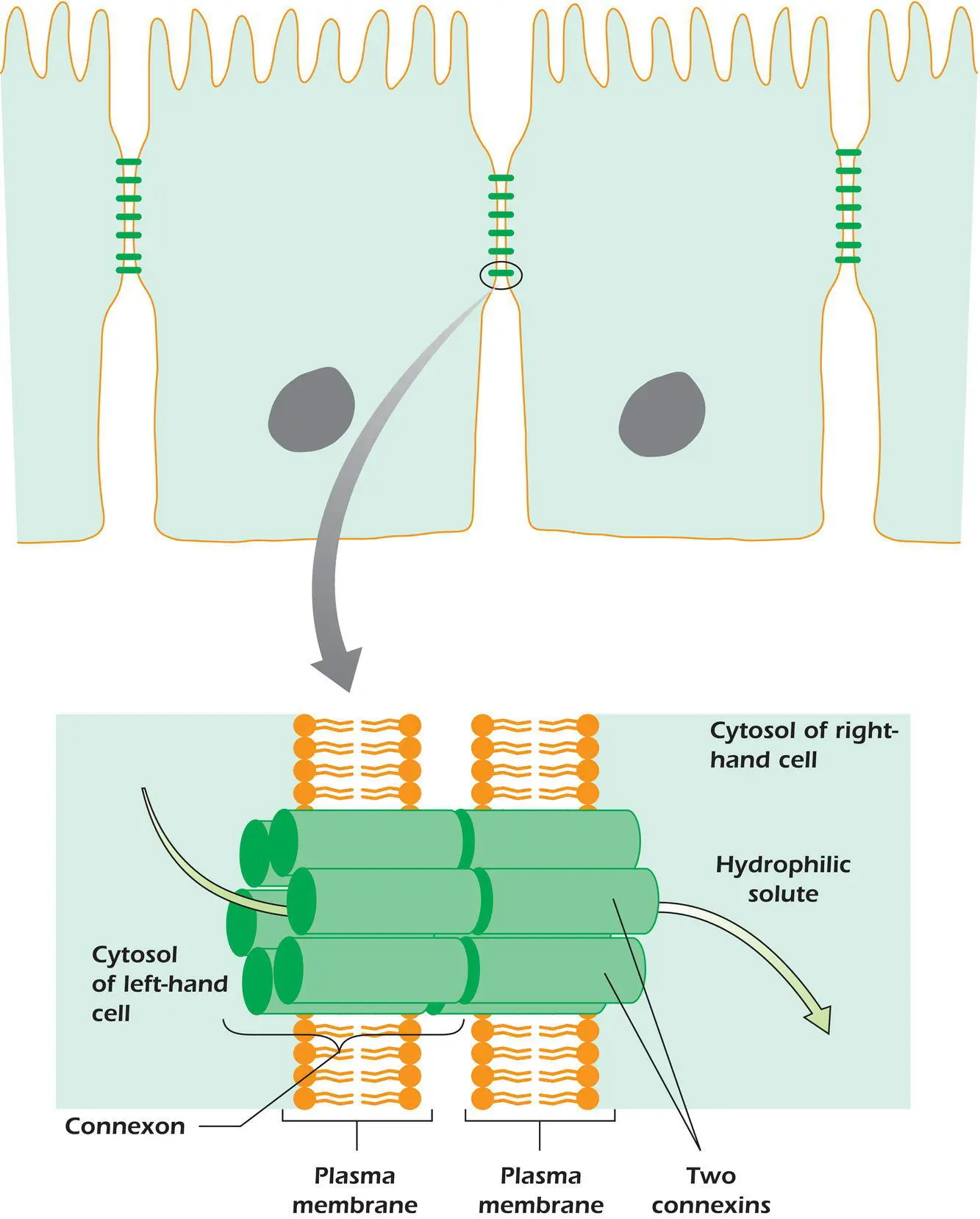
Figure 2.6.Gap junctions allow solute and electrical current to pass from the cytosol of one cell to the cytosol of its neighbor.
Example 2.2 Gap Junctions Keep Eggs Healthy
In the days leading up to ovulation, oocytes develop within structures called follicles, in which they are connected to surrounding granulosa cells by gap junctions. During their development oocytes are not yet themselves capable of performing several fundamental homeostatic processes, such as regulating intracellular pH. However, the surrounding granulosa cells have ample ability to regulate pH, and H +ions can pass through the gap junctions, such that the granulosa cells effectively regulate the pH of the oocyte on its behalf. By the time the oocyte is fully grown and ready to be ovulated it can finally regulate its own pH, at which time it jettisons the granulosa cells and becomes ready to be fertilized by a spermatozoon.
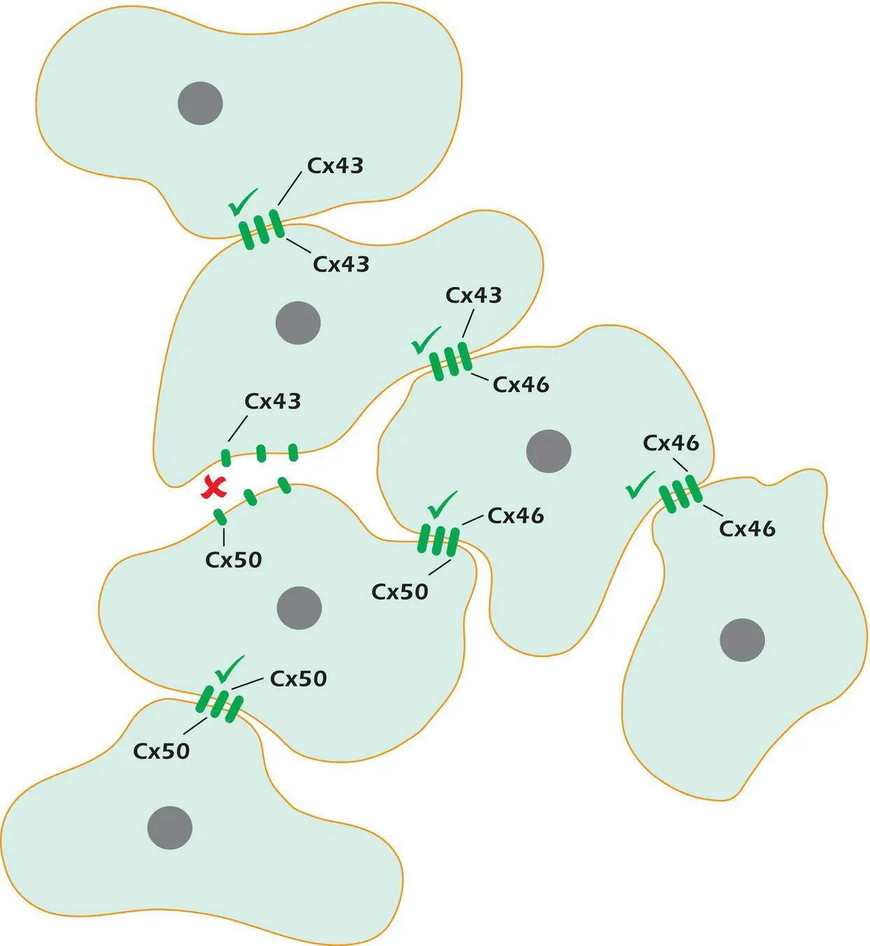
Figure 2.7.Not all connexins are compatible. A √ indicates a working gap junction, x indicates that gap junction channels cannot form.
There are over 20 different members of the connexin gene family in humans. Each can, if made in a population of cells, generate complete, working gap junction channels of 12 identical connexins. However, not all connexin isoformsare compatible. A cell with connexin 43 cannot form gap junctions with another cell that makes connexin 50 ( Figure 2.7). However, a connexon made from connexin 43 is able to form a good working channel if it meets a connexon made from connexin 46, and a connexon made from connexin 46 is in turn compatible with connexin 50.
Anchoring junctions bind cells tightly together and are found in tissues such as the skin and heart that are subjected to mechanical stress. They are described later (page 229).
Answer to thought question:This can only be speculation, but one obvious effect of cells having incompatible connexin isoforms is that the intracellular route for cell–cell communication is lost. Consider a tissue whose cells make connexin 43 that lies adjacent to another tissue whose cells use connexin 50. The cells of each tissue can coordinate their activity by passing signals via gap junctions, for example by intracellular messengers (page 166) or as voltage changes. However, the signals will remain private to each tissue and will not be shared with the neighboring tissue. The two tissues can still communicate when necessary, for instance by transmitter chemicals that the cells release into the extracellular medium (see Chapters 10and 11).
SUMMARY
1 Membranes are made of phospholipids, protein, and cholesterol.
2 Cells are bounded by membranes, while cell functions are compartmentalized into membrane‐bound organelles.
3 Solutes with a significant solubility in hydrophobic solvents can pass across biological membranes by simple diffusion. Charged molecules cannot.
4 The nucleus and mitochondrion are bounded by double‐membrane envelopes. In the case of the nucleus, this is perforated by nuclear pores. Both organelles contain DNA.
Читать дальшеИнтервал:
Закладка:
Похожие книги на «Cell Biology»
Представляем Вашему вниманию похожие книги на «Cell Biology» списком для выбора. Мы отобрали схожую по названию и смыслу литературу в надежде предоставить читателям больше вариантов отыскать новые, интересные, ещё непрочитанные произведения.


Обсуждение, отзывы о книге «Cell Biology» и просто собственные мнения читателей. Оставьте ваши комментарии, напишите, что Вы думаете о произведении, его смысле или главных героях. Укажите что конкретно понравилось, а что нет, и почему Вы так считаете.