Stephen R. Bolsover - Cell Biology
Здесь есть возможность читать онлайн «Stephen R. Bolsover - Cell Biology» — ознакомительный отрывок электронной книги совершенно бесплатно, а после прочтения отрывка купить полную версию. В некоторых случаях можно слушать аудио, скачать через торрент в формате fb2 и присутствует краткое содержание. Жанр: unrecognised, на английском языке. Описание произведения, (предисловие) а так же отзывы посетителей доступны на портале библиотеки ЛибКат.
- Название:Cell Biology
- Автор:
- Жанр:
- Год:неизвестен
- ISBN:нет данных
- Рейтинг книги:3 / 5. Голосов: 1
-
Избранное:Добавить в избранное
- Отзывы:
-
Ваша оценка:
Cell Biology: краткое содержание, описание и аннотация
Предлагаем к чтению аннотацию, описание, краткое содержание или предисловие (зависит от того, что написал сам автор книги «Cell Biology»). Если вы не нашли необходимую информацию о книге — напишите в комментариях, мы постараемся отыскать её.
Cell Biology: A Short Course
Cell Biology: A Short Course
Cell Biology: A Short Course
Cell Biology — читать онлайн ознакомительный отрывок
Ниже представлен текст книги, разбитый по страницам. Система сохранения места последней прочитанной страницы, позволяет с удобством читать онлайн бесплатно книгу «Cell Biology», без необходимости каждый раз заново искать на чём Вы остановились. Поставьте закладку, и сможете в любой момент перейти на страницу, на которой закончили чтение.
Интервал:
Закладка:
Fluorescent Proteins
Instead of introducing fluorescent dyes into fixed or living cells, we can cause cells to make them. This technology began with the description in 1962 of Green Fluorescent Protein (GFP ),a protein from the jellyfish Aequorea victoria that glows green when excited with blue light. Since the original description of GFP, a family of fluorescent proteins of different colors has become available, some through artificially mutating the original GFP, some found in other organisms. Using recombinant DNA technology ( Chapter 8), the gene for a fluorescent protein can be introduced into a living cell, which then makes the protein.
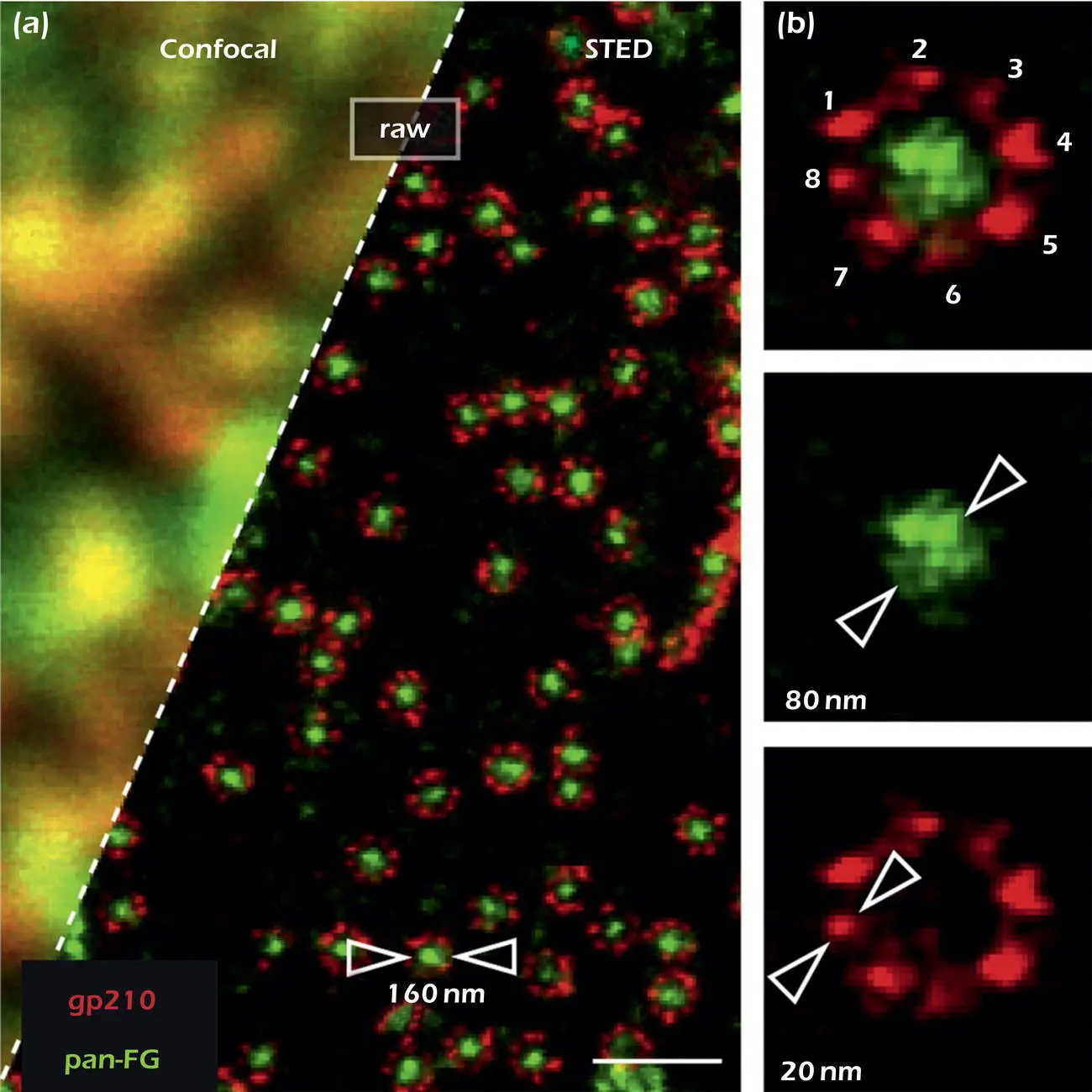
 Figure 1.14.Super‐resolution microscopy. Fluorescence image of the surface of a eukaryotic nucleus by standard confocal microscopy and by STED.
Source: Göttfert et al. (2013). Coaligned Dual‐Channel STED Nanoscopy and Molecular Diffusion Analysis at 20 nm Resolution. Biophysical Journal, 105(1), L01 ‐L03. doi:10.1016/j.bpj.2013.05.029
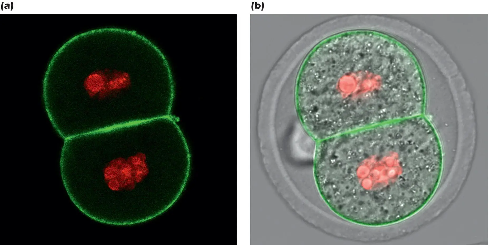
Figure 1.15.(a) Fluorescence microscope image of two‐cell mouse embryo expressing GFP and RFP chimeras. Two images were acquired in succession using optical configurations suitable for GFP and RFP respectively. The two images were then combined in a computer. (b) Image (a) combined with a phase contrast transmitted light image; the clear envelope around the embryo is the zona pellucida.
Source: Images by Lia Paim and Adelaide Allais, University of Montreal.
This basic technique is useful in itself for labeling a population of cells. However a battery of more and more sophisticated techniques has been developed from this starting point. Most use the approach of fusing the gene for a fluorescent protein with the gene for another protein of interest so that the cell makes a chimera – a single protein comprising the protein of interest plus the fluorescent protein. For example, Figure 1.15shows a fluorescence image of a mouse embryo at the two‐cell stage engineered to express a GFP chimera that concentrates at the plasma membrane together with a chimera of red fluorescent protein (RFP, from coral) and histone H2B (page 39) that concentrates in the nuclei.
In this example the two parts of the chimeras worked independently, so that the GFP and RFP simply showed where their respective partners were located. However, clever protein design has created more complex chimeras of the calcium‐binding protein calmodulin (page 115) with GFP mutants so that the fluorescence changes according to the concentration of calcium. Calcium concentrations change dramatically as cells respond to stimuli ( Chapter 10) and these fluorescent calmodulin chimeras can be used to report these changes. Even more clever, if the calcium‐measuring chimera is fused to a third protein with a known specific location in the cell, then the protein can be used to report the calcium concentration in that specific location.
 BrainBox 1.1 Osamu Shimomura, Martin Chalfie, and Roger Tsien
 Osamu Shimomura, Martin Chalfie, and Roger Tsien.
Source: The Nobel Foundation. Photo: U. Montan.
Many proteins are colored, but in most cases the color is generated by a prosthetic group (for example the heme group in hemoglobin (page 118) and in chlorophyll). However, in 1979 Osamu Shimomura, working at Princeton University in the USA, showed that the colored moiety in a GFP made by the jellyfish Aequorea aequorea was a reaction product of the amino acids themselves. This opened up the possibility of using the cell's own machinery to make genetically encoded labeling proteins that could be targeted to precise tissues and even specific sites within the cell. However, the suspicion was that one or more specialized enzymes in the jellyfish cells would be needed to carry out the conversion of the amino acids to the fluorophore, so that simply introducing the gfp gene would do nothing. In 1994 Martin Chalfie, working at Columbia University in New York, showed that this was not the case: the gfp gene product, on its own, converted itself into fluorescent GFP. The next leap in technology was to engineer GFP and GFP chimeras to be more than markers, and instead to be reporters of cell behavior. From 1992 onward, working at the University of California at San Diego, Roger Tsien and his lab engineered an ever‐increasing family of fluorescent proteins that are now used universally by cell biologists and drug companies to study almost all aspects of cell behavior, creating both beautiful science and beautiful images, such as Figure 1.15. Shimomura, Chalfie, and Tsien were awarded the Nobel Prize in Chemistry in 2008.
Answer to thought question:Only transmission electron microscopy reveals all the structures present in a particular volume of the cell at sufficient resolution to determine whether it is malformed. Super‐resolution microscopy has the resolution to reveal individual molecules on or within the Golgi, but only those individual molecules that the scientist chose to study are revealed, not the overall structure. Malformation of the endoplasmic reticulum and Golgi apparatus is thought to underlie one type of inherited spastic paraplegia.
SUMMARY
1 All living organisms are made of cells.
2 There are only two types of cells, prokaryotic and eukaryotic.
3 Prokaryotic cells have little visible internal organization. They are usually 1–2 μm in size.
4 Eukaryotic cells usually measure from 5–100 μm. They contain a variety of specialized internal organelles, the largest of which, the nucleus, contains the genetic material.
5 The endosymbiotic theory proposes that some eukaryotic organelles, such as mitochondria and chloroplasts, originated as free‐living prokaryotes.
6 In multicellular organisms, cells are organized into tissues. In animals there are four tissue types: epithelium, connective tissue, nervous tissue, and muscle.
7 The extracellular matrix is found on the outside of animal cells.
8 In tissues, specialized cells arise from unspecialized stem cells.
9 Light microscopy revealed the diversity of cell types and the existence of the major organelles, such as the nucleus and mitochondria.
10 The electron microscope revealed the detailed structure of the larger organelles and resolved the cell ultrastructure, the fine detail, at the nanometer scale.
11 Fluorescence microscopy can reveal the location of specific molecules within the cell by using antibodies or genetically encoded fluorescent chimeras.
12 Super‐resolution microscopy is an advanced form of fluorescence microscopy that can reveal objects tens of nanometers in size.
13 Cells can be engineered to express fluorescent proteins that reveal many aspects of cell organization and behavior.
Читать дальшеИнтервал:
Закладка:
Похожие книги на «Cell Biology»
Представляем Вашему вниманию похожие книги на «Cell Biology» списком для выбора. Мы отобрали схожую по названию и смыслу литературу в надежде предоставить читателям больше вариантов отыскать новые, интересные, ещё непрочитанные произведения.


Обсуждение, отзывы о книге «Cell Biology» и просто собственные мнения читателей. Оставьте ваши комментарии, напишите, что Вы думаете о произведении, его смысле или главных героях. Укажите что конкретно понравилось, а что нет, и почему Вы так считаете.