Stephen J. Bourke - Respiratory Medicine
Здесь есть возможность читать онлайн «Stephen J. Bourke - Respiratory Medicine» — ознакомительный отрывок электронной книги совершенно бесплатно, а после прочтения отрывка купить полную версию. В некоторых случаях можно слушать аудио, скачать через торрент в формате fb2 и присутствует краткое содержание. Жанр: unrecognised, на английском языке. Описание произведения, (предисловие) а так же отзывы посетителей доступны на портале библиотеки ЛибКат.
- Название:Respiratory Medicine
- Автор:
- Жанр:
- Год:неизвестен
- ISBN:нет данных
- Рейтинг книги:4 / 5. Голосов: 1
-
Избранное:Добавить в избранное
- Отзывы:
-
Ваша оценка:
Respiratory Medicine: краткое содержание, описание и аннотация
Предлагаем к чтению аннотацию, описание, краткое содержание или предисловие (зависит от того, что написал сам автор книги «Respiratory Medicine»). Если вы не нашли необходимую информацию о книге — напишите в комментариях, мы постараемся отыскать её.
, a team of distinguished physicians delivers a comprehensive and accessible overview of the essentials of respiratory medicine, including a review of respiratory anatomy and physiology, as well as the aetiology, epidemiology, symptoms, and management of a wide range of respiratory diseases.
This edition offers self-assessment exercises in each chapter and a range of clinical images and scans showing the critical features of each disease. The book also offers:
A thorough introduction to history taking, examination, and investigations Comprehensive explorations of respiratory diseases, including upper respiratory tract infections and influenza, pneumonia, and tuberculosis Practical discussions of bronchiectasis, lung abscess, cystic fibrosis, asthma, and chronic obstructive pulmonary disease In-depth examinations of lung transplantation A companion website featuring figures, key points, and interactive self-assessment questions Perfect for medical students and respiratory nurses,
will also earn a place in the libraries of early-career medical doctors and residents with an interest in respiratory medicine.
Respiratory Medicine — читать онлайн ознакомительный отрывок
Ниже представлен текст книги, разбитый по страницам. Система сохранения места последней прочитанной страницы, позволяет с удобством читать онлайн бесплатно книгу «Respiratory Medicine», без необходимости каждый раз заново искать на чём Вы остановились. Поставьте закладку, и сможете в любой момент перейти на страницу, на которой закончили чтение.
Интервал:
Закладка:
Examination
Some physical signs in medicine are difficult to assess, and examination skills may take years to refine (e.g. identifying the nature of a heart murmur). By contrast, most of the signs in respiratory disease are easy to elicit and interpret. Despite this, evidence of respiratory disease is often entirely overlooked.
Answer to question in Fig. 2.1: (b) has airway obstruction – note the high position of the shoulders.
The expertise in respiratory examination lies in knowing what to look for. Read the following and you will become expert. You will discover the insightful experience that is respiratory examination; ordinary doctors will be in awe of your deductive abilities.
General examination
Be alert to clues to respiratory disease that may be evident from the moment the patient is first seen ( Fig. 2.1) or that become apparent during history taking. These include the rate and character of breathing, signs of respiratory distress such as use of accessory musclesof respiration (e.g. sternocleidomastoids), the shape of the chest, spine and shoulders and the character of any cough. Hoarsenessof the voice may be a clue to recurrent laryngeal nerve damage by a carcinoma. Wheezemay be audible. Stridoris most commonly picked up during history taking, rather than examination.
Avoid proceeding directly to examination of the chest; first pause and ask the patient to cough.
Cough
A voluntarily produced cough is an extremely useful but much neglected sign in respiratory examination. It is probably wise to start with it, lest it be missed. The clues it provides to later examination findings are so useful it can almost feel like cheating.
From the explanation of cough given earlier, it can be seen that when the vocal cords are not opposed, cough will no longer have its normal distinct, crisp start. Such a cough is known as bovine cough(cows don’t have vocal cords). It is a sign not to be missed, as it may result from tumour in the left side of the chest, causing recurrent laryngeal nerve palsy. The hoarse voicethat accompanies this abnormality may be missed, particularly if the doctor and patient are meeting for the first time.
If airway obstruction is present, the cough will have a wheezy quality(listen out for it). As described already, the expiratory flow rate during a cough is greater than that generated in a normal forced expiration. Therefore, if airway obstruction is mild, it may be that wheeze is only heard during a cough.
When reporting a cough, a patient is usually asked if they produce sputum. The answer ‘no’ normally results in the doctor documenting that the cough is ‘dry’. Beware: an inability to ‘bring up’ sputum doesn’t imply it isn’t there. Consciously listening to the quality of a cough will avoid such a mistake.
Knowing whether a cough is wet or drycan be invaluable when it comes to determining the nature of the crackles heard later in the examination. Whilst in theory the ‘coarse’ crackles of bronchiectasis are different from the ‘fine’ crackles of fibrosis, in practice – on sound quality alone – it can be a difficult call. If you’ve already listened to the quality of the cough by the time of auscultation, your ability to distinguish fine from coarse will be uncannily good.
A loud, booming cough heard from one end of the ward to the other is unlikely to be as serious as it sounds.
Next, start with the hands and look for signs en route to the chest.
Hands
Look for clubbing, tar stainingor features of rheumatoid arthritis. Signs of CO 2retention include peripheral vasodilationand asterixis: a flapping tremor detected by asking the patient to spread their fingers, cock their wrists back and close their eyes. CO 2retention dulls proprioception and the hands tend to drift forward, particularly when the eyes are closed. Eventually, an awareness that the hands are no longer in position leads to a sudden corrective movement. In understanding the underlying mechanism of this sign, it should be clear why a doctor holding the patient’s hands in place (as many do to ‘feel’ for asterixis) will always miss it. Count the pulse rateand note any abnormalities in rhythm (e.g. atrial fibrillation) or character (e.g. a bounding pulse of carbon dioxide retention).
Count the respiratory rateover a period of at least 30 seconds. The respiratory rate is best counted surreptitiously, perhaps whilst feeling the pulse, as patients tend to breathe faster if they are aware that a doctor is focusing on their breathing. The respiratory rate is an incredibly easy observation to make and is a highly sensitive index of physiological derangement (as such, it is an integral part of all early‐warning systems in hospital), yet it is all too frequently missed. Do it. Do it properly.
Clubbing
Clubbing is increased curvature of the nail, with loss of the angle between the nail and the nail bed ( Fig. 2.2). It is a very important sign that is associated with a number of diseases ( Table 2.3), most notably bronchial carcinoma, bronchiectasis and fibrotic lung disease, such as idiopathic pulmonary fibrosis. Clubbing also occurs in asbestosis, although usually only in more advanced disease. Advanced clubbing is sometimes associated with hypertrophic pulmonary osteoarthropathy, in which there is new bone formation in the subperiosteal region of the long bones of the arms and legs, which is detectable on X‐ray and is associated with pain and tenderness.
Next examine the head and neck. Check for elevation of jugular venous pressureor lymph node enlargement. In the face, seek signs of cyanosisand anaemia(pallor of conjunctiva). Be alert for uncommon signs such as Horner syndrome(ptosis, meiosis, enophthalmos, anhydrosis), which indicates damage to the sympathetic nerves by a tumour situated at the lung apex (see Chapter 4).
Jugular veins
The jugular veins are examined with the patient in a semi‐reclining position, with the trunk at an angle of about 45° from the horizontal. The head is turned slightly to the opposite side and fully supported so that the sternocleidomastoid muscles are relaxed. The jugular venous pulse is seen as a diffuse superficial pulsation of multiple waveform that is distinct from the carotid arterial pulse. The height of the pulse wave is measured as the vertical heightof the top of the oscillating column of blood above the sternal angle. The jugular venous pressure normally falls during inspiration. It is elevated in right heart failure, which may occur as a result of pulmonary embolism or cor pulmonale in COPD, for example. Other signs of right heart failure, such as hepatomegaly and peripheral oedema, may also be present.
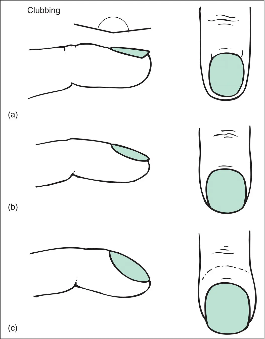
Figure 2.2 Clubbing. (a) Normal: the ‘angle’ is shown. (b) Early: the angle is absent. (c) Advanced: the nail shows increased curvature in all directions, the angle is absent, the base of the nail is raised up by spongy tissue and the end of the digit is expanded.
Cyanosis
This is a bluish discolouration of the skin and mucous membranes that results from an excessive amount of reduced (deoxygenated) haemoglobin (usually >5 g/dL in patients of European descent; the sign may be harder to detect in darker skin). As it relates to the quantity of deoxygenated blood rather than the proportion, it follows that it will be more readily observed in those with polycythaemia than anaemia. Central cyanosisis best seen on the tip of the tongue and is the cardinal sign of hypoxaemia, although it is not a sensitive sign because it is not usually detectable until the oxygen saturation has fallen to well below 85%, corresponding to a PO 2of <8 kPa (60 mmHg). Because of the poor sensitivity of cyanosis, it is essential to measure oxygenation by oximetry or arterial blood gas sampling in patients at risk for hypoxaemia. Peripheral cyanosiswill be present if there is central cyanosis, but it may also be caused by local circulatory slowing in the peripheries, resulting in more complete extraction of oxygen from the blood (e.g. blue hands and ears in cold weather).
Читать дальшеИнтервал:
Закладка:
Похожие книги на «Respiratory Medicine»
Представляем Вашему вниманию похожие книги на «Respiratory Medicine» списком для выбора. Мы отобрали схожую по названию и смыслу литературу в надежде предоставить читателям больше вариантов отыскать новые, интересные, ещё непрочитанные произведения.


Обсуждение, отзывы о книге «Respiratory Medicine» и просто собственные мнения читателей. Оставьте ваши комментарии, напишите, что Вы думаете о произведении, его смысле или главных героях. Укажите что конкретно понравилось, а что нет, и почему Вы так считаете.