X-Ray Fluorescence in Biological Sciences
Здесь есть возможность читать онлайн «X-Ray Fluorescence in Biological Sciences» — ознакомительный отрывок электронной книги совершенно бесплатно, а после прочтения отрывка купить полную версию. В некоторых случаях можно слушать аудио, скачать через торрент в формате fb2 и присутствует краткое содержание. Жанр: unrecognised, на английском языке. Описание произведения, (предисловие) а так же отзывы посетителей доступны на портале библиотеки ЛибКат.
- Название:X-Ray Fluorescence in Biological Sciences
- Автор:
- Жанр:
- Год:неизвестен
- ISBN:нет данных
- Рейтинг книги:5 / 5. Голосов: 1
-
Избранное:Добавить в избранное
- Отзывы:
-
Ваша оценка:
X-Ray Fluorescence in Biological Sciences: краткое содержание, описание и аннотация
Предлагаем к чтению аннотацию, описание, краткое содержание или предисловие (зависит от того, что написал сам автор книги «X-Ray Fluorescence in Biological Sciences»). Если вы не нашли необходимую информацию о книге — напишите в комментариях, мы постараемся отыскать её.
Discover a comprehensive exploration of X-ray fluorescence in chemical biology and the clinical and plant sciences X-Ray Fluorescence in Biological Sciences: Principles, Instrumentation, and Applications
X-Ray Fluorescence in Biological Sciences: Principles, Instrumentation, and Applications
X-Ray Fluorescence in Biological Sciences — читать онлайн ознакомительный отрывок
Ниже представлен текст книги, разбитый по страницам. Система сохранения места последней прочитанной страницы, позволяет с удобством читать онлайн бесплатно книгу «X-Ray Fluorescence in Biological Sciences», без необходимости каждый раз заново искать на чём Вы остановились. Поставьте закладку, и сможете в любой момент перейти на страницу, на которой закончили чтение.
Интервал:
Закладка:
In addition to the aforementioned XRF configurations, there are other X‐ray systems which have important roles in special applications in the field of vegetation analysis. A good example for that is for instance the use of total reflection X‐ray spectrometry (TXRF) for analysis of vegetal mass‐limited samples. To perform analysis under total reflection conditions, samples must be provided as thin films by deposition of a small amount of sample (few μg or μl) on a reflector. For this reason, TXRF is especially suited when the amount of vegetation sample available to perform the analysis is scarce such as xylem sap [20]. Another interesting application, which has been the topic of research of some scientific contributions, is the use of TXRF for the determination of minor and trace elements in biofilms [21]. In addition to trace metals, carbon content is also important to determine for a better understanding of the biofilm's growth. However, to detect such a low atomic number element, a specially designed TXRF spectrometer with a Cr X‐ray source, a vacuum chamber, and a detector with an ultra‐thin window is required [22]. It is interesting to note that, in the last years, other publications have highlighted the potential of TXRF as a reliable technique for multi‐elemental analysis of other types of samples such as mosses [23] and vegetal foodstuff [24].
In some studies, in addition to elemental determination content in vegetation samples is also of significance to get information about the element distribution and location within vegetal tissues. For this purpose, imaging techniques with an adequate lateral resolution are required, such as μ‐EDXRF. A major advantage of μ‐EDXRF over many other microscope techniques is the possibility of sample analysis with minimal sample preparation. For instance, the possibility of analyzing non‐conductive samples without the need of a vacuum has been exploited in nearly all areas of plant science.
Usually, most of the studies dealing with μ‐EDXRF in plant sciences are using the combination of high‐performance X‐ray micro‐focusing optics with high‐brilliance synchrotron radiation (SR‐μ‐XRF). Using this approach, very fast bulk analysis on small areas (spot smaller than 1 mm) with very low detection limits are assessed, allowing the investigation of different aspects of plant sciences (i.e. plant physiology, morphology, ecology, biochemistry, etc.), even at the cellular level, as it has been reported by Vijayan and colleagues [25]. Analytical methods for elemental mapping in vegetation tissues using laboratory μ‐EDXRF systems have also been described despite the limited resolution and sensibility in comparison with that achievable with SR [26]. Nevertheless, since the development and use of polycapillary focusing optics in laboratory μ‐EDXRF systems, spot sizes less than 25 μm for Mo‐K lines are possible with a reasonable sensibility. This fact has promoted their use in different applications such as the mapping of macro and micro nutrients in biofortified wheat grains [27] as well as in carrot sections grown in soils irrigated with municipal treated wastewater [10]. In a recent contribution, Fittschen and co‐workers [28] used a laboratory‐made μ‐XRF spectrometer with a special sample holder developed by 3D printing for in vivo μ‐XRF measurements in vegetation samples. The spot size was less than 14 μm at Rh Kα line (20.214 keV) and detection limits were similar to those obtained in a previous work performed at a second generation synchrotron facility.
2.3 General Sample Treatment Procedures used for Vegetation Sample Analysis using XRF Techniques
As aforementioned, one of the main advantages of XRF in comparing with other atomic spectroscopic techniques is the potential to obtain multielemental information of vegetation samples with a relatively simple sample treatment. At present, there are different sample treatment strategies to be used in combination with XRF techniques [29]. The choice of the most adequate one depends on the purpose of the analysis, the amount of sample available as well as the requirements inherent to each XRF configuration. In Figure 2.3, a summary of the main sample treatment procedures used in vegetation analysis by XRF is displayed.
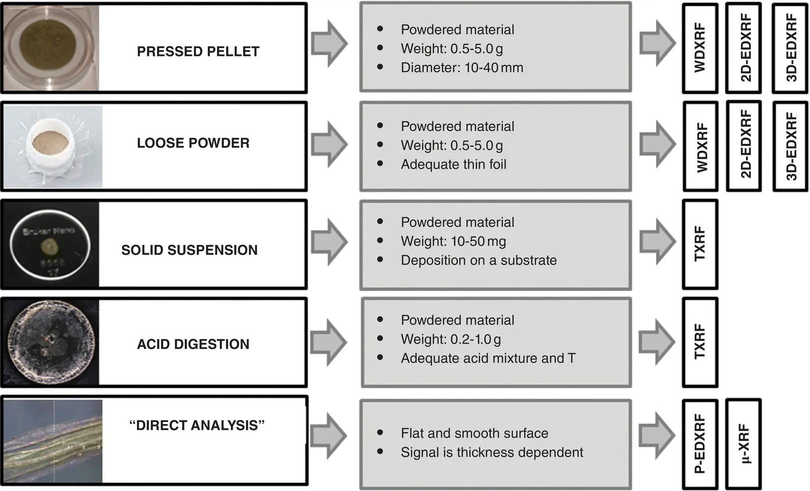
Figure 2.3 Most commonly used sample treatment strategies for vegetation sample analysis using XRF techniques.
One of the most widespread sample preparation strategies for vegetation bulk‐mass sample analysis when using WDXRF, 2D‐EDXRF, and 3D‐EDXRF systems is the fabrication of pressed pellets from the powdered plant material. Prior to the grinding of the sample into fine powder, vegetation specimens are usually freeze dried or oven dried at T ≤ 60° to remove water content of the matrix. Then, to reduce the vegetal material to a fine powder a milling procedure is employed using a grinder (i.e. ball mill). In this sense it is of significance to choose a suitable container (i.e. agate, silicon carbide, boron carbide, tungsten carbide…) in the grinding process in order to overcome contamination problems arising from the grinding material. This fact is of special relevance when elements at trace levels should be determined. For plant sample analysis by XRF, grain size effects are relatively small in comparison with other environmental solid samples such as soils. In fact, a study performed by Omote and co‐workers showed that when the grain size is ≤710 μm, the measured X‐ray intensity became constant [30]. In view of the morphology of plant powder and the capacity to be compacted together, the fabrication of pressed pellets can be performed without the addition of a binder [29]. Nevertheless, in some applications a binding agent (free of contaminant elements and with a low X‐ray absorption coefficient) is added before pelletizing to make pressed pellets more stable and to elude distortion of the pellet's flat surface over time. The most commonly used binding agnets include boric acid, cellulose and wax, and the amount of these substances to be used depends strongly on the vegetal matrix, the amount of sample used to prepare the pellet, and the pellet size. For instance, to make solid pellets of 3.2 cm in diameter, it was necessary to add 150–200 mg of cellulose to 500 mg of savannah grass powder [31]. To press the pellets usually requires a hydraulic or manual press and an adequate die set, which includes a die body, base, a plunger and two polished metal disks. These disks are usually made of a metal alloy such as tungsten carbide which proved to be useful for pressing this type of samples. At present, there are a broad variety of analytical methods in the literature to prepare vegetation pressed pellets involving different sample amounts (usually 0.5–5 g) and pellet dimensions (10–40 mm).
If the amount of plant sample available is limited (i.e. roots), the option to make a stable pellet is unpractical and in this case the vegetation sample can be presented to the XRF spectrometer as a loose powder by packing it in a cell or by distributing the powder on an adhesive tape or X‐ray foil (made of polyester, polypropylene, polycarbonate, etc.). Using this latter approach, a plant layer ranging from 0.8–2.5 mg/cm 2can be obtained [32]. However, in view of the inherent X‐ray absorption by the foil, the sensitivity for low Z elements such as Na is reduced in comparison with the analysis of pressed pellets. Another shortcoming of loose powder preparation is the worse precision of the results obtained due to the bulk density and grain size differences between replicate measurements. In any case, depending on the analytical purpose, the loose powder preparation method can be a fast analytical alternative above all when the amount of sample is limited and WDXRF, 2D‐EDXRF, and 3D‐EDXRF systems are used.
Читать дальшеИнтервал:
Закладка:
Похожие книги на «X-Ray Fluorescence in Biological Sciences»
Представляем Вашему вниманию похожие книги на «X-Ray Fluorescence in Biological Sciences» списком для выбора. Мы отобрали схожую по названию и смыслу литературу в надежде предоставить читателям больше вариантов отыскать новые, интересные, ещё непрочитанные произведения.


Обсуждение, отзывы о книге «X-Ray Fluorescence in Biological Sciences» и просто собственные мнения читателей. Оставьте ваши комментарии, напишите, что Вы думаете о произведении, его смысле или главных героях. Укажите что конкретно понравилось, а что нет, и почему Вы так считаете.