X-Ray Fluorescence in Biological Sciences
Здесь есть возможность читать онлайн «X-Ray Fluorescence in Biological Sciences» — ознакомительный отрывок электронной книги совершенно бесплатно, а после прочтения отрывка купить полную версию. В некоторых случаях можно слушать аудио, скачать через торрент в формате fb2 и присутствует краткое содержание. Жанр: unrecognised, на английском языке. Описание произведения, (предисловие) а так же отзывы посетителей доступны на портале библиотеки ЛибКат.
- Название:X-Ray Fluorescence in Biological Sciences
- Автор:
- Жанр:
- Год:неизвестен
- ISBN:нет данных
- Рейтинг книги:5 / 5. Голосов: 1
-
Избранное:Добавить в избранное
- Отзывы:
-
Ваша оценка:
X-Ray Fluorescence in Biological Sciences: краткое содержание, описание и аннотация
Предлагаем к чтению аннотацию, описание, краткое содержание или предисловие (зависит от того, что написал сам автор книги «X-Ray Fluorescence in Biological Sciences»). Если вы не нашли необходимую информацию о книге — напишите в комментариях, мы постараемся отыскать её.
Discover a comprehensive exploration of X-ray fluorescence in chemical biology and the clinical and plant sciences X-Ray Fluorescence in Biological Sciences: Principles, Instrumentation, and Applications
X-Ray Fluorescence in Biological Sciences: Principles, Instrumentation, and Applications
X-Ray Fluorescence in Biological Sciences — читать онлайн ознакомительный отрывок
Ниже представлен текст книги, разбитый по страницам. Система сохранения места последней прочитанной страницы, позволяет с удобством читать онлайн бесплатно книгу «X-Ray Fluorescence in Biological Sciences», без необходимости каждый раз заново искать на чём Вы остановились. Поставьте закладку, и сможете в любой момент перейти на страницу, на которой закончили чтение.
Интервал:
Закладка:
23 23 Haschke, M. and Boehm, S. (2017). Micro‐XRF in scanning electron microscopes. Adv. Imaging Electron Phys. 199: 1–60.
24 24 Bjeoumikhov, A., Arkadiev, V., Eggert, F. et al. (2005). A new microfocus X‐ray source, iMOXS, for highly sensitive XRF analysis in scanning electron microscopes. X‐Ray Spectrom. 34: 493.
25 25 Haschke, M., Eggert, F., and Elam, W.T. (2007). Micro‐XRF excitation in an SEM. X‐Ray Spectrom. 36: 254–259.
26 26 Bonvin, D. (2019). Analysis of clinker and cement with Thermo Scientific ARL OPTIM’X WDXRF sequential spectrometer. Switzerland: Thermo Fisher Scientific https://assets.thermofisher.com/TFS‐Assets/CAD/Application‐Notes/Analysis‐Clinker‐Cement‐ARL‐OPTIMX‐WDXRF‐41732.pdf
27 27 Fiege, A. and Behrens, K. (2020). Cement: Process‐related analysis of all materials up to the finished product with XRF. https://www.bruker.com/de/news‐and‐events/webinars/2020/process‐related‐analysis‐of‐all‐materials‐up‐to‐the‐finished‐product‐with‐XRF.html.
28 28 Yearly, R. (2015). Better Together: XRF and XRD [internet]. United States: ThermoFisher Scientific https://www.thermofisher.com/blog/mining/better‐together‐xrf‐and‐xrd/.
29 29 Aibéo, C.L., Goffin, S., Schalm, O. et al. (2008). Micro‐Raman analysis for the identification of pigments from 19th and 20th century paintings. J. Raman Spectrosc. 8: 1091–1098.
30 30 Presser, V., Keuper, M., Berthold, C., and Nickel, K.G. (2009). Experimental determination of the Raman sampling depth in zirconia ceramics. Appl. Spectrosc. 63–11: 1288–1292.
2 X‐Ray Fluorescence for Multi‐elemental Analysis of Vegetation Samples
Eva Marguí1 and Ignasi Queralt2
1 Department of Chemistry, University of Girona, Girona, Spain
2 Institute of Environmental Assessment and Water Research, IDAEA‐CSIC, Barcelona, Spain
2.1 Introduction
Presently, the development of analytical methods to determine elemental composition of vegetation samples is of significance in many different fields. For instance, vegetal species are commonly used in environmental studies as bioindicators due to their ability to accumulate metal elements. Vegetables are also an essential part of human diet and thus, the determination of multi‐elemental composition of vegetal foodstuff is also relevant for safety and nutritional purposes. Finally, it is also important to note the considerable number of studies published comparing edible plant growth and development under different fertilizers or farming regimes through laboratory‐controlled and field experiments.
An analytical method's fitness for purpose for the analysis of vegetation samples in the aforementioned fields is related to the fulfillment of some “desired” characteristics ( Figure 2.1). Additionally, in some applications such as the analysis of mass‐limited vegetation samples (i.e. biofilms), or the study of element distribution within the vegetation tissue, more specific analytical characteristics such as microanalytical or imaging capabilities can be also of special interest.
In fact, there exists not one single analytical technique which combines all these features. Multi‐elemental atomic spectrometry techniques such as inductively coupled plasma‐optical emission spectrometry (ICP‐OES) or inductively coupled plasma mass spectrometry (ICP‐MS) are usually employed to measure major, minor and trace elements in vegetal samples [1, 2]. The main drawback of these techniques in the field of vegetation analysis is the need to bring the solid sample into the liquid state by means of a decomposition procedure. Vegetal matrices seem to be very similar to other biological ones which can be decomposed easily using mixtures of strong acids (i.e. nitric acid) and oxidant agents (i.e. hydrogen peroxide). However, there is a clear difference between these types of matrices, namely a higher silicon content (up to 10%) in the case of plant material. This factor complicates the dissolution step and sometimes the addition of stronger acids such as hydrofluoric acid is mandatory to enable a total destruction of the matrix [3]. In such cases, the use of alternative analytical methods that obviate matrix destruction is tempting. Therefore, the development of other methods for direct and multi‐elemental analysis of vegetal samples has been increased in the last years [4]. In this field, it is interesting and needed to highlight the potential of X‐ray fluorescence (XRF) spectrometry.
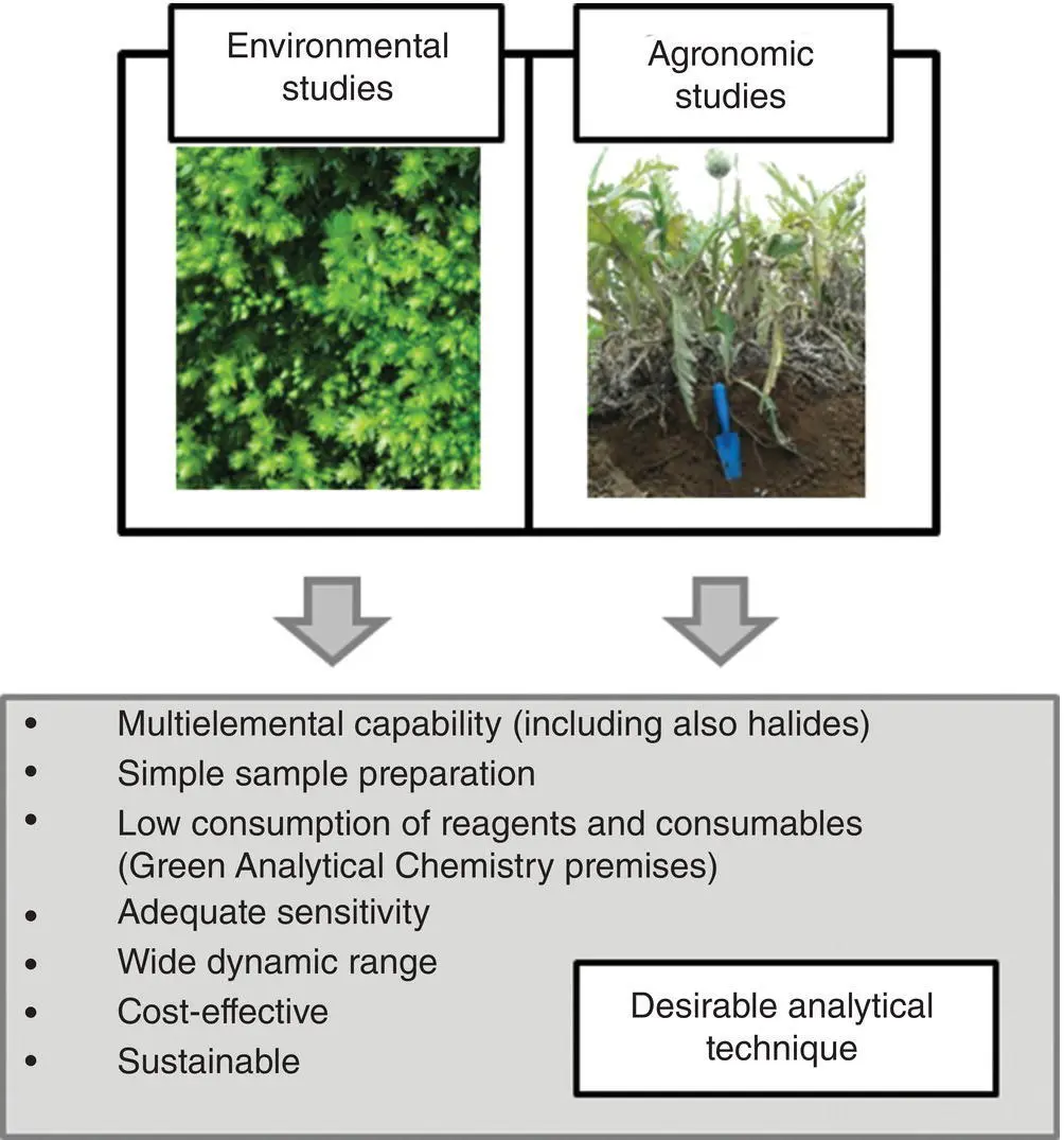
Figure 2.1 Desirable analytical features of a technique to be used for vegetation sample analysis.
Most of XRF configurations comply with the desired features for analysis of vegetal specimens ( Figure 2.1). Perhaps the main drawback, that has restricted a more frequent use of XRF systems in some applications, is the limited sensitivity for trace element determination including some environmentally relevant elements such as Cd and Pb. But still, multi‐elemental XRF is playing an important role in many applications dealing with multi‐elemental analysis of vegetation samples in environmental and agronomic studies.
The present chapter discusses the details and potentials of different XRF configurations in the field of multi‐elemental analysis of vegetation samples. Most commonly used sample treatment procedures have also been described. This chapter includes some of the many interesting applications of XRF in environmental and agronomic studies dealing with the analysis of vegetation samples. Finally, concluding remarks and future perspectives are highlighted.
2.2 Features and Analytical Capabilities of XRF Configurations used in Vegetation Sample Analysis
At present, different types of XRF systems are commercially available. The selection of the most suitable spectrometer is based on the demands for a given purpose. Table 2.1present a summary of the features and analytical capabilities of XRF systems used in vegetation sample analysis. More detailed information about basic principles and components of XRF spectrometers can be found elsewhere [5].
Table 2.1 Features and analytical capabilities of XRF systems used in vegetation sample analysis.
| Feature | WDXRF | 2D‐EDXRF | 3D‐EDXRF | Portable EDXRF | TXRF | μ‐EDXRF |
|---|---|---|---|---|---|---|
| Elements (L: low Z, M: Mid Z, H: High Z) | L, M, H | L, M, H | L, M, H | M, H | M, H | L, M, H |
| Amount of sample | few g | few g | few g | n.a. a | few mg/μl | n.a. a |
| Bulk analysis | Yes | Yes | Yes | Yes | Yes | No |
| Element distribution information | No | No | No | No | No | Yes |
| Limits of detection | mg/kg‐μg/kg | mg/kg | mg/kg‐μg/kg | mg/kg | μg/kg‐ng/kg | ng‐pg |
| Critical moving parts | Crystals | None | Targets | No | Incidence angle | No |
| Portability (in situ analysis) | No | No | No | Yes | No | No |
| Capital costs | High | Medium | High | Low | High/medium | Medium |
| Consumables | Medium | Low | Medium | Low | Low | Low/medium |
an.a.: not applicable (direct analysis of the solid sample).
WDXRF: Wavelength dispersive XRF, 2D‐EDXRF: Two‐dimensional energy dispersive XRF, 3D‐EDXRF: Three‐dimensional energy dispersive XRF or polarized energy dispersive XRF, TXRF: total reflection XRF, μ‐EDXRF: micro‐energy dispersive XRF.
Читать дальшеИнтервал:
Закладка:
Похожие книги на «X-Ray Fluorescence in Biological Sciences»
Представляем Вашему вниманию похожие книги на «X-Ray Fluorescence in Biological Sciences» списком для выбора. Мы отобрали схожую по названию и смыслу литературу в надежде предоставить читателям больше вариантов отыскать новые, интересные, ещё непрочитанные произведения.


Обсуждение, отзывы о книге «X-Ray Fluorescence in Biological Sciences» и просто собственные мнения читателей. Оставьте ваши комментарии, напишите, что Вы думаете о произведении, его смысле или главных героях. Укажите что конкретно понравилось, а что нет, и почему Вы так считаете.