Successful Training in Gastrointestinal Endoscopy
Здесь есть возможность читать онлайн «Successful Training in Gastrointestinal Endoscopy» — ознакомительный отрывок электронной книги совершенно бесплатно, а после прочтения отрывка купить полную версию. В некоторых случаях можно слушать аудио, скачать через торрент в формате fb2 и присутствует краткое содержание. Жанр: unrecognised, на английском языке. Описание произведения, (предисловие) а так же отзывы посетителей доступны на портале библиотеки ЛибКат.
- Название:Successful Training in Gastrointestinal Endoscopy
- Автор:
- Жанр:
- Год:неизвестен
- ISBN:нет данных
- Рейтинг книги:3 / 5. Голосов: 1
-
Избранное:Добавить в избранное
- Отзывы:
-
Ваша оценка:
Successful Training in Gastrointestinal Endoscopy: краткое содержание, описание и аннотация
Предлагаем к чтению аннотацию, описание, краткое содержание или предисловие (зависит от того, что написал сам автор книги «Successful Training in Gastrointestinal Endoscopy»). Если вы не нашли необходимую информацию о книге — напишите в комментариях, мы постараемся отыскать её.
Teaches trainee gastroenterologists the endoscopic skills needed to meet the medical training requirements to practice gastroenterology and helps clinical specialists refresh their skills to pass their recertification Successful Training in Gastrointestinal Endoscopy, Second Edition
Successful Training in Gastrointestinal Endoscopy, Second Edition
Successful Training in Gastrointestinal Endoscopy — читать онлайн ознакомительный отрывок
Ниже представлен текст книги, разбитый по страницам. Система сохранения места последней прочитанной страницы, позволяет с удобством читать онлайн бесплатно книгу «Successful Training in Gastrointestinal Endoscopy», без необходимости каждый раз заново искать на чём Вы остановились. Поставьте закладку, и сможете в любой момент перейти на страницу, на которой закончили чтение.
Интервал:
Закладка:
The next important skill is the development of a slow, careful inspection pattern. Inspection is carried out by developing a circular inspection pattern as the scope is slowly pulled back. This circular pattern does not necessarily need to be done with the scope tip but more with the eyes and only augmented by minor deflections of the scope tip as needed to see the entire circumference of the lumen. Scope readjustments are an ongoing process involving not only the use of the dial controls but also torque of the scope to keep the tip in the center of the lumen. As the scope passes larger folds, it is often necessary to readvance the scope just above the fold and use greater deflection of the scope tip with the dials to view behind the fold and ensure pathology is not missed. In experienced endoscopists, it is felt that a minimum of 6–7 minutes is needed to examine the entire colon adequately [9, 10]. For trainees, this process initially takes much longer due to their developing skills of scope control, inspection behind folds, and pathology recognition. As skills advance, this inspection time will gradually decline. Trainees must clearly understand that while average withdrawal time is a surrogate marker for a careful exam, the key objective is complete mucosal inspection; areas poorly seen due to the colonoscope “jumping” past folds or due to puddles must be reexamined, even if it means reinserting the scope as needed to reinspect.
The final maneuver of a colonoscopy is retroflexion within the rectum. This is performed to better inspect the distal rectum for very low‐lying polyps, internal hemorrhoids, or other perianal pathology. This is best accomplished by returning the patient to their left side if they have been repositioned during the exam. The scope is then inserted to the first (or most distal) semilunar valve in the rectum at roughly 10–12 cm from the anal verge. The large dial is then deflected maximally upward while at the same time the shaft of the scope is torqued in either direction roughly 180° and the scope inserted another few centimeters. When these three steps are done simultaneously, it should result in a view of the distal rectum with the scope shaft entering the rectal vault ( Figure 6.17). The scope is then torqued in either direction to obtain views of the entire circumference of the distal rectum. On some occasions, maximal deflection of the large dial is not enough and the addition of small dial deflection in one direction or the other is needed in order to successfully retroflex the scope. This maneuver should be done with care as it is often uncomfortable for the patient and can result in perforation of the rectum if too much force or torque is used against resistance. If difficulty is encountered, maximally bending the knees toward the chest can also aid in retroflexion, though this is usually not necessary.
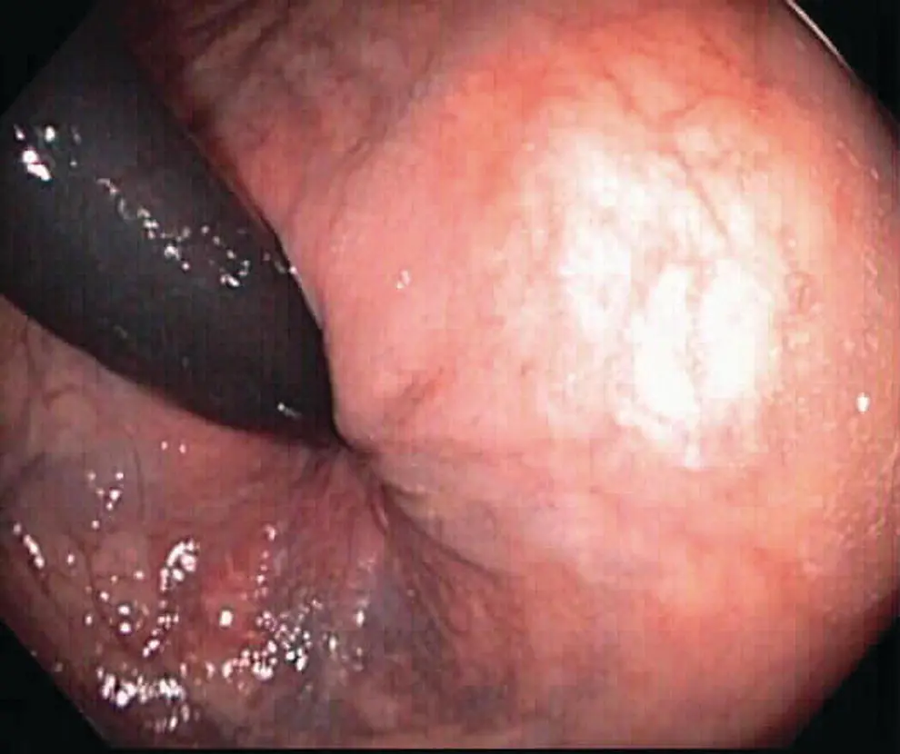
Figure 6.17 Retroflex views in rectum. Retroflexion in the rectum allows for better visualization of the distal rectum where polyps or other pathology such as internal hemorrhoids can often be found.
Intermediate skills
In this section, the focus will be on those cognitive and motor skills required to be proficient at routine colonoscopy. Specifically, this section will address the cognitive skills of pathology recognition, the selection and settings of basic therapeutic devices, and the management of complications. The motor skills addressed here will include the basic management of loops, difficult turns, TI intubation, and the use of the basic biopsy cable and snare. More advanced skills such as those needed in complex or therapeutic endoscopy will be covered in later chapters.
Intermediate cognitive skills
Pathology recognition
Colonoscopy is not simply reaching the cecum and performing a careful inspection during withdrawal; it also involves the ability to identify and manage potential pathologic findings such as polyps or inflammation. Skilled detection of abnormalities requires a careful inspection process and rapid recognition of variances in the mucosa. The method of inspection was discussed earlier in this chapter. Here, the stress will be on the development of recognition of abnormalities. This skill of rapid pathology identification by experienced endoscopists is a result of the cognitive skill of pattern recognition. Pattern recognition is the ability to quickly identify small changes and understand their meaning, much like a master chess player can look at a complex arrangement of chess pieces on a board and quickly understanding the implications, such as which player will eventually win the game and how it can be done. This pattern recognition is the key factor that allows an experienced endoscopist to quickly scan the mucosa and continue the withdrawal with greater speed than trainees. This skill typically is acquired only with experiencing firsthand the broad spectrum of normal and abnormal findings with considerable repetition but can be augmented with the self‐study of photo atlases or online resources (especially for rare endoscopic findings) ( Figure 6.18) (Video 6.4). Over time and with careful supervision and guidance by instructors, this skill is eventually acquired to a sufficient level to operate independently (competence), but skills will continue to be honed throughout the lifetime of the endoscopist. Education researchers have suggested that it takes roughly 10,000 hours of experience to become a “master” at whatever it is one wishes to do, be it playing chess, sports, or performing endoscopy [11]. Basic competence, however, occurs much earlier, yet what defines basic competence in this skill is difficult to assess. Later in this chapter, we will discuss how best to teach these skills of pathology recognition as well as how best to assess these skills as the trainee works toward competence.
Device selection and settings
As fellows begin to identify pathology such as polyps, the next cognitive skill that must be acquired is how to best manage the abnormality. Part of this management is the hands‐on motor skills of applying therapy and will be covered later in this chapter. The cognitive components of this skill include selection of the ideal device, such as a biopsy forceps or cold/electrocautery snare. Additionally, if electrocautery is used, one must also understand what settings to use on the current generator to ensure ablation of the pathologic findings yet minimize risks of post‐treatment ulcerations, bleeding, or perforation. As with all skills that require coordination with an assistant, trainees must become facile with communication of directions. This section will focus on these basic issues as they pertain to simple polyp removal.
The goal of polyp removal is for both diagnostic purposes (histology) as well as therapeutic to ensure no residual adenomatous tissue remains. Very small polyps ( < 3 mm) can typically be removed effectively with simple cold biopsy (i.e., no electrocautery). This is performed by grasping the polyp with a biopsy forceps. The open forceps is placed over the polyp and closed to grasp the entire polyp. With a quick tugging maneuver, the polyp is plucked off the mucosal surface and the cable withdrawn. The tissue is saved for diagnostic microscopic examination. This process results in only a small amount of oozing at the biopsy site and rarely results in any immediate or delayed complications.
Slightly larger polyps pose a different problem. Simple cold biopsy tends not to completely remove the polyp. Some endoscopists may take multiple cold biopsies until the polyp appears removed. This approach is safe but runs the risk of leaving some small amount of adenoma behind and is generally discouraged. Hot biopsy is also a technique that has fallen out of favor due to increased risks for post polypectomy ulceration and bleeding. Instead, the use of cold snare is the generally accepted method for en bloc polypectomy for lesions ranging up to 10 mm in size [12]. This technique is performed by placing an open snare around the polyp with the snare's catheter near the base of the polyp ( Figure 6.19). The snare is then slowly closed by an endoscopy assistant until the wire loop is snuggly around the base of the polyp. Care must be taken to ensure as little normal surrounding tissue is caught within the loop of the snare but also that the entire polyp is included. The assistant is then instructed to apply greater force to cause the wire loop to close further and cut through the tissue at the base of the polyp. The snare is then removed and the polyp tissue is then suctioned up through the scope and collected in a trap placed in the suction circuit. Cold snare polyp removal is quite effective for these slightly larger (3–9 mm) polyps and does not result in much immediate bleeding despite their increased size. Cautery is typically not needed for polyps in this size but can be employed if needed. For lesions ranging from 10 to 20 mm in size, en bloc resection is typically performed with an electrocautery snare.
Читать дальшеИнтервал:
Закладка:
Похожие книги на «Successful Training in Gastrointestinal Endoscopy»
Представляем Вашему вниманию похожие книги на «Successful Training in Gastrointestinal Endoscopy» списком для выбора. Мы отобрали схожую по названию и смыслу литературу в надежде предоставить читателям больше вариантов отыскать новые, интересные, ещё непрочитанные произведения.


Обсуждение, отзывы о книге «Successful Training in Gastrointestinal Endoscopy» и просто собственные мнения читателей. Оставьте ваши комментарии, напишите, что Вы думаете о произведении, его смысле или главных героях. Укажите что конкретно понравилось, а что нет, и почему Вы так считаете.