Magnetic Resonance Microscopy
Здесь есть возможность читать онлайн «Magnetic Resonance Microscopy» — ознакомительный отрывок электронной книги совершенно бесплатно, а после прочтения отрывка купить полную версию. В некоторых случаях можно слушать аудио, скачать через торрент в формате fb2 и присутствует краткое содержание. Жанр: unrecognised, на английском языке. Описание произведения, (предисловие) а так же отзывы посетителей доступны на портале библиотеки ЛибКат.
- Название:Magnetic Resonance Microscopy
- Автор:
- Жанр:
- Год:неизвестен
- ISBN:нет данных
- Рейтинг книги:4 / 5. Голосов: 1
-
Избранное:Добавить в избранное
- Отзывы:
-
Ваша оценка:
Magnetic Resonance Microscopy: краткое содержание, описание и аннотация
Предлагаем к чтению аннотацию, описание, краткое содержание или предисловие (зависит от того, что написал сам автор книги «Magnetic Resonance Microscopy»). Если вы не нашли необходимую информацию о книге — напишите в комментариях, мы постараемся отыскать её.
Explore the interdisciplinary applications of magnetic resonance microscopy in this one-of-a-kind resource Magnetic Resonance Microscopy: Instrumentation and Applications in Engineering, Life Science and Energy Research,
Magnetic Resonance Microscopy: Instrumentation and Applications in Engineering, Life Science and Energy Research
Magnetic Resonance Microscopy: Instrumentation and Applications in Engineering, Life Science and Energy Research
Magnetic Resonance Microscopy — читать онлайн ознакомительный отрывок
Ниже представлен текст книги, разбитый по страницам. Система сохранения места последней прочитанной страницы, позволяет с удобством читать онлайн бесплатно книгу «Magnetic Resonance Microscopy», без необходимости каждый раз заново искать на чём Вы остановились. Поставьте закладку, и сможете в любой момент перейти на страницу, на которой закончили чтение.
Интервал:
Закладка:
Currently, the only other means known to further increase resolution in cell biology, is based on the higher sensitivity and simultaneously higher localization that arises from proximity to the spin. When detector and spin are separated on the order of 1 nm, the dipolar coupling is strong. The techniques, such as magnetic resonance force microscopy [7], or nitrogen-vacancy (NV) centers in nanodiamonds [10], are either invasive (magnetic resonance force microscope [MRFM]) or require similar sample preparation as for STED (NV centers), namely to introduce a mobile quantum emitter that is tagged to a molecule to wander through the cell.
1.1.3 Limit of Imaging Resolution
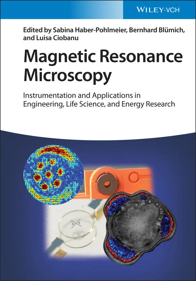
Paul Callaghan [1] and others [11–14] showed that the imaging resolution of MR microscopy is fundamentally limited by three factors: the diffusion coefficient of molecules within the sample, the line broadening due to magnetic susceptibility effects, and the specified SNR per voxel. While the limits placed by the first two factors can be pushed by stronger field gradients and dedicated pulse sequences [15], the rather poor SNR of the MR signal remains as the ultimate fundamental limit of resolution. This can be clearly seen from the following equation [16,17], which summarizes the factors that determine the achievable resolution for a specified SNR of the image:
 (1.2)
(1.2)
where V voxelis the voxel volume, d is the coil diameter, t acqis the total acquisition time, and B 0is the field strength. According to this equation, maintaining the image SNR while, for instance, halving the isotropic resolution (reducing the voxel volume by a factor of 8) necessitates either miniaturizing the coil by a factor of 8, increasing the acquisition time by a factor of 64, or increasing the B 0field by a factor of 3.28. This explains why high-resolution MR images take excruciatingly long to acquire, and why most groups decrease coil diameter. However, at room temperature, coil diameter cannot be reduced indefinitely without disadvantageously increasing coil resistance, so that quality factor Q will ultimately limit this strategy.
1.2 High Resolution From Enhanced Sensitivity
1.2.1 Coil Miniaturization
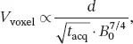
In Equation 1.2, d represents the sensitivity of the NMR detector, which has been proven to increase with decreasing d [18]. Numerous reports have been published on how to improve the NMR detection sensitivity by miniaturizing the detection coil [19–22]. The vast majority of those papers targeted NMR spectroscopy applications, and far fewer talked about microscopy [23–25]. One of the papers that targeted both applications though is the work reported by [26]. In this paper a novel high-resolution NMR/MRI Helmholtz microcoil was introduced (Figure 1.1). The coil that was manufactured using a combination of standard and home-developed micro fabrication technologies featured an extremely user-friendly sample-handling approach that allows easy loading/unloading of the sample.
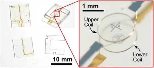
Figure 1.1 A micro Helmholtz coil manufactured using a mixture of standard and home-developed micro-manufacturing technologies. [26] N. Spengler et al. (2014), figure 04 [p.05]/with permission from IOP Publishing Ltd.
The coil was manufactured by stacking three layers, the top and the bottom of which are made of glass, each featuring a wire-bonded 1.5-winding coil using a 25-µm diameter copper wire. The coils were encapsulated in SU-8 epoxy-based photoresist. The glass layers also contained copper traces for the feed and return paths of the current. These layers were spaced by a poly(methyl methacrylate) (PMMA) layer, which was U-shaped to allow the sample-handling microfluidic chip to slide in the sensor. The Helmholtz microcoil was designed and optimized to achieve an extremely uniform B 1field (92% ratio of signal intensity at flip angles of 810/90) while maintaining the high B 0homogeneity (1.79 Hz achieved linewidth of a water sample).
The exceptional performance of the microcoil in terms of B 1uniformity and local field homogeneity allowed high-resolution microimaging. Figure 1.2 shows the optical (left) and MR (right) microimages of a deionized water sample of 154 nl volume. The sample contains 50-µm diameter polymer beads to show some contrast. The MR image that is a sum of 80 acquisition was recorded over a total scan time of 11 h, 22 min, and 40 s. With a matrix size of 256 × 256 and covering a 2.5 × 2.5 mm field of view (FoV), the MR image exhibited an in-plane resolution of 10 × 10 µm for a slice with a 100-µm thickness.
Figure 1.2 MR microimaging of a 154-nl deionized water sample with 50-µm diameter polymer beads. (left) Optical micrograph. (right) MR image. [26] N. Spengler et al. (2014), figure 10 [p.07]/with permission from IOP Publishing Ltd.
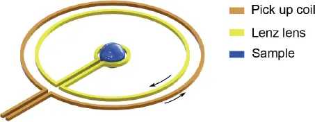
1.2.2 The Lenz Lens: A Tool to Boost Sensitivity
The quest for higher coil sensitivity, SNRcoil ∝ B1/(iR), made researchers invest huge efforts in the design and optimization of the NMR detector. These efforts resulted in numerous publications covering a wide diversity of coil geometries and applications. Nevertheless, none of these efforts went beyond two major pathways: (i) optimizing the coil geometry to better confine the sample and thus achieve a higher filling factor, which, in turn, results in a higher B 1per unit current seen by the sample; and (ii) reducing coil resistance, R , and thus its noise contribution either by a careful selection of coil materials, or by cooling the coil windings. In certain cases, however, these strategies might not be applicable as, for example, if one wants to study the inner body parts. For this particular case, implantable inductively coupled inductor-capacitor (LC) resonators have been shown to enhance the imaging SNR remarkably [27–32]. Nevertheless, despite their advantages, LC resonators suffer from limited frequency accessibility due to their narrow-band nature, which originates from the resonance phenomena, and the difficulty in tunability due to their sensitivity to both the sample and the pick-up coil [33].
An alternative to the LC resonator is the Lenz lens (LL) shown in Figure 1.3. The LL consists of two electrically connected loops: the inner and outer loop, where the outer loop collects the magnetic flux of the pick-up coil and converts it to a current. As this current passes through the inner loop of the LL, it generates a larger magnetic field in the sample than the field that would otherwise be produced by the pick-up coil only. The LL, though less powerful in terms of field amplification compared with the LC resonator, is superior to it in terms of tunability and wide frequency accessibility [33]. Figure 1.4 depicts a simulated comparison of the SNR as the coil geometry increases with respect to the sample volume of a single-loop planar wired coil (blue), a single-loop planar coil with a LL (red), and a single-loop planar coil with an LC resonator (green).
Интервал:
Закладка:
Похожие книги на «Magnetic Resonance Microscopy»
Представляем Вашему вниманию похожие книги на «Magnetic Resonance Microscopy» списком для выбора. Мы отобрали схожую по названию и смыслу литературу в надежде предоставить читателям больше вариантов отыскать новые, интересные, ещё непрочитанные произведения.


Обсуждение, отзывы о книге «Magnetic Resonance Microscopy» и просто собственные мнения читателей. Оставьте ваши комментарии, напишите, что Вы думаете о произведении, его смысле или главных героях. Укажите что конкретно понравилось, а что нет, и почему Вы так считаете.