Magnetic Resonance Microscopy
Здесь есть возможность читать онлайн «Magnetic Resonance Microscopy» — ознакомительный отрывок электронной книги совершенно бесплатно, а после прочтения отрывка купить полную версию. В некоторых случаях можно слушать аудио, скачать через торрент в формате fb2 и присутствует краткое содержание. Жанр: unrecognised, на английском языке. Описание произведения, (предисловие) а так же отзывы посетителей доступны на портале библиотеки ЛибКат.
- Название:Magnetic Resonance Microscopy
- Автор:
- Жанр:
- Год:неизвестен
- ISBN:нет данных
- Рейтинг книги:4 / 5. Голосов: 1
-
Избранное:Добавить в избранное
- Отзывы:
-
Ваша оценка:
Magnetic Resonance Microscopy: краткое содержание, описание и аннотация
Предлагаем к чтению аннотацию, описание, краткое содержание или предисловие (зависит от того, что написал сам автор книги «Magnetic Resonance Microscopy»). Если вы не нашли необходимую информацию о книге — напишите в комментариях, мы постараемся отыскать её.
Explore the interdisciplinary applications of magnetic resonance microscopy in this one-of-a-kind resource Magnetic Resonance Microscopy: Instrumentation and Applications in Engineering, Life Science and Energy Research,
Magnetic Resonance Microscopy: Instrumentation and Applications in Engineering, Life Science and Energy Research
Magnetic Resonance Microscopy: Instrumentation and Applications in Engineering, Life Science and Energy Research
Magnetic Resonance Microscopy — читать онлайн ознакомительный отрывок
Ниже представлен текст книги, разбитый по страницам. Система сохранения места последней прочитанной страницы, позволяет с удобством читать онлайн бесплатно книгу «Magnetic Resonance Microscopy», без необходимости каждый раз заново искать на чём Вы остановились. Поставьте закладку, и сможете в любой момент перейти на страницу, на которой закончили чтение.
Интервал:
Закладка:
Using radio waves taken for convenience at 300 MHz, a thus interpreted refractive MRI system would have a resolution of ~500 mm, which is a dire prospect for applications of MRI. In a seminal paper, Mansfield et al. [2] reported on a form of nuclear magnetic resonance (NMR) diffraction, in which they considered a solid-state periodic lattice of spins in a macroscopically sized lattice, revealing diffraction patterns on the order of the lattice. As a follow-up to this idea, Blümler et al. [3] and Bernhard Blümich [4] reported (the latter in a paper dedicated to Paul Callaghan) on an interesting intertwining of concepts of the k-space vector of refractive MRI and the spatial periodicity of a lattice-like diffractive structure, further exploring diffractive imaging. Blümich’s paper contains a few more gems worth discussing, but would distract us too far from the optical viewpoint we are considering here.
Near-field effects can be further exploited to increase the resolution of an imaging system. At optical wavelengths, one is hardly able to extend beyond 200 nm of resolution with currently available light sources. “The diffraction limit of light is 100 times the size of structures that cell biologists study as they characterise events in organelles or membranes,” Hari Schroff (NIH/NIBIB) is quoted to say in [5], yet below 200 nm “is where most cellular action is,” the author notes. The alternative is to avoid scattering as an imaging paradigm, instead, to image photon sources (also known as quantum emitters).
Interestingly, deep space astronomy always worked this way around by observing photon emitters, so that astronomers only consider objects that were once themselves sources of radiation, such as stars and their predecessors and descendants. In astronomy, the limit of resolution is therefore not dominated by the wavelength of the radiation, which can be very small when compared to the size and distance of the astronomical objects, but rather by the measuring instrument’s principle of operation, its detection sensitivity, and in particular, its effective aperture.
When imaging radiation sources, such as single photon emitters in molecules, we now know that we can greatly improve on the Abbe limit, by about a factor of 10, especially when combined with techniques of stimulated emission and depletion, and one of the numerous variations based on fluorophore emission dynamics. These techniques, which have revolutionized cellular biology and won its inventor Stefan Hell the Nobel Prize in 2014, are of course not accessible to astronomers, who would have to wait too long for excitation signals to pass from observer to object and back again. But for cell biology this is not problematic. Although at currently ~30 nm, the resolution is still far from the desired 1 nm limit, advances in image processing present a feasible route to achieve further improvements. But the technique also raises some questions. Sample preparation is very difficult, and imaging is indirect as fluorophores have to be invasively attached to interesting molecules, almost certainly modifying their behavior.
Magnetic resonance microimaging is a noninvasive technique that is clearly more closely related to stimulated emission depletion (STED) microscopy than to conventional scattering light microscopy. A localized atomic nucleus’ spin is a quantum absorber/emitter. By localizing the excitation field spatially or by frequency, a sub-selection of the spins in a sample can be prepared to absorb radiation. Further localization can ensure that emission of radiation energy is again sub-selected, for example along the geometrical intersection of two orthogonal manifold slices, the one for excitation, the other for emission. A range of further techniques, such as available through relaxation contrast, or phase accumulation, can again further sub-select spins before readout, thereby improving resolution in direct analogy to the techniques of fluorophore emission dynamics. Noninvasive Faraday-detected MRI has been reported down to ~3 µm resolution [6], which is already five orders of magnitude below the radiation wavelength. Nevertheless, even though MRI records radio frequency emissions, this is done almost exclusively through near-field interactions, i.e. by Faraday induction, and not from a beam or ray that requires a lens for focusing.
One of the limitations in NMR is certainly that a single quantum emission event is not yet readily observable as it would be in photonics, even though Dan Rugar showed that a single spin can be observed [7]. Thus all Faraday induction-acquired MRI images have to resort to averaging of a vast number of emission events, and over extended time, to yield useful information. If detection sensitivity were to be increased, fewer emitters could be used, and could perhaps be averaged over shorter times.
1.1.2 Limit of Detection
The statistical polarization level, i.e. that proportion of the total spin population that is available for quantum emission, is an additional cause of lack of signal. Proton spins for example are indistinguishable fermions, with a level occupation that follows
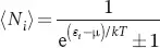 (1.1)
(1.1)
and which collapses to Maxwell–Boltzmann statistics when e (εi−µ)/kT≫ 1, because the energy of a proton flip γħB 0= 3.3 × 10 −25J is tiny compared with the thermal energy kT = 4.11 × 10 −21J. Thus at typical equilibrium polarization levels at 11.7 T, a factor of only 10 −4in excess in population difference with respect to the Fermi level µ can contribute to the signal. An imaging voxel size is therefore principally limited by polarization, because we find – for microcoils at their limit of detection – a sample containing around 10 13spins is needed to form an observable signal. Clearly, this sets a lower concentration limit once the voxel size is specified. For example, at the average size of a single eukaryotic cell of (10 µm) 3, containing the required nuclei, implies a concentration of at least 1.66 µM. By increasing polarization, the voxel size is thus principally reduced, or the lower concentration limit is reduced, which could be achieved by resorting to out-of-equilibrium polarization techniques such as parahydrogen-induced polarization (PHiP), signal amplification by reversible exchange (SABRE), or dynamic nuclear polarization (DNP), all of which are rather hard to perform noninvasively, and hard to selectively localize too. We will return to this point shortly. One of the key advantages of MR-based microscopy is the ability to noninvasively reveal molecular composition, correlated with morphology. From the perspective of biological systems, this can be leveraged to monitor, for example, spatially resolved metabolism. To estimate the best achievable spatial resolution (voxel size), signal-to-noise ratio (SNR) should be considered in the context of the metabolically active system. Key parameters are the molecule abundances (concentrations) and timescale that are targeted. Consider a spatially resolved fluxomic investigation: can one estimate a realistic MRI spatial resolution taking into consideration the expected biological dynamics? Alternatively, what is the smallest biological structure with which metabolic flux can be measured – thus, is it possible to monitor flux at the level of an organelle, single cell, cell cluster, or tissue?
Water is the most abundant molecule in biosystems and can be used as a useful reference from which scaling based on metabolite concentrations can be made. Using only the physical volume of a water molecule (0.03 nm 3), and an optimistic limit of detection (LOD) of 10 13spins, then an order of magnitude estimate of the smallest voxel is 0.1 pl (approximately 4.5 µm isotropic resolution). This is approximately the volume of a single red blood cell. Intracellular metabolite concentrations vary over several orders of magnitude, with the most abundant molecules typically in the tens of millimolar regime. The best-case scenario scaling factor is then 10 4relative to water (assuming [water] = 55 M), and thus the smallest voxel volume increases to 1000 pl (100 µm isotropic resolution). For reference, this would correspond to 10 000 red blood cells or 2 fat cells (volume 600 pl per cell). Can the resolution be improved by signal averaging as a means to enhance SNR? Assuming metabolism is active during the measurement then one must consider the turnover rate of the target metabolite(s) relative to the time over which signal averaging is performed. Enzyme catalytic (second-order) rate constants span several orders of magnitude ( k cat/ K M~10 1–10 9s −1M −1), with a median of ~10 5s −1M −1[8]. If the metabolite concentration is 100 mM, then the metabolite will encounter the “median enzyme” with a rate of 10 4s −1. At this concentration and a volume of 1000 pl, the metabolite concentration would drop below the LOD (~10 mM) in 6000 days giving more than sufficient time for signal averaging. At the diffusion limit 10 9s −1M −1then, 6 days are required before the signal is not observable. This rough estimate takes many liberties in the assumptions (catabolic and anabolic reactions, multiple pathways, enzyme performance, cell cycle, etc. are neglected) and simply suggests that signal averaging is reasonable, most likely limited by technical factors like long-term sample maintenance and spectral resolution accounting for magnetic susceptibility effects. Interesting, it is revealed that single cell metabolic monitoring is challenging yet possible as long as (i) large cells are selected; (ii) the metabolite is among the most abundant in the cell at millimolar concentration; and (iii) the cell can be maintained in an active state during the measurement. Spatial resolution and/or detected concentrations can be improved if hyperpolarization strategies are used where the effective LOD can be improved by orders of magnitude with percent (instead of ppm) levels of polarization. Using the same set of assumptions, now with percent levels of polarization, it is estimated that on the order of a few minutes is required (“median enzyme” kinetics) before the observation of hyperpolarized metabolic products, consistent with observations [9].
Читать дальшеИнтервал:
Закладка:
Похожие книги на «Magnetic Resonance Microscopy»
Представляем Вашему вниманию похожие книги на «Magnetic Resonance Microscopy» списком для выбора. Мы отобрали схожую по названию и смыслу литературу в надежде предоставить читателям больше вариантов отыскать новые, интересные, ещё непрочитанные произведения.


Обсуждение, отзывы о книге «Magnetic Resonance Microscopy» и просто собственные мнения читателей. Оставьте ваши комментарии, напишите, что Вы думаете о произведении, его смысле или главных героях. Укажите что конкретно понравилось, а что нет, и почему Вы так считаете.