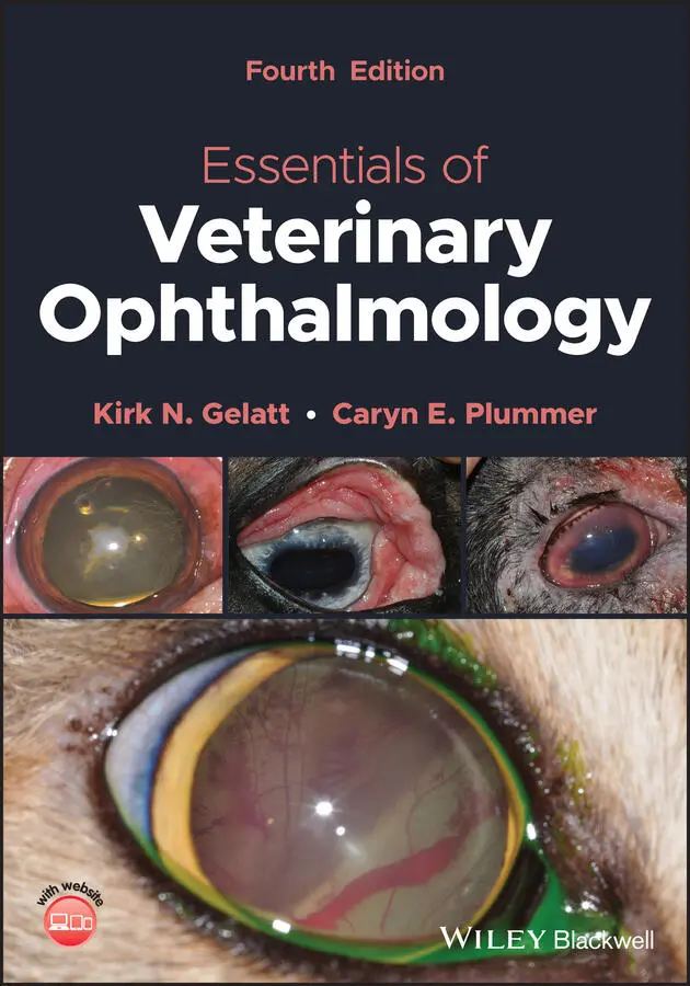
Kirk N. Gelatt - Essentials of Veterinary Ophthalmology
Здесь есть возможность читать онлайн «Kirk N. Gelatt - Essentials of Veterinary Ophthalmology» — ознакомительный отрывок электронной книги совершенно бесплатно, а после прочтения отрывка купить полную версию. В некоторых случаях можно слушать аудио, скачать через торрент в формате fb2 и присутствует краткое содержание. Жанр: unrecognised, на английском языке. Описание произведения, (предисловие) а так же отзывы посетителей доступны на портале библиотеки ЛибКат.
- Название:Essentials of Veterinary Ophthalmology
- Автор:
- Жанр:
- Год:неизвестен
- ISBN:нет данных
- Рейтинг книги:4 / 5. Голосов: 1
-
Избранное:Добавить в избранное
- Отзывы:
-
Ваша оценка:
Essentials of Veterinary Ophthalmology: краткое содержание, описание и аннотация
Предлагаем к чтению аннотацию, описание, краткое содержание или предисловие (зависит от того, что написал сам автор книги «Essentials of Veterinary Ophthalmology»). Если вы не нашли необходимую информацию о книге — напишите в комментариях, мы постараемся отыскать её.
Ophthalmic foundations: ophthalmic development and structure, physiology of the eye and vision, and ocular pharmacology and therapeutics Canine ophthalmology: canine orbit (disease and surgery), canine eyelids (disease and surgery), canine lacrimal apparatus (tear secretion and drainage), canine cornea (diseases and surgery) and canine glaucoma Other species: feline ophthalmology, equine ophthalmology, and food and fiber animal ophthalmology Ophthalmic and systemic diseases: comparative neuro-ophthalmology and systemic disease and the eye
is a useful guide for veterinary students and practitioners looking to build out their core foundations of knowledge within their specific programs of study and disciplines.
Essentials of Veterinary Ophthalmology — читать онлайн ознакомительный отрывок
Ниже представлен текст книги, разбитый по страницам. Система сохранения места последней прочитанной страницы, позволяет с удобством читать онлайн бесплатно книгу «Essentials of Veterinary Ophthalmology», без необходимости каждый раз заново искать на чём Вы остановились. Поставьте закладку, и сможете в любой момент перейти на страницу, на которой закончили чтение.
Интервал:
Закладка:
Scotopic Vision
Rods and Rod Pathways
Cones are inactive in scotopic conditions, and in such an environment our fovea becomes a relative blind spot. Instead, scotopic vision is possible because of the molecular and anatomical characteristics of both rods and the rod pathway. The unique features make an individual rod more sensitive than an individual cone. Another important feature that enables sensitive scotopic vision is the converging nature of the rod pathways. In cats, it has been estimated that in the peripheral retina, the output of approximately 75 000 rod photoreceptors converges on about 5000 rod bipolar cells, which output to 250 amacrine cells, that converge on one ganglion cell.
In many animal species, increased number and density of rods enhance scotopic vision. It can be appreciated that dogs have a higher maximal rod concentration than cats, even though most people associate the latter with greater scotopic sensitivity. This discrepancy may be explained by the structure of the tapetum, which is less reflective in dogs than in cats.
Tapetum
One of the most fascinating adaptations for enhanced scotopic vision is the evolution of a reflective tapetum in the choroid. Light photons striking this layer bounce back onto the retina, thus giving them a second chance to be absorbed by the photoreceptors. This second opportunity is not significant in daytime, as cones absorb enough photons during their “first pass” through the retina. In fact, the tapetum has a detrimental effect on visual acuity in broad daylight, as the light is reflected onto a photoreceptor different from the one in the original trajectory. However, at night this detrimental effect on visual resolution is insignificant as cones are inactive. Instead, the retina benefits from the increased probability that rods will absorb the few photons entering the eye in a dim environment, thus enhancing scotopic vision.
Globe Size
The dimensions of the ocular tissues also contribute to improved scotopic sensitivity in many species. For example, the mean diameter of the cornea in cats and humans is 16.5 and 11.7 mm, respectively. Consequently, much more light enters the cat's eye. Next, light must pass through the pupil. The diameter of a mydriatic pupil in cats and humans is about 12 and 8 mm, respectively, translating into a pupillary aperture of 113 and 50 mm 2, respectively. As a result, far more light passes through the cornea and pupil to reach the feline retina at night, when the pupil is fully dilated. Indeed, it has been calculated that a fully dilated pupil increases the amount of light reaching the retina by 135‐fold in the cat, compared to an 80‐fold increase in humans.
Dark Adaptation
Dark adaptation is a process in which sensitivity of photoreceptors increases, and their threshold decreases. It takes place in darkness following prolonged exposure to bright light, which causes bleaching of a substantial portion of the photopigment and includes several processes. One mechanism of dark adaptation is biochemical and revolves around the re‐synthesis of rhodopsin from free opsin and from recycled (or newly available) 11‐ cis ‐retinal ( Figure 2.16).
Photopic Vision
Light Adaptation
Light adaptation is a process in which cone (and rod) sensitivity decreases, and threshold increases, in response to increased background light intensity and the resulting increased photopigment bleaching. This is a much faster process than dark adaptation. Our eyes begin light adaptation within seconds to a sudden increase in background light intensity, such as that experienced when exiting a dark room. Indeed, it is suggested that the reason for the initial photophobia exhibited when exiting a dark room is an attempt by the eye to preserve the dark‐adapted state of the retina. Several mechanisms account for light adaptation. One is the increased activity of phosphodiesterase, resulting in shorter turnover time for cGMP and accelerating the response kinetics of cones.
Pupil
As with scotopic vision, the pupil also contributes to photopic vision because miosis protects the retina from excessive and harmful amounts of light. It is proposed that the large corpora nigra found on the superior (and often the inferior pupil also) border of the iris in some species provides additional protection as it decreases the amount of light entering the eye from the superior visual field (where the Sun is located), further reducing glare and improving vision in bright light. Moreover, because a miotic slit pupil can block light more efficiently than a miotic circular one, it is suggested that slit pupils have evolved in nocturnal or crepuscular species such as cats and geckos that need to function in daytime.
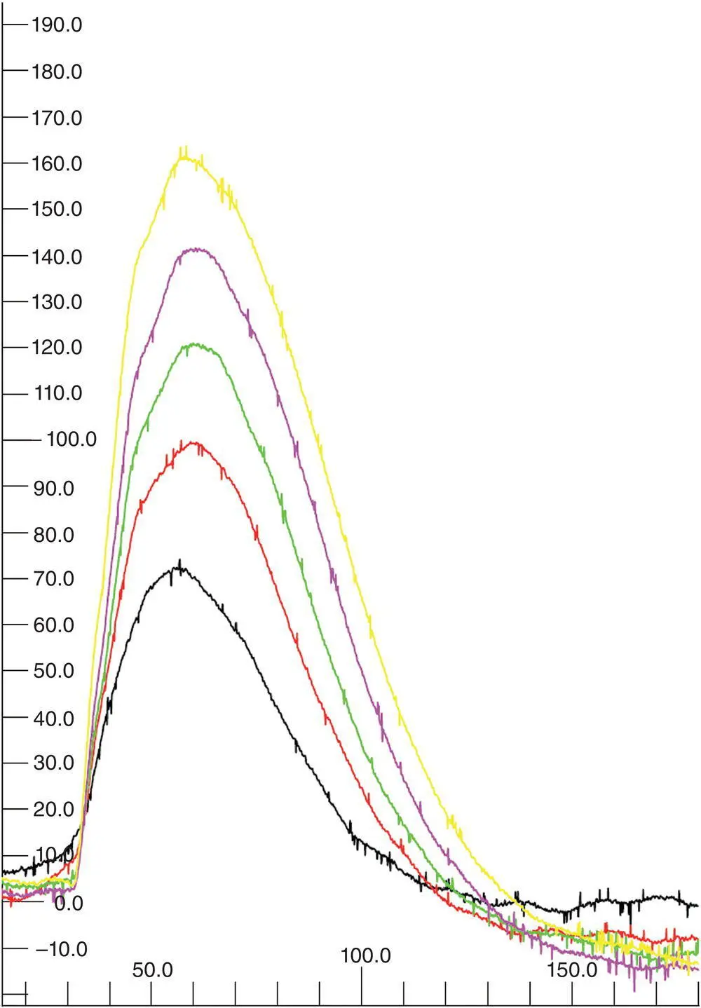
Figure 2.16 In a comprehensive canine ERG protocol, following preparation of the animal in ambient light, the light is turned off. During the next 20 min, the retina is stimulated with a dim flash every 4 min, thus generating a dark adaptation curve. In a normal animal, signal amplitude will increase from one flash to the next as the retina dark adapts (black, red, green, pink, and yellow traces represent the respective responses recorded after 4, 8, 12, 16, and 20 min in the dark). Failure of the signal to increase with time in the dark may be an early sign of rod dysfunction.
Flicker Detection
Temporal responsiveness of the retina has two aspects: motion detection and flicker detection. The retina responds to flashes of light as long as there is sufficient interval between two consecutive flashes, allowing the retina to recover from one response before the next flash is presented. However, as the frequency of these flashes increases, a point is reached at which the retina does not have enough time to recover between flashes and therefore it can no longer distinguish the individual flashes. At this point, which is termed the critical flicker frequency (CFF) or flicker fusion frequency, the eye perceives a steady light even though this light is made up of numerous flickers. There are separate CFFs for rod‐driven responses and signals generated from the different cone types, just as there are separate adaptation mechanisms for the rod and cone systems.
Motion Perception
Moving through the environment produces a flow of images projected onto the photoreceptors, resulting in huge amounts of visual information necessary for navigating through a complex environment. Perception of motion is required for both directing visual attention to a certain location in the visual field and segmenting moving objects from their background. Psychophysically, there are various ways of discerning motion: (i) movement can be perceived if an object is moving across our visual field while our eyes and head are stationary; and (ii) motion can also be perceived when either the head or the eyes are moved to pursue a moving object.
Visual Fields, Binocular Vision, and Depth Perception
It is rather extraordinary to note that all vertebrate species, and many invertebrates, have two eyes. Having more than one eye is required for a large visual field, virtually 360° in some species, and for stereopsis , or depth perception. However, no more than two eyes are required to achieve these aims.
Visual Fields
The extent of the visual field depends largely on placement of the orbits within the skull ( Figure 2.17). Many mammalian prey, avian, and fish species have lateral eyes, providing almost 360° field of view. These animals have a small, frontal binocular field; two large peripheral monocular fields; and a small blind spot behind their head.
Читать дальшеИнтервал:
Закладка:
Похожие книги на «Essentials of Veterinary Ophthalmology»
Представляем Вашему вниманию похожие книги на «Essentials of Veterinary Ophthalmology» списком для выбора. Мы отобрали схожую по названию и смыслу литературу в надежде предоставить читателям больше вариантов отыскать новые, интересные, ещё непрочитанные произведения.


Обсуждение, отзывы о книге «Essentials of Veterinary Ophthalmology» и просто собственные мнения читателей. Оставьте ваши комментарии, напишите, что Вы думаете о произведении, его смысле или главных героях. Укажите что конкретно понравилось, а что нет, и почему Вы так считаете.