Kirk N. Gelatt - Essentials of Veterinary Ophthalmology
Здесь есть возможность читать онлайн «Kirk N. Gelatt - Essentials of Veterinary Ophthalmology» — ознакомительный отрывок электронной книги совершенно бесплатно, а после прочтения отрывка купить полную версию. В некоторых случаях можно слушать аудио, скачать через торрент в формате fb2 и присутствует краткое содержание. Жанр: unrecognised, на английском языке. Описание произведения, (предисловие) а так же отзывы посетителей доступны на портале библиотеки ЛибКат.
- Название:Essentials of Veterinary Ophthalmology
- Автор:
- Жанр:
- Год:неизвестен
- ISBN:нет данных
- Рейтинг книги:4 / 5. Голосов: 1
-
Избранное:Добавить в избранное
- Отзывы:
-
Ваша оценка:
Essentials of Veterinary Ophthalmology: краткое содержание, описание и аннотация
Предлагаем к чтению аннотацию, описание, краткое содержание или предисловие (зависит от того, что написал сам автор книги «Essentials of Veterinary Ophthalmology»). Если вы не нашли необходимую информацию о книге — напишите в комментариях, мы постараемся отыскать её.
Ophthalmic foundations: ophthalmic development and structure, physiology of the eye and vision, and ocular pharmacology and therapeutics Canine ophthalmology: canine orbit (disease and surgery), canine eyelids (disease and surgery), canine lacrimal apparatus (tear secretion and drainage), canine cornea (diseases and surgery) and canine glaucoma Other species: feline ophthalmology, equine ophthalmology, and food and fiber animal ophthalmology Ophthalmic and systemic diseases: comparative neuro-ophthalmology and systemic disease and the eye
is a useful guide for veterinary students and practitioners looking to build out their core foundations of knowledge within their specific programs of study and disciplines.
Essentials of Veterinary Ophthalmology — читать онлайн ознакомительный отрывок
Ниже представлен текст книги, разбитый по страницам. Система сохранения места последней прочитанной страницы, позволяет с удобством читать онлайн бесплатно книгу «Essentials of Veterinary Ophthalmology», без необходимости каждый раз заново искать на чём Вы остановились. Поставьте закладку, и сможете в любой момент перейти на страницу, на которой закончили чтение.
Интервал:
Закладка:
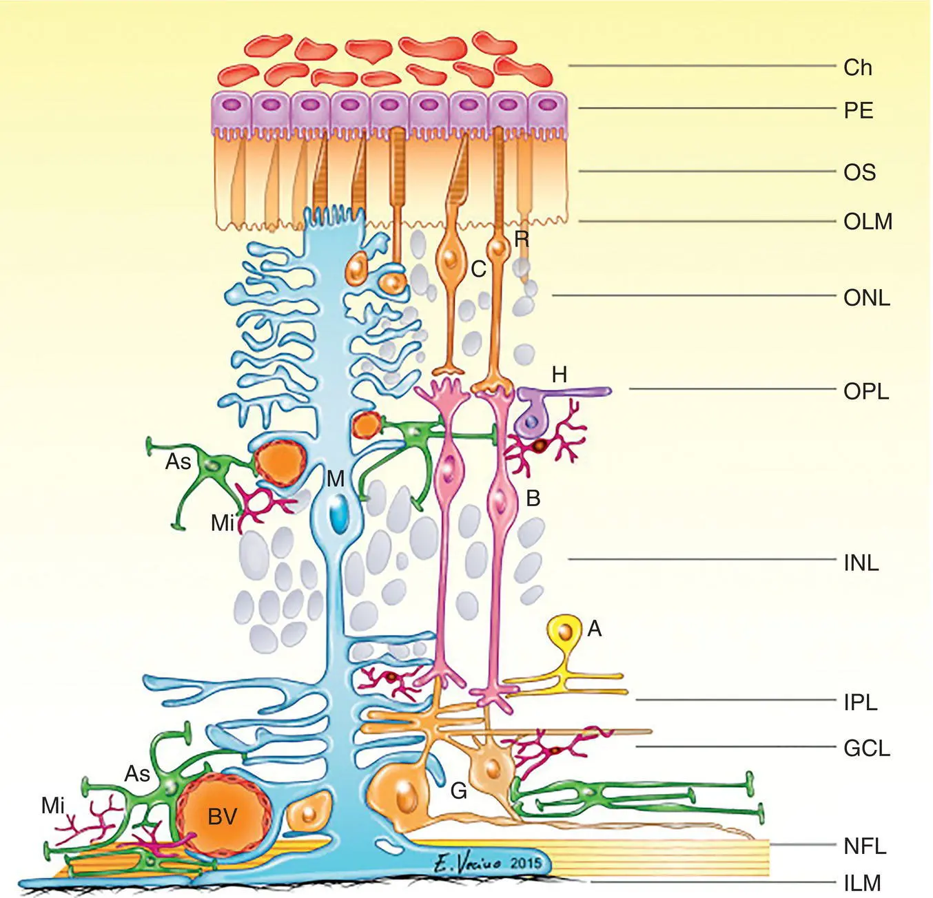
Figure 2.13 Schematic drawing of the mammalian retina with part of the choroid (Ch) on top. Below the retinal pigment epithelium (PE) are the layers of the neuroretina: the outer segments of the photoreceptors (OS), the outer limiting membrane (OLM), the outer nuclear layer with nuclei of cones (C) and rods (R), the neuropil of the OPL, the INL with nuclei of horizontal (H), bipolar (B), and amacrine (A) cells, the inner plexiform layer with synapses in strata, the ganglion cell layer (G), their axons in the nerve fiber layer, and finally the inner limiting membrane (ILM) facing the vitreous. The glial elements of the retina, Müller cells (M) and microglia (Mi), as well as astrocytes (As) embracing retinal blood vessels, are shown.
Optic Chiasm and Optic Tract
As the optic nerve approaches the optic chiasm, the location of fibers within the nerve gradually shifts in preparation for decussation at the optic chiasm. Generally, fibers from the temporal retina remain in the ipsilateral hemisphere, and fibers from the nasal retina cross over to the contralateral side. The amount of decussation varies between species, perhaps representing a broad evolutionary scale. Birds, as well as many amphibian and reptilian species, have complete crossover of fibers to the contralateral side. A greater proportion of fibers remain on the ipsilateral side in mammals that have developed binocular vision. In the horse, 15% of the fibers stay on the ipsilateral side, as do 25% in the dog and 33% in the cat. In humans, only half of the fibers cross over.
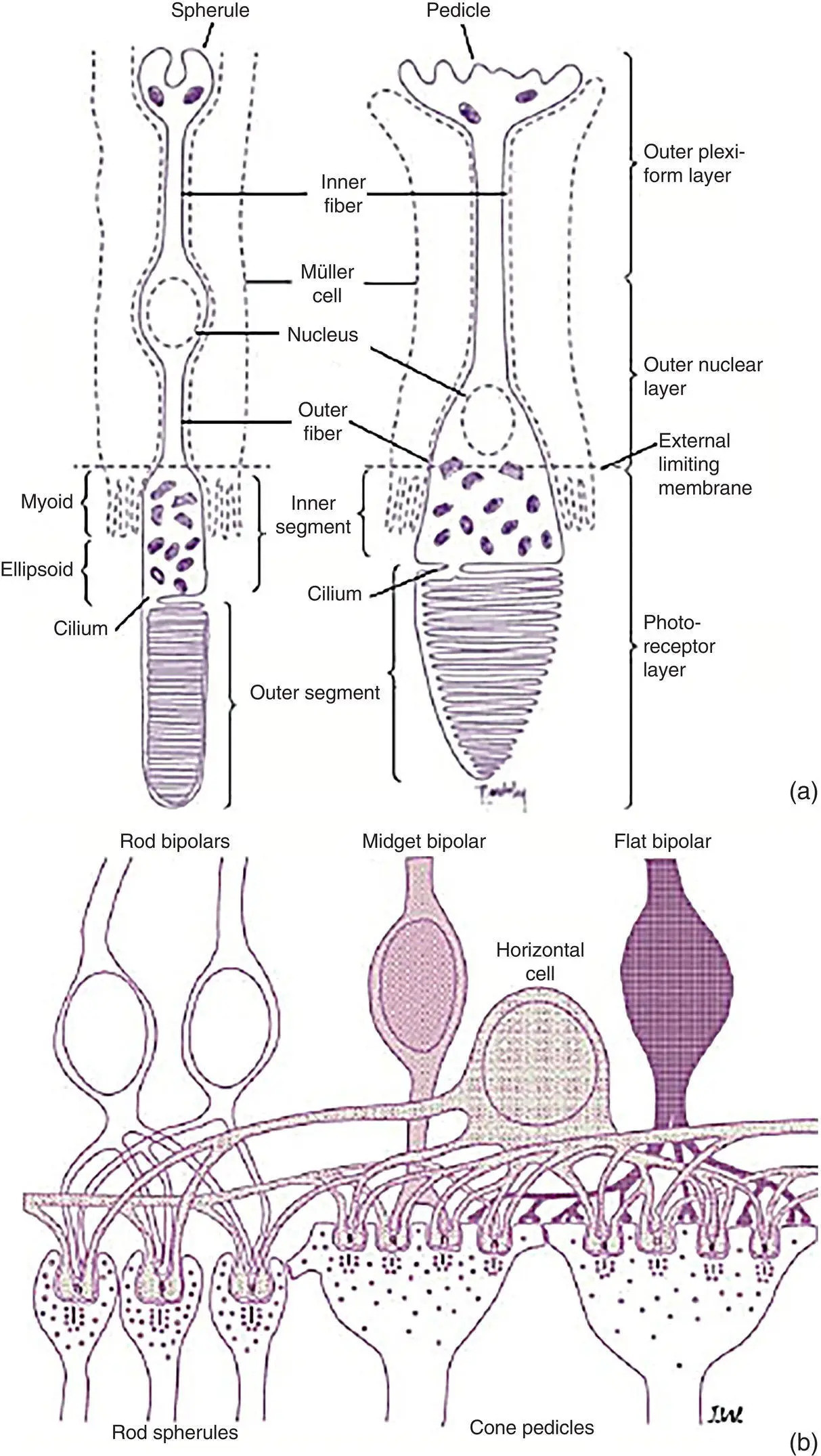
Figure 2.14 (a) The discs of the outer segments (facing the retinal pigment epithelium) of the photoreceptors contain the photopigment required for vision. The photoreceptors' inner segments contain the mitochondria, and together with the outer segments constitute the photoreceptor layer. The rod spherule and cone pedicle are synaptic expansions where their axons synapse with dendrites of bipolar and horizontal cells in the OPL. Portions of Müller's cells (dotted lines) are shown adjoining the rods and cones. (b) Rod and cone bipolar cells show extensive contacts. Horizontal cells also make synapses with both rods and cones. Interconnections are shown between spherules and pedicles.
The optic tract runs from the optic chiasm to the lateral geniculate nucleus (LGN). Because of decussation at the chiasm, fibers of the optic tract conduct information from the opposite visual field of both eyes. In humans, where roughly 50% of the axons decussate in the chiasm, the left optic tract relays the right visual hemifield of both eyes, and the right optic tract relays both left visual hemifields. In animals, where a greater percentage of fibers cross over, the left optic tract will relay a greater proportion of the right visual field from the right eye and a smaller proportion of the right visual field from the left eye.
Lateral Geniculate Nucleus
For most RGC axons, the first synapse occurs in the LGN, which is one of about 10 targets of RGCs in the thalamus. The axons maintain their retinotopic arrangement through the optic nerves, chiasm, and tracts and as they enter the LGN. Here, the RGC axons synapse with dendrites of LGN interneurons (which provide for signal processing) and projecting cells in synaptic glomeruli. In the LGN, RGC axons segregate by eye and functional group, usually forming layers where they terminate in discrete clusters, generating a retinotopic map of the contralateral visual hemifield (with receptive fields similar in size and response properties to the retinal receptive fields).
Primary Visual Cortex
Brodmann demonstrated that the primary visual cortex (i.e., area 17 ) receiving the input from the LGN is located in the posterior part of the occipital lobe in a number of species. This area is now usually called V1 (visual area 1) or the striate cortex , after the striae of Gennari. In contrast, all other visual areas in the cortex lacking the stria (which is a myelinated stripe where the LGN axons enter the gray matter of the V1) are termed extrastriate .
V1 has been mapped in several species. In the cat, it occupies the posteromedial portion of the cortex, extending from the crown of the lateral gyrus on the dorsal surface to the superior bank of the splenial sulcus on the medial surface. In the dog, it is located at the junction of the marginal and endomarginal gyri. The striate cortex has also been identified in the horse.
Section III: Vision
Ability to detect light is, of course, fundamental for vision, but other aspects, such as detection of motion and determining other qualities of an object, such as shape and details, color, size, and distance, help to form our visual percept. The processing of the output from the photoreceptors starts in the retina ( Figure 2.15), but countless neurons in the brain finally shape and interpret the image of the world around us. Animal visual perception is a subject of great fascination to researchers, clinicians and animal owners. Unfortunately, we can neither tell exactly what an animal sees because they usually cannot tell, nor do we know precisely what there is to see because of the limitations of our own visual system.
Scotopic and Photopic Vision
Photoreceptors can respond to changes in levels of background luminance by processes of adaptation and this results in an extended operating range, allowing the eye optimal performance at a given illumination level. A decrease in background illumination to below 0.03 cd/m 2will deactivate the cone system, resulting in increased light sensitivity (i.e., lower threshold ) and scotopic rod vision. An increase in background illumination, to 0.03–3 cd/m 2, will lead to mesopic vision in which both the rod and cone systems are active, for example, before dawn or after sunset. Further increase in background illumination above 3 cd/m 2, to photopic levels, will result in rod saturation. In such an environment, cones will continue to function, albeit with a higher threshold, or with lower sensitivity.
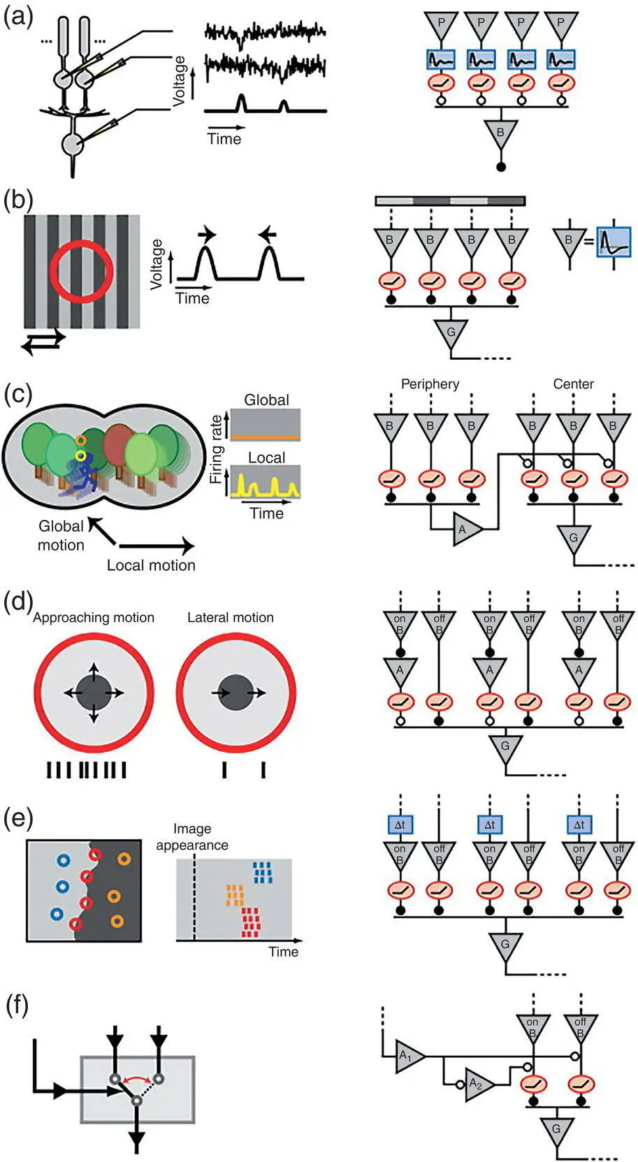
Figure 2.15 A considerable amount of processing of data from the photoreceptors is performed already in the neuroretina. Left‐hand panels briefly describe the purpose of the computations performed, and the right‐hand column illustrates important elements of the underlying circuits schematically (triangle – neuron; A – amacrine cell; B – bipolar cell; G – RGC; P – photoreceptor; rectangle – temporal filter function; oval – instantaneous rectifier; closed/open circle – sign‐preserving/sign‐inverting synapse. (a) The rod‐to‐rod pathway detects single photons. The output of each rod (noisy tracings) is sent through a bandpass temporal filter followed by a thresholding operation. Signals from several rods are then pooled to and summed by one rod bipolar cell, which shows distinct activations (tracings without noise). (b) The Y‐RGC is activated by texture motion in either direction over its receptive field (red circle). Each movement elicits either transient ON or OFF responses in the bipolar cells, but only the depolarized bipolar cells signal to the ganglion cell that fires transiently to each shift in the grating. (c) An RGC sensitive to local motion fires when the object in its central receptive field moves in different direction that from the background, thus detecting differential motion. This RGC is silent when the object in the center moves in the same direction as the background because the excitatory input in the center is counteracted by inhibitory input from the surround via the amacrine cell. (d) A RGC responds strongly (several spikes) to an approaching dark object, but only weakly to lateral motion. More OFF bipolar cells are excited when a larger part of the receptive field is dark. When the object only moves laterally, the RGC receives both excitatory signals from the OFF bipolar cells and inhibitory signals from amacrine cells activated by ON bipolar cells. (e) Specific RGCs use differences in spike latencies to rapidly encode the structure of an image. RGCs with receptive fields (circles) in the dark part of the image have short latencies, and those in the light part have long latencies. RGCs with receptive fields containing both dark and light areas fire in between, thus indicating the position of the border. Here, signals from both ON and OFF bipolar cells are individually rectified, and the timing difference follows from a delay (Δ t ) in the ON pathway. (f) Wide‐field amacrine cells (A 1) are activated during rapid shifts of the image in the retinal periphery, which suppresses the OFF bipolar cell signal and disinhibits the ON bipolar cell through a local amacrine cell (A 2). Hence, this circuit acts like a switch, in this case enabling a signal in the more central part of the retina.
Читать дальшеИнтервал:
Закладка:
Похожие книги на «Essentials of Veterinary Ophthalmology»
Представляем Вашему вниманию похожие книги на «Essentials of Veterinary Ophthalmology» списком для выбора. Мы отобрали схожую по названию и смыслу литературу в надежде предоставить читателям больше вариантов отыскать новые, интересные, ещё непрочитанные произведения.


Обсуждение, отзывы о книге «Essentials of Veterinary Ophthalmology» и просто собственные мнения читателей. Оставьте ваши комментарии, напишите, что Вы думаете о произведении, его смысле или главных героях. Укажите что конкретно понравилось, а что нет, и почему Вы так считаете.