Kirk N. Gelatt - Essentials of Veterinary Ophthalmology
Здесь есть возможность читать онлайн «Kirk N. Gelatt - Essentials of Veterinary Ophthalmology» — ознакомительный отрывок электронной книги совершенно бесплатно, а после прочтения отрывка купить полную версию. В некоторых случаях можно слушать аудио, скачать через торрент в формате fb2 и присутствует краткое содержание. Жанр: unrecognised, на английском языке. Описание произведения, (предисловие) а так же отзывы посетителей доступны на портале библиотеки ЛибКат.
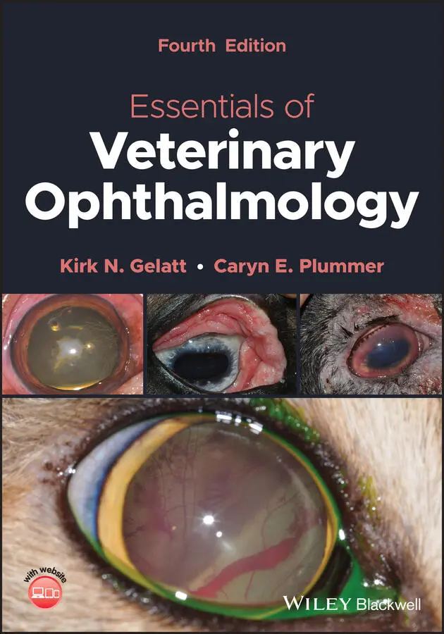
- Название:Essentials of Veterinary Ophthalmology
- Автор:
- Жанр:
- Год:неизвестен
- ISBN:нет данных
- Рейтинг книги:4 / 5. Голосов: 1
-
Избранное:Добавить в избранное
- Отзывы:
-
Ваша оценка:
Essentials of Veterinary Ophthalmology: краткое содержание, описание и аннотация
Предлагаем к чтению аннотацию, описание, краткое содержание или предисловие (зависит от того, что написал сам автор книги «Essentials of Veterinary Ophthalmology»). Если вы не нашли необходимую информацию о книге — напишите в комментариях, мы постараемся отыскать её.
Ophthalmic foundations: ophthalmic development and structure, physiology of the eye and vision, and ocular pharmacology and therapeutics Canine ophthalmology: canine orbit (disease and surgery), canine eyelids (disease and surgery), canine lacrimal apparatus (tear secretion and drainage), canine cornea (diseases and surgery) and canine glaucoma Other species: feline ophthalmology, equine ophthalmology, and food and fiber animal ophthalmology Ophthalmic and systemic diseases: comparative neuro-ophthalmology and systemic disease and the eye
is a useful guide for veterinary students and practitioners looking to build out their core foundations of knowledge within their specific programs of study and disciplines.
Essentials of Veterinary Ophthalmology — читать онлайн ознакомительный отрывок
Ниже представлен текст книги, разбитый по страницам. Система сохранения места последней прочитанной страницы, позволяет с удобством читать онлайн бесплатно книгу «Essentials of Veterinary Ophthalmology», без необходимости каждый раз заново искать на чём Вы остановились. Поставьте закладку, и сможете в любой момент перейти на страницу, на которой закончили чтение.
Интервал:
Закладка:
Nystagmus is usually characterized by an REM in one direction and a slow movement in the opposite direction. Nystagmus can be either horizontal or vertical. The types of nystagmus are categorized on the basis of their causes, and they include optokinetic, rotatory, postrotatory, ocular, caloric, galvanic, anesthetic, brain stem, cerebellar, and vestibular. In optokinetic nystagmus, the eyelids must be open, and the fast phase is opposite in direction to the movement of the visual stimuli. However, the visual stimuli can be moving with the head stationary, or the head and body can be moving with the visual stimuli stationary. In the latter case, the fast phase is in the same direction as the movement of the head. This can be used as an objective means of detecting vision in animals. Optokinetic nystagmus usually occurs in the horizontal plane and is less well substantiated in the vertical plane. In rotatory nystagmus, the fast phase is in the same direction as the rotation of the head. In postrotatory nystagmus, which is seen after rotation stops, the fast phase is opposite to the direction of the rotation of the head. Postrotatory nystagmus lasts approximately 10 s after rotation stops. In rotatory and postrotatory nystagmus, the stimuli are acceleration and deceleration, respectively. At a constant rate of rotation, nystagmus does not occur if the lids are closed. Optokinetic nystagmus occurs if the eyelids are open. Ocular nystagmus is associated with congenital blindness and has a wandering or searching movement of the eyes rather than the distinct fast and slow phases (see Chapter 18for more details).
There are several other types of eye movements. During certain stages of sleep, REM occurs, usually in bursts lasting from 5 to 60 min. Numerous REM bursts, which are traditionally associated with dreaming, may occur during a single sleep. Sleep patterns vary with age, however. Discrete eye movement bursts during certain stages of sleep are infrequent in newborn kittens, but after three weeks of age adult patterns of sleep develop.
Another important class of movements, microsaccades or micronystagmus, are those that maintain eye position while gazing at a stationary target. These movements are required to maintain fixation on the object even when both the observer and the target are immobile. Though slow drifts are also used for this purpose, position maintenance movements are usually characterized by their low magnitude (several minutes/arc) and high frequency (1–50/s).
Kittens are born with a divergent strabismus that is evident following eyelid opening at approximately 12–14 days postnatally. Normal interocular alignment, which depends on visual stimuli, develops during the second postnatal month. Crossed eyes (i.e., convergent strabismus), which are commonly seen in adult Siamese cats and certain albino mammals, result from a genetic neuroanatomical defect in the primary visual pathway that involves the retinogeniculate and geniculocortical projections.
Oculocardiac Reflex
The oculocardiac reflex can cause reflexive slowing of the heart and can be stimulated by pressure on the globe, tension on the EOMs or iris, or increased intraorbital pressure caused by injection, hemorrhage, or a foreign body. The most common effect of the reflex is bradycardia, but other clinically significant effects are cardiac arrest and ventricular fibrillation. The oculocardiac reflex has been reported in humans, dogs, cats, horses, rabbits, mice, and a cockatiel. The afferent arc of the oculocardiac reflex begins with the long and short ciliary nerves to the ciliary ganglion. The ophthalmic division of the trigeminal nerve (CN V) continues to the trigeminal ganglion to its sensory nucleus. The afferent arc continues along short internuncial fibers in the reticular formation to connect with the efferent pathway in the motor nucleus of the vagus nerve (CN X) to the myocardium. Sensory stimulation of the eye and orbital areas results in stimulation of the vagal nucleus in the brain stem, thus causing a reflexive slowing of the heart. Conscious, healthy rabbits and dogs do not show clinically significant decreases in heart rate with globe compression of 1 min. Endotracheal intubation can cause vagal stimulation as well, resulting in similar reflexive cardiac alterations. In the dog, as the IOP increases, the heart rate may also increase, thus indicating the possibility of an intraocular‐sympathetic‐cardiac reflex as well as a trigeminovagal reflex.
Section II: Optics and Physiology of Vision
Introduction
The goal of this section is to describe the physical, anatomical, and physiological aspects of the process of vision, and it has been divided into two parts. The first part is devoted to light and vision and the different refractive structures of the eye. It covers the physical changes that light undergoes during its passage from the cornea, through the various structures of the eye, until it reaches the retina. The second part is devoted to the visual processing and describes what happens once light reaches the retina. This part is dedicated to the neuronal processes of vision and describes the generation, processing, and propagation of the visual signal in the retina, the visual pathways, and the cortex. Both these parts lay the foundations for understanding how optical and neuronal processes enable the detection of movement, details, and color that create the rich experience of vision described in last section of this chapter.
Visual Optics
Physical Optics
Light
Light has alternately been described as a wave or as photon particles. However, these descriptions are not mutually exclusive. Both models also are applicable in the eye: the wave theory explains the physical changes light undergoes during its passage through the eye, and the particle theory explains the energy transformation that occurs when light strikes the outer segments of the photoreceptors. Hence, the first part of this chapter discusses light as a wave, while the second part discusses it as a particle.
Light behaves as a wave as it passes through transparent media such as air, vacuum, or the visual axis of the eye. Much like a wave of water, a wave of light has two principal characteristics ( Figure 2.8). Its amplitude, A , is the maximum value of the field generated by the propagating wave; it determines the wave's intensity. The wavelength, λ , is the distance between adjacent wave crests; it determines the wave's location in the electromagnetic spectrum. Light, which is the visible portion of the electromagnetic spectrum, occupies a small fraction of that spectrum, which ranges from cosmic and gamma rays ( λ < 10 −10m) to radio transmission ( λ > 10 3m). In humans, visible light normally ranges in wavelength from 380 nm (i.e., deep blue) to 780 nm (i.e., deep red). However, additional wavelengths, outside the 380–780 nm spectrum, can be seen by other species. Many nonmammalian species, and some mammals, possess ultraviolet (UV) vision that allows them to detect light with a wavelength shorter than 380 nm, enabling them to see hues that are not perceived by humans; this capability is used in both foraging and courting behavior. The cat retina has also been shown to respond to infrared (IR) light (826–875 nm), though the functional and behavioral implications of this capability are not clear. This is not to be confused with the IR “vision” of snakes, which relies on the heat detection properties of the pit organs.
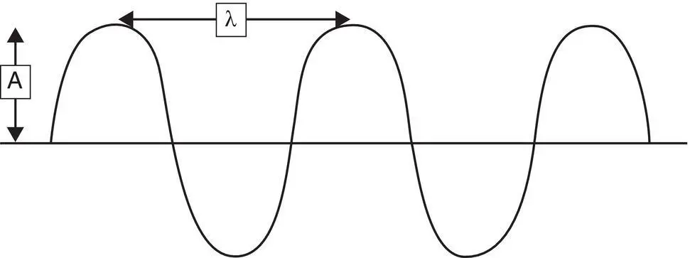
Figure 2.8 Representation of light as a wave, which is characterized by two parameters. Its amplitude ( A ) is the maximum value the wave obtains as it propagates. Its wavelength ( λ ) is the distance between two consecutive peaks.
Читать дальшеИнтервал:
Закладка:
Похожие книги на «Essentials of Veterinary Ophthalmology»
Представляем Вашему вниманию похожие книги на «Essentials of Veterinary Ophthalmology» списком для выбора. Мы отобрали схожую по названию и смыслу литературу в надежде предоставить читателям больше вариантов отыскать новые, интересные, ещё непрочитанные произведения.


Обсуждение, отзывы о книге «Essentials of Veterinary Ophthalmology» и просто собственные мнения читателей. Оставьте ваши комментарии, напишите, что Вы думаете о произведении, его смысле или главных героях. Укажите что конкретно понравилось, а что нет, и почему Вы так считаете.