Poly(lactic acid)
Здесь есть возможность читать онлайн «Poly(lactic acid)» — ознакомительный отрывок электронной книги совершенно бесплатно, а после прочтения отрывка купить полную версию. В некоторых случаях можно слушать аудио, скачать через торрент в формате fb2 и присутствует краткое содержание. Жанр: unrecognised, на английском языке. Описание произведения, (предисловие) а так же отзывы посетителей доступны на портале библиотеки ЛибКат.

- Название:Poly(lactic acid)
- Автор:
- Жанр:
- Год:неизвестен
- ISBN:нет данных
- Рейтинг книги:5 / 5. Голосов: 1
-
Избранное:Добавить в избранное
- Отзывы:
-
Ваша оценка:
Poly(lactic acid): краткое содержание, описание и аннотация
Предлагаем к чтению аннотацию, описание, краткое содержание или предисловие (зависит от того, что написал сам автор книги «Poly(lactic acid)»). Если вы не нашли необходимую информацию о книге — напишите в комментариях, мы постараемся отыскать её.
Poly(lactic acid) Synthesis, Structures, Properties, Processing, Applications, and End of Life
Poly(lactic acid) Synthesis, Structures, Properties, Processing, Applications, and End of Life, Second Edition
Poly(lactic acid) — читать онлайн ознакомительный отрывок
Ниже представлен текст книги, разбитый по страницам. Система сохранения места последней прочитанной страницы, позволяет с удобством читать онлайн бесплатно книгу «Poly(lactic acid)», без необходимости каждый раз заново искать на чём Вы остановились. Поставьте закладку, и сможете в любой момент перейти на страницу, на которой закончили чтение.
Интервал:
Закладка:
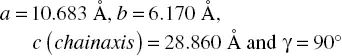
In the X‐ray structure analysis of the α form, the total number of the adjustable parameters of one asymmetric unit (i.e., the coordinates and thermal factors of the atoms in one asymmetric unit) is 101 for C and O atoms of isotropic thermal factors, and 306 for C, H, and O atoms of anisotropic thermal factors. So, for the determination of the accurate crystal structure of the α form, we need to collect the X‐ray diffraction spots of about two to three times larger than the number of the parameters, i.e., 600–900 spots. The usage of an X‐ray beam of a shorter wavelength is useful for this purpose. For example, the X‐ray beam of 0.328 Å wavelength, generated from the synchrotron radiation facility, was incident to the ultra‐drawn PLLA α form sample [14]. From the collected 2D‐WAXD pattern, 700 independent diffraction spots were recognized, which are high enough when compared with the above‐mentioned number of the adjustable parameters. The quantitative analysis was performed manually by reading the positions and integrated intensities of all the observed diffraction peaks. By taking the above‐mentioned problems into consideration, the space group was reduced to the P 12 11 of the monoclinic system, which is lower than the space group P 2 12 12 1. The following unit cell parameters were obtained.
The finally refined crystal structure, which can be assumed as the most accurate crystal structure of the α form, is shown in Figure 6.3. The two antiparallel L‐helices are packed in the unit cell. The chain conformation is approximately a repetition of TTG sequences. But, the individual chain is not symmetric. The two chains are connected by the 2 1screw symmetry along the b axis to give the structure of the alternately upward and downward chains along the c axis. This model can reproduce the observed X‐ray diffraction profiles along all the layer lines quite well as shown in Figures 6.4and 6.5a.
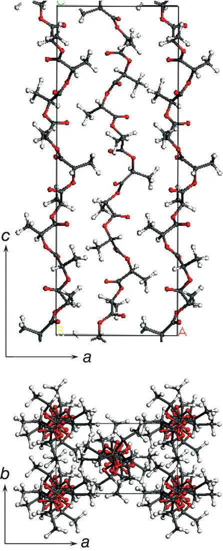
FIGURE 6.3 Crystal structure of PLLA α form. The space group is P 12 11. No symmetry is existent along the chain axis. The helical chains are packed upward and downward along the chain axis alternately.
Source: Reproduced from Wasanusuk et al., Macromolecules 2011, 44, 6441–6452.
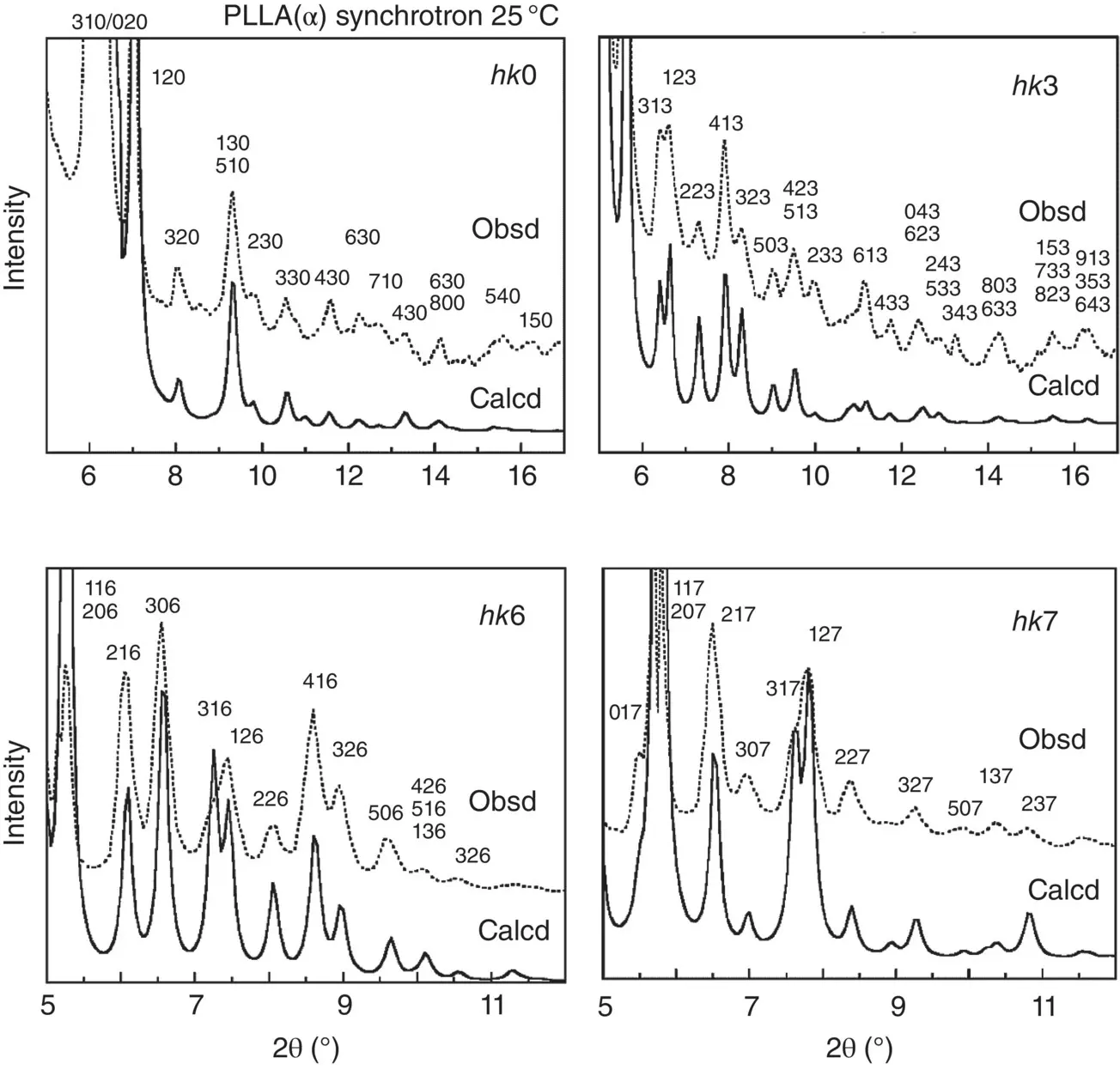
FIGURE 6.4 Comparison between the observed (broken line) and calculated (solid line) X‐ray diffraction profiles of PLLA αform for the several layer lines (25°C).
Source: Reproduced from Wasanasuk et al., Macromolecules 2011, 44, 6441–6552.
The H atom positions, which are important for the theoretical calculation of the mechanical properties, can be determined from the quantitative analysis of the wide‐angle neutron diffraction data, since the neutron beam is scattered almost equally by C, O, and H (and D) atomic species, which is quite in contrast to the X‐ray scattering, in which the scattering amplitude is extremely small for H atoms compared with the C and O atoms [49].
As mentioned above, the finally‐determined space group is P 2 1. This symmetry reduction is needed to generate the 00 l diffraction peaks of the odd l values (see Figure 6.5a). Strictly speaking, even this model could not reproduce the observed 00 l profile perfectly, although most of the hkl diffraction peaks were reproduced satisfactorily enough ( Figure 6.4). This situation requires introducing some structural disorder that was not considered in the structural analyses mentioned above. After many trials, the disordered domain model was found to reproduce the data quite well. Figure 6.5b shows that the domains of a specific size are gathered together with the relative height disorder. The relative‐height shift between the domains is small (0.1–0.2 c ), but it gives a good reproduction of the 00 l diffraction profile as shown in Figure 6.5a. Since the domain size is large enough, the observed X‐ray diffraction profiles of the general hkl peaks are not seriously affected [14].
6.2.3 Crystal Structure of the δ Form
The existence of the δ form (or the old name, α′ form) had been controversial for a long time. Historically, the suggestion of the δ form came from the measurement of the melt‐isothermal crystallization rate, which showed an anomalous peak at around 110–118°C [1, 50]. The DSC thermogram showed double melting peaks in the cold‐crystallization process [51]. Simultaneous DSC and WAXD measurement was performed as shown in Figure 6.6. The unoriented δ form sample was heated gradually, during which the 1D X‐ray diffraction profile was measured stepwise. When the temperature increased to 148–165°C, the 203 and 116 diffraction peaks, which were originally single peaks, changed to double peaks and the relative intensity of these two peaks was exchanged, as indicated by the arrows. This indicates that these two phases are coexistent during the transition, implying a thermodynamically first‐order transition. Besides, several peaks (210 and 213) started to appear, which were assigned to the diffraction peaks of the α form. In this way, the δ form is the crystal form thermodynamically independent of the α form.
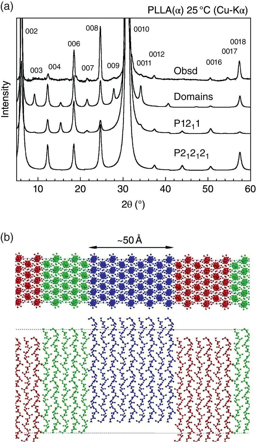
FIGURE 6.5 (a) Comparison of the observed 00 l reflection profile with the calculated curves for both of P 2 12 12 1and P 12 11 models and for the disordered domain model.
Source: Reproduced from Wasanusuk et al., Macromolecules 2011, 44, 6441–6452.
(b) PLLA domain model (several tens Å size along the a axis).
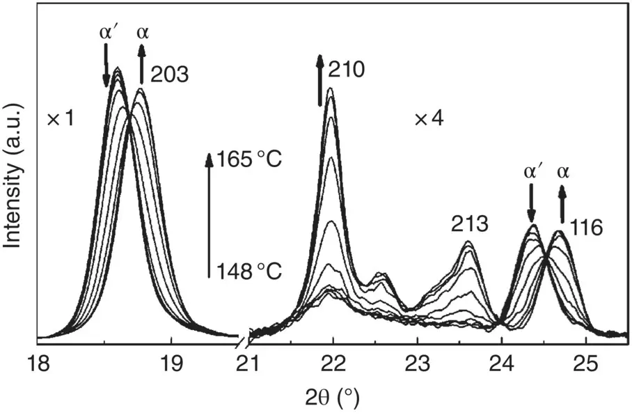
FIGURE 6.6 Temperature dependence of the 1D WAXD profile measured in the phase transition from the δ( α’) to αform of the unoriented PLLA sample (heating process).
Source: Reproduced from Zhang et al., Macromolecules 2008, 41, 1352–1357.
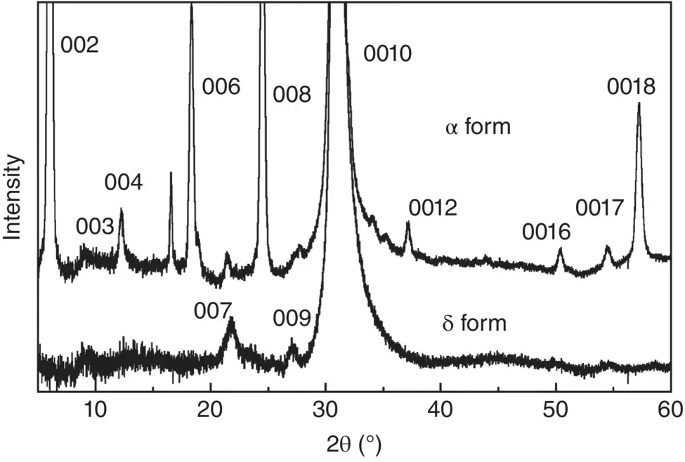
FIGURE 6.7 Observed X‐ray 00 l reflection profiles of PLLA αand δforms.
Source: Reproduced from Wasanasuk and Tashiro, Polymer 2011, 52, 6097–6109.
The crystal structure of the δ form was proposed by analyzing the 2D X‐ray diffraction pattern shown in Figure 6.2b, where the sample was prepared by stretching the melt‐quenched sample followed by annealing at ca . 100°C. In contrast to the α form, the X‐ray diffraction pattern is not very clear and consists of only the 41 broad diffraction spots. In addition, the diffuse streaks are detected relatively strongly along the layer lines, suggesting the existence of the remarkable disorder of the unit cell structure [9]. The orthogonal unit cell was proposed with the parameters a = 10.80 Å, b = 6.20 Å, and c (chain axis) = 28.80 Å. As seen in the 00 l profile ( Figure 6.7), the 0010 peak is quite strong, but the others are appreciably broad and diffuse, which is different from the sharp 00 l peaks observed for the α form. This indicates that the chains takes the 10/3 helical conformation, but they are deformed to more extent than the α form ( Figure 6.3). The detailed analysis of the X‐ray diffraction data revealed that not only the conformational disorder but also the chain packing disorder occurs in the crystal lattice of the δ form. In particular, the relative height between the neighboring chains is appreciably random compared with the α form, as seen from the more remarkable streaks along the layer lines of the 2D X‐ray diffraction pattern [9]. Besides, the aggregation of the domains is also more highly disordered than the α form (see Figure 6.5b). The X‐ray‐coherent crystallite sizes, as evaluated from the half width of the various diffraction peaks using Scherrer’s equation [52], are appreciably smaller than those of the α form (see Section 6.3.1).
Читать дальшеИнтервал:
Закладка:
Похожие книги на «Poly(lactic acid)»
Представляем Вашему вниманию похожие книги на «Poly(lactic acid)» списком для выбора. Мы отобрали схожую по названию и смыслу литературу в надежде предоставить читателям больше вариантов отыскать новые, интересные, ещё непрочитанные произведения.


Обсуждение, отзывы о книге «Poly(lactic acid)» и просто собственные мнения читателей. Оставьте ваши комментарии, напишите, что Вы думаете о произведении, его смысле или главных героях. Укажите что конкретно понравилось, а что нет, и почему Вы так считаете.