Melissa B. Miller - Cases in Medical Microbiology and Infectious Diseases
Здесь есть возможность читать онлайн «Melissa B. Miller - Cases in Medical Microbiology and Infectious Diseases» — ознакомительный отрывок электронной книги совершенно бесплатно, а после прочтения отрывка купить полную версию. В некоторых случаях можно слушать аудио, скачать через торрент в формате fb2 и присутствует краткое содержание. Жанр: unrecognised, на английском языке. Описание произведения, (предисловие) а так же отзывы посетителей доступны на портале библиотеки ЛибКат.
- Название:Cases in Medical Microbiology and Infectious Diseases
- Автор:
- Жанр:
- Год:неизвестен
- ISBN:нет данных
- Рейтинг книги:5 / 5. Голосов: 1
-
Избранное:Добавить в избранное
- Отзывы:
-
Ваша оценка:
Cases in Medical Microbiology and Infectious Diseases: краткое содержание, описание и аннотация
Предлагаем к чтению аннотацию, описание, краткое содержание или предисловие (зависит от того, что написал сам автор книги «Cases in Medical Microbiology and Infectious Diseases»). Если вы не нашли необходимую информацию о книге — напишите в комментариях, мы постараемся отыскать её.
This new
includes:
an entirely new section, «Advanced Cases,» which includes newly recognized disease agents as well as highly complex cases where the interaction of the immune system and human pathogens can be more closely examined a revised «Primer on the Laboratory Diagnosis of Infectious Diseases» section that reflects the increasing importance of molecular-based assays Forty-two new cases that explore the myriad advances in the study of infectious disease in the past decade Thirty-two updated cases that reflect the current state of the art as it relates to the organism causing the infection This textbook also include specific tools to assist students in solving the cases, including a table of normal values, glossary of medical terms, and figures illustrating microscopic organism morphology, laboratory tests, and clinical symptoms.
is a proven resource for preparing for Part I of the National Board of Medical Examiners Exam and an excellent reference for infectious disease rotations.
Cases in Medical Microbiology and Infectious Diseases — читать онлайн ознакомительный отрывок
Ниже представлен текст книги, разбитый по страницам. Система сохранения места последней прочитанной страницы, позволяет с удобством читать онлайн бесплатно книгу «Cases in Medical Microbiology and Infectious Diseases», без необходимости каждый раз заново искать на чём Вы остановились. Поставьте закладку, и сможете в любой момент перейти на страницу, на которой закончили чтение.
Интервал:
Закладка:
Trichrome stain
The trichrome stain is used to visualize protozoans in fecal specimens. This stain is particularly effective at staining internal structures, the examination of which is important in determining the identity of certain protozoans, such as Entamoeba histolytica . Modification of the trichrome stain is used in the detection and identification of microsporidia.
Direct fluorescent-antibody stains
The development of monoclonal antibodies has enhanced both the sensitivity and the specificity of staining techniques that use antibodies to detect microbes in clinical specimens. The most widely used staining technique that incorporates the use of antibodies is the direct fluorescent-antibody (DFA) stain. In this technique, a highly specific antibody is coupled to a fluorochrome, typically fluorescein, which emits an “apple-green” fluorescence. The antibody binds specifically either to antigens on the surface of the microbes or to viral antigens expressed by virally infected cells, which can be visualized under the fluorescent microscope ( Fig. 7). This technique is rapid, usually requiring 1 to 2 hours. In the hands of a skilled operator, the test is highly specific, although it frequently has a sensitivity of only 60 to 70% compared with bacterial culture. Because of its rapidity, the test has been used to detect some relatively slow-growing or difficult-to-grow bacteria, such as Bordetella pertussis and Legionella pneumophila . For respiratory viruses and herpesviruses, the sensitivity of this technique approaches 90% of the sensitivity of culture. However, the development of molecular amplification techniques for the detection of viral agents has demonstrated that DFA sensitivities can be as low as 50%, but may range up to 80% for some viruses. As result, many laboratories have replaced DFA with molecular amplification for organisms such as B. pertussis , herpesviruses, and respiratory viruses.
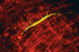
Figure 7
DFA staining is frequently used for the detection of microbes that cannot be cultured. DFA is the method of choice for detection of the nonculturable fungus Pneumocystis jirovecii , a common cause of pneumonia in people with AIDS. DFA is much more sensitive than other commonly used staining techniques, such as silver, Giemsa, or toluidine blue O staining. Likewise, for the gastrointestinal protozoans Giardia lamblia and Cryptosporidium parvum , DFA staining has been found to be much more sensitive than examination of wet mounts or the use of trichrome (for Giardia ) or modified acid-fast stain (for Cryptosporidium ). Molecular amplification techniques are also beginning to be deployed to detect these organisms as well and may soon replace DFA testing.
Infectious disease diagnosis from peripheral blood smears and tissue sections
Not all staining used in the diagnosis of infectious disease is done in the microbiology laboratory. The hematologist and the anatomical pathologist can play important roles in the diagnosis of certain infectious diseases.
The peripheral blood smear is the method of choice for detection of one of the most important infectious diseases in the world, malaria, which is caused by protozoans within the genus Plasmodium . The various developmental stages of these parasites are detected in red blood cells. Other, less frequently encountered parasites seen in a peripheral blood smear include Babesia species, trypanosomes, and the microfilariae.
Bacterial and fungal pathogens may be seen in peripheral smears on occasion. The most likely of these is Histoplasma capsulatum , which is seen as small, intracellular yeasts in peripheral white blood cells. Ehrlichia and Anaplasma can produce characteristic inclusions (morulae), which can be seen in peripheral mononuclear cells and granulocytic cells, respectively.
Examination of tissue by the anatomical pathologist is an important technique for detecting certain infectious agents. Tissue cysts due to toxoplasmosis can be detected in brain biopsy material from patients with encephalitis. The diagnosis of Creutzfeldt-Jakob disease is based on the finding of typical lesions on brain biopsy. The finding of hyphal elements in lung tissue is an important tool in the diagnosis of invasive aspergillosis and pulmonary zygomycosis. The observation of ribbon-like elements in a sinus biopsy is pathognomonic for the diagnosis of rhinocerebral zygomycosis, a potentially fatal disease most frequently seen in diabetic patients.
Antigen detection
Visual examination of a clinical specimen is not the only means by which an infectious agent can be directly detected. A variety of tests have been developed that, like DFA, are dependent on the availability of highly specific antibodies to detect antigens of specific bacteria, fungi, viruses, and parasites. The most widely used antigen detection tests are various formats of the enzyme immunoassay or the latex agglutination assay. These tests take anywhere from 10 minutes to 2 hours. The test most widely used is a 10- to 15-minute enzyme immunoassay for the detection of group A streptococci. The sensitivity of these various formats has been reported to be 80 to 90%, with specificity usually greater than 95%. In the United States, there are more than 50 different test formats marketed for the detection of this organism. The test is done in a wide variety of laboratories, clinics, and physicians’ offices. Antigen detection tests are widely used in the United States to detect a variety of infectious agents, including Cryptococcus neoformans , Clostridium difficile toxin, respiratory syncytial virus, rotavirus, influenza virus, and Giardia and Cryptosporidium spp. It should be noted, however, that as more molecular tests become commercially available and are used as reference methods, the sensitivities of many of the rapid antigen tests deteriorate. For example, published sensitivities for rapid antigen tests for influenza are as low as 10% and those for respiratory syncytial virus are as low as 59%.
MOLECULAR DIAGNOSTICS
In addition to standard methods of culturing and identifying pathogenic microorganisms, there are now a number of molecular methods available that are able to detect the presence of the specific nucleic acid of these organisms. These methods are used in demonstrating the presence of the organism in patient specimens as well as in determining the identification of an isolated organism. In some cases, these methods are able to determine the quantity of the nucleic acid.
As an example, bacteria of a particular species will have a chromosomal nucleic acid sequence significantly different from that of another bacterial species. On the other hand, the nucleic acid sequence within a given species has regions that are highly conserved. For example, the base sequence of the Mycobacterium tuberculosis rRNA differs significantly from the base sequence in the Mycobacterium avium complex rRNA, yet the sequence of bases in this region among members of the M. tuberculosis complex is highly conserved. These properties form the basis for the use of genetic probes to identify bacteria to the species level. There are a number of commercially available genetic probes that can detect specific sequences in bacteria, mycobacteria, and fungi.
Nucleic acid hybridization is a method by which there is the in vitro association of two complementary nucleic acid strands to form a hybrid strand. The hybrid can be a DNA-RNA hybrid, a DNA-DNA hybrid, or, less commonly, an RNA-RNA hybrid. To do this, one denatures the two strands of a DNA molecule by heating to a temperature above which the complementary base pairs that hold the two DNA strands together are disrupted and the helix rapidly dissociates into two single strands. A second nucleic acid sequence is introduced that will bind to regions that are complementary to its sequence. The stringency, or specificity, of the reaction can be varied by reaction conditions such as the temperature.
Читать дальшеИнтервал:
Закладка:
Похожие книги на «Cases in Medical Microbiology and Infectious Diseases»
Представляем Вашему вниманию похожие книги на «Cases in Medical Microbiology and Infectious Diseases» списком для выбора. Мы отобрали схожую по названию и смыслу литературу в надежде предоставить читателям больше вариантов отыскать новые, интересные, ещё непрочитанные произведения.


Обсуждение, отзывы о книге «Cases in Medical Microbiology and Infectious Diseases» и просто собственные мнения читателей. Оставьте ваши комментарии, напишите, что Вы думаете о произведении, его смысле или главных героях. Укажите что конкретно понравилось, а что нет, и почему Вы так считаете.