Melissa B. Miller - Cases in Medical Microbiology and Infectious Diseases
Здесь есть возможность читать онлайн «Melissa B. Miller - Cases in Medical Microbiology and Infectious Diseases» — ознакомительный отрывок электронной книги совершенно бесплатно, а после прочтения отрывка купить полную версию. В некоторых случаях можно слушать аудио, скачать через торрент в формате fb2 и присутствует краткое содержание. Жанр: unrecognised, на английском языке. Описание произведения, (предисловие) а так же отзывы посетителей доступны на портале библиотеки ЛибКат.
- Название:Cases in Medical Microbiology and Infectious Diseases
- Автор:
- Жанр:
- Год:неизвестен
- ISBN:нет данных
- Рейтинг книги:5 / 5. Голосов: 1
-
Избранное:Добавить в избранное
- Отзывы:
-
Ваша оценка:
Cases in Medical Microbiology and Infectious Diseases: краткое содержание, описание и аннотация
Предлагаем к чтению аннотацию, описание, краткое содержание или предисловие (зависит от того, что написал сам автор книги «Cases in Medical Microbiology and Infectious Diseases»). Если вы не нашли необходимую информацию о книге — напишите в комментариях, мы постараемся отыскать её.
This new
includes:
an entirely new section, «Advanced Cases,» which includes newly recognized disease agents as well as highly complex cases where the interaction of the immune system and human pathogens can be more closely examined a revised «Primer on the Laboratory Diagnosis of Infectious Diseases» section that reflects the increasing importance of molecular-based assays Forty-two new cases that explore the myriad advances in the study of infectious disease in the past decade Thirty-two updated cases that reflect the current state of the art as it relates to the organism causing the infection This textbook also include specific tools to assist students in solving the cases, including a table of normal values, glossary of medical terms, and figures illustrating microscopic organism morphology, laboratory tests, and clinical symptoms.
is a proven resource for preparing for Part I of the National Board of Medical Examiners Exam and an excellent reference for infectious disease rotations.
Cases in Medical Microbiology and Infectious Diseases — читать онлайн ознакомительный отрывок
Ниже представлен текст книги, разбитый по страницам. Система сохранения места последней прочитанной страницы, позволяет с удобством читать онлайн бесплатно книгу «Cases in Medical Microbiology and Infectious Diseases», без необходимости каждый раз заново искать на чём Вы остановились. Поставьте закладку, и сможете в любой момент перейти на страницу, на которой закончили чтение.
Интервал:
Закладка:
Fungi are typically divided into two groups based on morphology. One is a yeast ( Fig. 2), which is a unicellular organism, and the other is a mold, which is a multicellular organism with complex ribbon-like structures called hyphae ( Fig. 3). Organisms that are referred to as parasites may be unicellular—the protozoans ( Fig. 4)—or highly complex—the nematodes and cestodes ( Fig. 5). Parasites are typically identified on the basis of morphology alone.
Because of their small size, viruses cannot be visualized by light microscopy. Alternative approaches described below are needed to detect these microbes in clinical specimens.
Wet mounts
The wet mount technique is extremely simple to perform. As the name implies, the clinical specimen is usually mixed with a small volume of saline, covered with a glass coverslip, and examined microscopically. It is most commonly utilized to examine discharges from the female genital tract for the presence of yeasts or the parasite Trichomonas vaginalis . Wet mounts are also used to make the diagnosis of oral thrush, which is caused by the yeast Candida albicans . Using a special microscopic technique—dark-field microscopy—scrapings from genital ulcers and certain skin lesions can be examined for the spirochete Treponema pallidum , the organism that causes syphilis. This technique is not particularly sensitive but is highly specific in the hands of an experienced microscopist. It is typically done in STI clinics where large numbers of specimens are available, enabling the microscopist to maintain his or her skill in detecting this organism.
The wet mount can be modified by replacing a drop of saline with a drop of a 10% KOH solution to a clinical specimen. This technique is used to detect fungi primarily in sputum or related respiratory tract specimens, skin scrapings, and tissues. The purpose of the KOH solution is to “clear” the background by “dissolving” tissue and bacteria, making it easier to visualize the fungi.
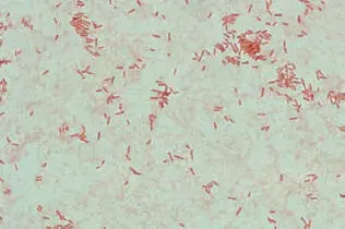
Figure 1a
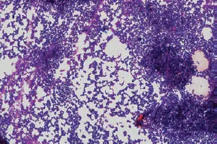
Figure 1b
Figure 2
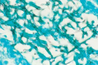
Figure 3
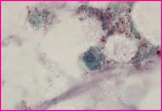
Figure 4
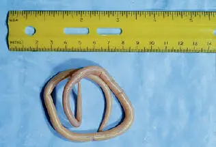
Figure 5
Another modification of the wet mount is to mix a drop of 5% Lugol’s iodine solution with feces. This stains any protozoans or eggs of various worms that may be present in the stool, making them easier to see and identify.
Gram stain
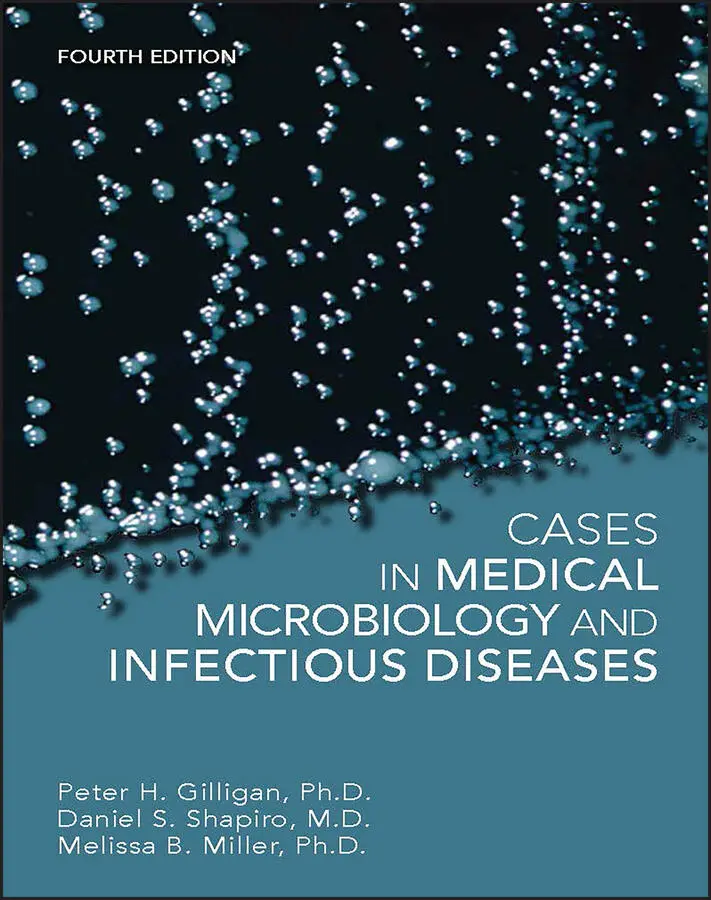
The most frequently utilized stain in the microbiology laboratory is the Gram stain. This stain differentiates bacteria into two groups. One is referred to as Gram positive because of its ability to retain crystal violet stain, while the other is referred to as Gram negative because it is unable to retain this stain (see Fig. 1). These organisms can be further subdivided based on their morphological characteristics.
The structure of the bacterial cell envelope determines an organism’s Gram stain characteristics. Gram-positive organisms have an inner phospholipid bilayer membrane surrounded by a cell wall composed of a relatively thick layer of the polymer peptidoglycan. Gram-negative organisms also have an inner phospholipid bilayer membrane surrounded by a peptidoglycan-containing cell wall. However, in the Gram-negative organisms, the peptidoglycan layer is much thinner. The cell wall in Gram-negative organisms is surrounded by an outer membrane composed of a phospholipid bilayer. Embedded within this bilayer are proteins and the lipid A portion of a complex molecule called lipopolysaccharide. Lipopolysaccharide is also referred to as endotoxinbecause it can cause a variety of toxic effects in humans.
Because of their size or cell envelope composition, certain clinically important bacteria cannot be seen on Gram stain. These include all species of the genera Mycobacterium , Mycoplasma , Rickettsia , Coxiella , Ehrlichia , Chlamydia , and Treponema . Yeasts typically stain as Gram-positive organisms, while the hyphae of molds may inconsistently take up stain but generally will be Gram positive.
Gram stains can be performed quickly, but attention to detail is important to get an accurate Gram reaction. One clue to proper staining is to examine the background of the stain. The presence of significant amounts of purple (Gram positive) in the epithelial cells, red or white blood cells, or proteinaceous material, all of which should stain Gram negative, suggests that the stain is under-decolorized and that the Gram reaction of the bacteria may not be accurate. This type of staining characteristic is frequently seen in “thick” smears. The detection of over-decolorization is much more difficult and is dependent on the observation skills of the individual examining the slide.
Staining of acid-fast organisms
Mycobacterium spp., unlike other bacteria, are surrounded by a thick mycolic acid coat. This complex lipid coat makes the cell wall of these bacteria refractory to staining by the dyes used in the Gram stain. As a result, bacteria within this genus usually cannot be visualized or, infrequently, may have a beaded appearance on Gram stain. Certain stains, such as carbol fuchsin or auramine-rhodamine, can form a complex with the mycolic acid. This stain is not washed out of the cell wall by acid-alcohol or weak acid solution, hence the term “acid-fast” bacterium.
Auramine and rhodamine are nonspecific fluorochromes. Fluorochromes are stains that “fluoresce” when excited by light of a specific wavelength. Bacteria that retain these dyes during the acid-fast staining procedure can be visualized with a fluorescent microscope ( Fig. 6). In clinical laboratories with access to a fluorescent microscope, the auramine-rhodamine stain is the method of choice because the organisms can be visualized at a lower magnification. By screening at lower magnification, larger areas of the microscope slide can be examined more quickly, making this method more sensitive and easier to perform than acid-fast stains using carbol fuchsin and light microscopy.
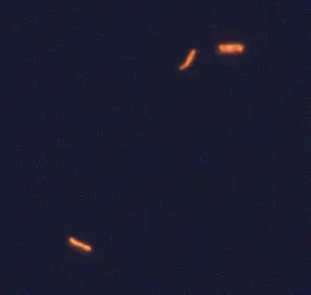
Figure 6
Several other organisms are acid-fast, although they typically are not alcohol-fast. As a result, they are stained using a modified acid-fast decolorizing step whereby a weak acid solution is substituted for an alcohol-acid one. This technique is frequently used to distinguish two genera of Gram-positive, branching rods from each other. Nocardia species are acid-fast when the modified acid-fast staining procedure is used, while Actinomyces species are not. Rhodococcus equi is a coccobacillus that may also be positive by modified acid-fast stain when first isolated. The modified acid-fast stain has also been effective in the detection of two gastrointestinal protozoan parasites, Cryptosporidium and Cyclospora . It should be noted that Cyclospora stains inconsistently, with some organisms giving a beaded appearance while others do not retain the stain at all.
Читать дальшеИнтервал:
Закладка:
Похожие книги на «Cases in Medical Microbiology and Infectious Diseases»
Представляем Вашему вниманию похожие книги на «Cases in Medical Microbiology and Infectious Diseases» списком для выбора. Мы отобрали схожую по названию и смыслу литературу в надежде предоставить читателям больше вариантов отыскать новые, интересные, ещё непрочитанные произведения.


Обсуждение, отзывы о книге «Cases in Medical Microbiology and Infectious Diseases» и просто собственные мнения читателей. Оставьте ваши комментарии, напишите, что Вы думаете о произведении, его смысле или главных героях. Укажите что конкретно понравилось, а что нет, и почему Вы так считаете.