S. Jane Flint - Principles of Virology, Volume 2
Здесь есть возможность читать онлайн «S. Jane Flint - Principles of Virology, Volume 2» — ознакомительный отрывок электронной книги совершенно бесплатно, а после прочтения отрывка купить полную версию. В некоторых случаях можно слушать аудио, скачать через торрент в формате fb2 и присутствует краткое содержание. Жанр: unrecognised, на английском языке. Описание произведения, (предисловие) а так же отзывы посетителей доступны на портале библиотеки ЛибКат.
- Название:Principles of Virology, Volume 2
- Автор:
- Жанр:
- Год:неизвестен
- ISBN:нет данных
- Рейтинг книги:3 / 5. Голосов: 1
-
Избранное:Добавить в избранное
- Отзывы:
-
Ваша оценка:
Principles of Virology, Volume 2: краткое содержание, описание и аннотация
Предлагаем к чтению аннотацию, описание, краткое содержание или предисловие (зависит от того, что написал сам автор книги «Principles of Virology, Volume 2»). Если вы не нашли необходимую информацию о книге — напишите в комментариях, мы постараемся отыскать её.
Volume I: Molecular Biology
Volume II: Pathogenesis and Control
Principles of Virology, Fifth Edition
Principles of Virology, Volume 2 — читать онлайн ознакомительный отрывок
Ниже представлен текст книги, разбитый по страницам. Система сохранения места последней прочитанной страницы, позволяет с удобством читать онлайн бесплатно книгу «Principles of Virology, Volume 2», без необходимости каждый раз заново искать на чём Вы остановились. Поставьте закладку, и сможете в любой момент перейти на страницу, на которой закончили чтение.
Интервал:
Закладка:
Respiratory Tract
Surfaces exposed to the environment but not covered by skin are lined by living cells and are at risk for infection despite the continuous actions of self-cleansing mechanisms. The most common route of viral entry is through the respiratory tract. In a human lung, there are about 300 million terminal sacs, called alveoli, which function in gaseous exchange between inspired air and the blood. Each sac is in close contact with capillary and lymphatic vessels. The combined surface area of the human lungs is ∼180 m 2, approximately the size of a tennis court! At rest, humans inspire ∼6 liters of air per minute. Together, the impressive surface area and large volumes of “miasma” that one inhales each minute imply that foreign particles, such as bacteria, allergens, and viruses, are likely introduced into the lungs with every breath.
Mechanical barriers play a significant role in antiviral defense in the respiratory tract. The tract is lined with a mucociliary blanket consisting of ciliated cells, mucus-secreting goblet cells, and subepithelial mucus-secreting glands ( Fig. 2.6). Foreign particles deposited in the nasal cavity or upper respiratory tract are often trapped in mucus, carried to the back of the throat, swallowed, and destroyed in the low-pH environment of the gut ( Box 2.3). In the lower respiratory tract, particles trapped in mucus are brought up from the lungs to the throat by ciliary action ( Fig. 2.7). Cold temperatures, cigarette smoke, and low humidity cause the cilia to stop functioning effectively, likely accounting for the association of these environmental conditions with increased incidence of respiratory tract infections. When coughing occurs, both the host and the virus benefit; the host expels virus-laden mucus with each productive cough, and the virus is carried out of the host, perhaps to infect another nearby. The lowest portions of the tract, the alveoli, lack cilia or mucus, but macro phages lining the alveoli ingest and destroy virus particles.
BOX 2.2
EXPERIMENTS
Dermal damage increases immunity and host survival
When it was still in use, the smallpox vaccine was delivered by a bifurcated (two-pronged) needle in a process referred to as scarification. Vaccination resulted in local damage to the skin and a subsequent (though quickly re solved) lesion in most individuals that often left a lifelong scar. Until recently, it was not appreciated that the scarification process itself was an important component of the vaccine’s efficacy. Experiments using the smallpox-related virus vaccinia virus showed that intra dermal inoculation of the virus into rabbits resulted in lethal disease by 8 days after infection, whereas delivery by scarification led to a protective host response and animal survival. Scarified rabbits also responded immunologically earlier than those inoculated by the intradermal route. Moreover, scarification in the absence of virus, followed immediately by a same-site intradermal challenge with virus, resulted in significant protection to the infected rabbits. This dramatic difference can be attributed to the rapid induction of a nonspecific host response caused by the scarification wound itself. Scarification damages skin cells and the underlying epidermis, inducing the release of cytokines and chemokines that help direct the host’s immune response to the site of infection and restrict the dissemination of the virus throughout the host.
Rice AD, Adams MM, Lindsey SF, Swetnam DM, Manning BR, Smith AJ, Burrage AM, Wallace G, MacNeill AL, Moyer RW. 2014. Protective properties of vaccinia virus-based vaccines: skin scarification promotes a nonspecific immune response that protects against orthopoxvirus disease. J Virol 88:7753–7763.
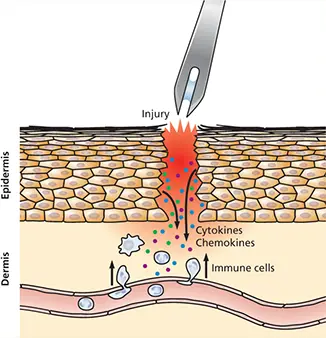
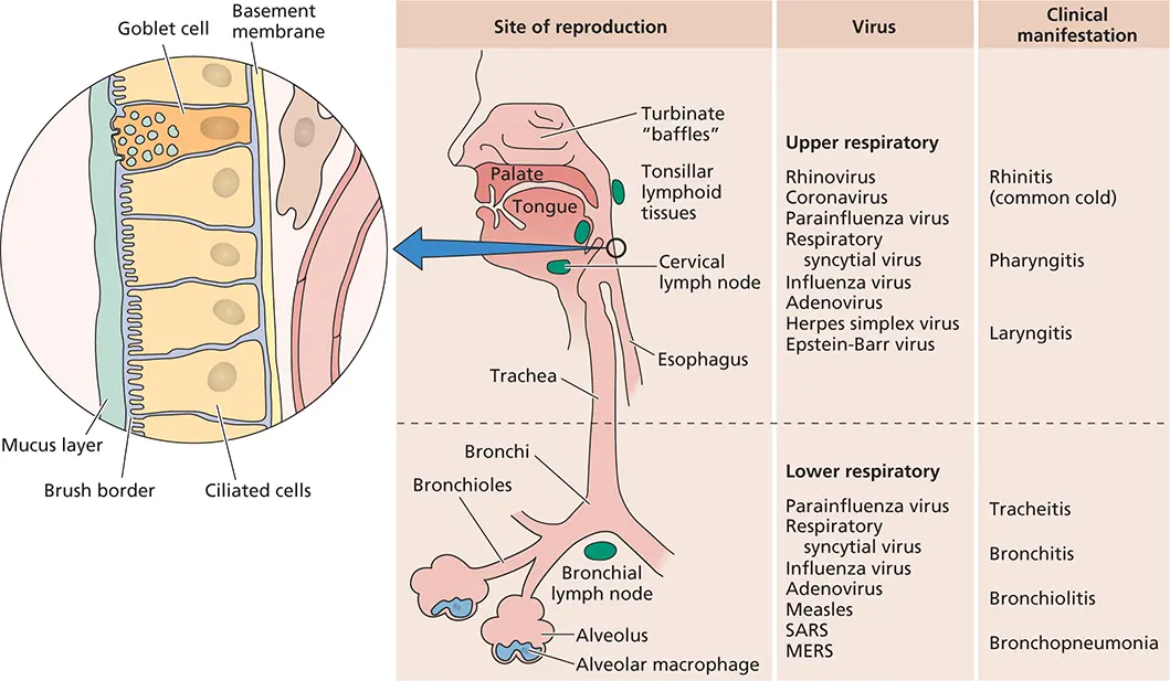
Figure 2.6 Sites of viral entry in the respiratory tract. (Left) A detailed view of the respiratory epithelium. A layer of mucus, produced by goblet cells, is a formidable barrier to virus particle attachment. Virus particles that traverse this layer may reproduce in ciliated cells or pass between them, reaching another physical barrier, the basement membrane. Beyond this extra cellular matrix are tissue fluids, from which particles may be taken into lymphatic capillaries and reach the blood. Local macrophages patrol the tissue fluids in search of foreign particles. (Right) Viruses that reproduce at different locations within the respiratory tract, noted with the associated clinical syndromes. SARS, severe acute respiratory syndrome; MERS, Middle East respiratory syndrome. Adapted from Mims CA et al. 1995. Mims’ Pathogenesis of Infectious Disease (Academic Press, Orlando, FL).
BOX 2.3
DISCUSSION
In praise of mucus
When you have a cold or sinus infection, it can be disconcerting to take a peek in your tissue after you have blown your nose. However, the thick, colorful mucus that accompanies many such infections actually serves an important purpose. Mucus-producing cells line the mouth, nose, sinuses, throat, lungs, vagina, and entire gastrointestinal tract. In addition to its lubricant function, mucus acts as a protective blanket over these surfaces, preventing the tissue underneath from dehydrating. Mucus also acts as a pathogen flypaper, trapping viruses and bacteria. More than being just a sticky goo, mucus contains antibodies, enzymes that destroy the invaders it traps, and a variety of immune cells poised to respond to pathogens that attach to it.
It is a common misconception that yellow or green mucus is directly due to the presence of bacteria or viruses. When an individual ac quires a respiratory tract infection, neutrophils, a key element of the host innate response, rush to the infected site. These cells contain an enzyme, myeloperoxidase, that is critical for the ability of neutrophils to eliminate pathogens, as individuals with a genetic loss of this enzyme are immunocompromised, especially for respiratory tract infections. Myeloperoxidase is stored in azurophilic granules prior to release; these granules are naturally green or tan. Thus, when neutrophils are present in large numbers, the mucus appears green. One may indeed assume that discolored mucus is a sign of infection, as recruitment of neutrophils often accompanies infection.
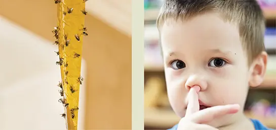
One final thought that you may wish you did not know: while it is not a very socially acceptable practice, eating one’s own nasal secretions (mucophagy), a habit of many young children, may have some evolutionary benefit. Some have argued that mucophagy pro vides benefits to the immune system, especially the underdeveloped host responses of children. As noted above, mucus destroys most of the pathogens that it tethers, so nasal secretions themselves are unlikely to be laden with infectious virus particles. Rather, introducing these crippled microorganisms into the gut, where antigen-presenting cells are abundant, may be a form of “low-tech” vaccination or immune memory booster.
Bellows A. 2009. A booger a day keeps the doctor away, p 28–30. In Alien Hand Syndrome and Other Too-Weird-Not-To-Be-True Stories. Workman Publishing, New York, NY.
Many viruses enter the respiratory tract in the form of aerosolized droplets expelled by an infected individual by coughing or sneezing ( Fig. 2.8). These include well-known vi ruses such as influenza and rhinoviruses that cause the common cold, but less-known pathogens, including adenoviruses, respiratory syncytial virus, measles, mumps, and hantaviruses, are also transmitted via respiratory droplets. Infection can also spread through contact with respiratory secretions or saliva from an infected individual. Larger, virus-containing droplets are deposited in the nasal mucosa, whereas smaller droplets can penetrate deeper into the airways or the alveoli. To infect the respiratory tract successfully, virus particles must not be captured or swept away by mucus, neutralized by antibody, or destroyed by alveolar macrophages. Attributes of some viruses, such as the neuraminidase protein of influenza virus, facilitate penetration of the thick mucus to enable access to permissive cells below. Influenza neuraminidase cleaves sialic acids that are abundant on the glycoproteins that form mucus; as a result, mucous membranes are degraded locally, affording access to the cells below. Oseltamivir (also known by the brand name Tamiflu), an antiviral that reduces influenza virus symptoms, blocks the function of neuraminidase and thus reduces the risk of infection, but this mechanism also explains why oseltamivir must be taken early after infection to be effective.
Читать дальшеИнтервал:
Закладка:
Похожие книги на «Principles of Virology, Volume 2»
Представляем Вашему вниманию похожие книги на «Principles of Virology, Volume 2» списком для выбора. Мы отобрали схожую по названию и смыслу литературу в надежде предоставить читателям больше вариантов отыскать новые, интересные, ещё непрочитанные произведения.


Обсуждение, отзывы о книге «Principles of Virology, Volume 2» и просто собственные мнения читателей. Оставьте ваши комментарии, напишите, что Вы думаете о произведении, его смысле или главных героях. Укажите что конкретно понравилось, а что нет, и почему Вы так считаете.