Anil Bangalore Shivappa - An Illustrated Atlas of Tooth Carving and Wax-Up Techniques
Здесь есть возможность читать онлайн «Anil Bangalore Shivappa - An Illustrated Atlas of Tooth Carving and Wax-Up Techniques» — ознакомительный отрывок электронной книги совершенно бесплатно, а после прочтения отрывка купить полную версию. В некоторых случаях можно слушать аудио, скачать через торрент в формате fb2 и присутствует краткое содержание. Жанр: unrecognised, на английском языке. Описание произведения, (предисловие) а так же отзывы посетителей доступны на портале библиотеки ЛибКат.
- Название:An Illustrated Atlas of Tooth Carving and Wax-Up Techniques
- Автор:
- Жанр:
- Год:неизвестен
- ISBN:нет данных
- Рейтинг книги:4 / 5. Голосов: 1
-
Избранное:Добавить в избранное
- Отзывы:
-
Ваша оценка:
An Illustrated Atlas of Tooth Carving and Wax-Up Techniques: краткое содержание, описание и аннотация
Предлагаем к чтению аннотацию, описание, краткое содержание или предисловие (зависит от того, что написал сам автор книги «An Illustrated Atlas of Tooth Carving and Wax-Up Techniques»). Если вы не нашли необходимую информацию о книге — напишите в комментариях, мы постараемся отыскать её.
Contains information on the pre-carving preparation of wax blocks Provides a description of anatomical landmarks Offers a complete and stepwise guide to the carving and wax-up of each tooth Includes video resources, located on the companion website, to assist students in the procedure
is perfect for undergraduate and graduate students in dentistry who aim to improve their cognitive and psychomotor skills.
An Illustrated Atlas of Tooth Carving and Wax-Up Techniques — читать онлайн ознакомительный отрывок
Ниже представлен текст книги, разбитый по страницам. Система сохранения места последней прочитанной страницы, позволяет с удобством читать онлайн бесплатно книгу «An Illustrated Atlas of Tooth Carving and Wax-Up Techniques», без необходимости каждый раз заново искать на чём Вы остановились. Поставьте закладку, и сможете в любой момент перейти на страницу, на которой закончили чтение.
Интервал:
Закладка:
Figures 3.5–3.8 Sequence of steps to divide the wax blocks before the actual carving steps.
Division of Wax Block
Divide each end of the wax block into four equal parts. Taking the guidance of the cross lines at the ends divide each side of the wax block into two equal parts. The midline drawn on the wax block would help in placing the incisal edge of a tooth either buccally or lingually or in the centre to the long axis (the midline drawn), depending on the tooth ( Figures 3.5–3.8).
References
1 1 Widjijono, Purwanto, A., and Dyah, I. (2009). Mechanical properties of carving wax with various Ca‐bentolite filter composition. Dental Journal 42 (3): 114–117.
2 2 Linek, H.A. (1949). Tooth Carving Manual, 1e. Long Island City, NY: Columbia Dentoform Corporation.
3 3 Rashmi, G.S. (2014). Textbook of Dental Anatomy, Physiology and Occlusion, 1e. New Delhi: Jaypee Brothers Medical Publishers Ltd.
4 Anatomical Landmarks
LEARNING OBJECTIVE
At the end of the chapter, the student should have knowledge of various anatomical landmarks of anterior and posterior teeth that enhances the psychomotor skills for carving and waxing techniques.
Crown
Anatomic crown: Part of the tooth covered by enamel ( Figure 4.1a) [1, 2].
Clinical crown: Part of the crown, visible in the oral cavity ( Figure 4.1a) [1].
Root/Radicular Part
Anatomic root: Part of a tooth covered by cementum ( Figure 4.1b) [1].
Clinical root: Part of a tooth covered with gingiva ( Figure 4.1b) [1].
Median Line
The imaginary line that runs through the centre of the face, between the central incisors at their point of contact both in the maxilla and the mandible ( Figure 4.1c) [1, 3].
Aspect
Labial Aspect
Features of the labial surface bordered by mesial, distal cervical, and incisal outlines ( Figure 4.1d).
Lingual/Palatal Aspect
Features of the lingual surface bordered by mesial, distal cervical, and incisal outlines ( Figure 4.1d).
Mesial Aspect
Features of the mesial surface bordered by labial, lingual/palatal, cervical, and incisal outlines ( Figure 4.1e).
Distal Aspect
Features of the distal surface bordered by labial, lingual/palatal, cervical, and incisal outlines ( Figure 4.1e).
Incisal Aspect
Features seen from a view bordered by labial, lingual/palatal, mesial, and distal outlines ( Figure 4.1f).
Surfaces
‘Facial surfaces’ is a collective term for the surfaces of teeth facing towards lips or buccal surfaces. Individually, the surfaces of the anterior teeth (incisors and canine) facing towards the lips are called labial surfaces. Those surfaces of posterior teeth facing towards the cheek are known as buccal surfaces. Those surfaces of mandibular teeth facing towards the tongue are called lingual surfaces. Lingual surfaces on maxillary teeth are also known as palatal surfaces. Surfaces of a tooth contacting adjacent teeth in the same dental arch are called proximal surfaces. The proximal surfaces of teeth facing towards the median line are called mesial surfaces (Greek mesos ; middle). The proximal surfaces facing away from the median line or midline of an arch are known as distal surfaces. The point at which the teeth contact each other is the contact area, which can be mesial or distal ( Figure 4.1g and h) [1–3].
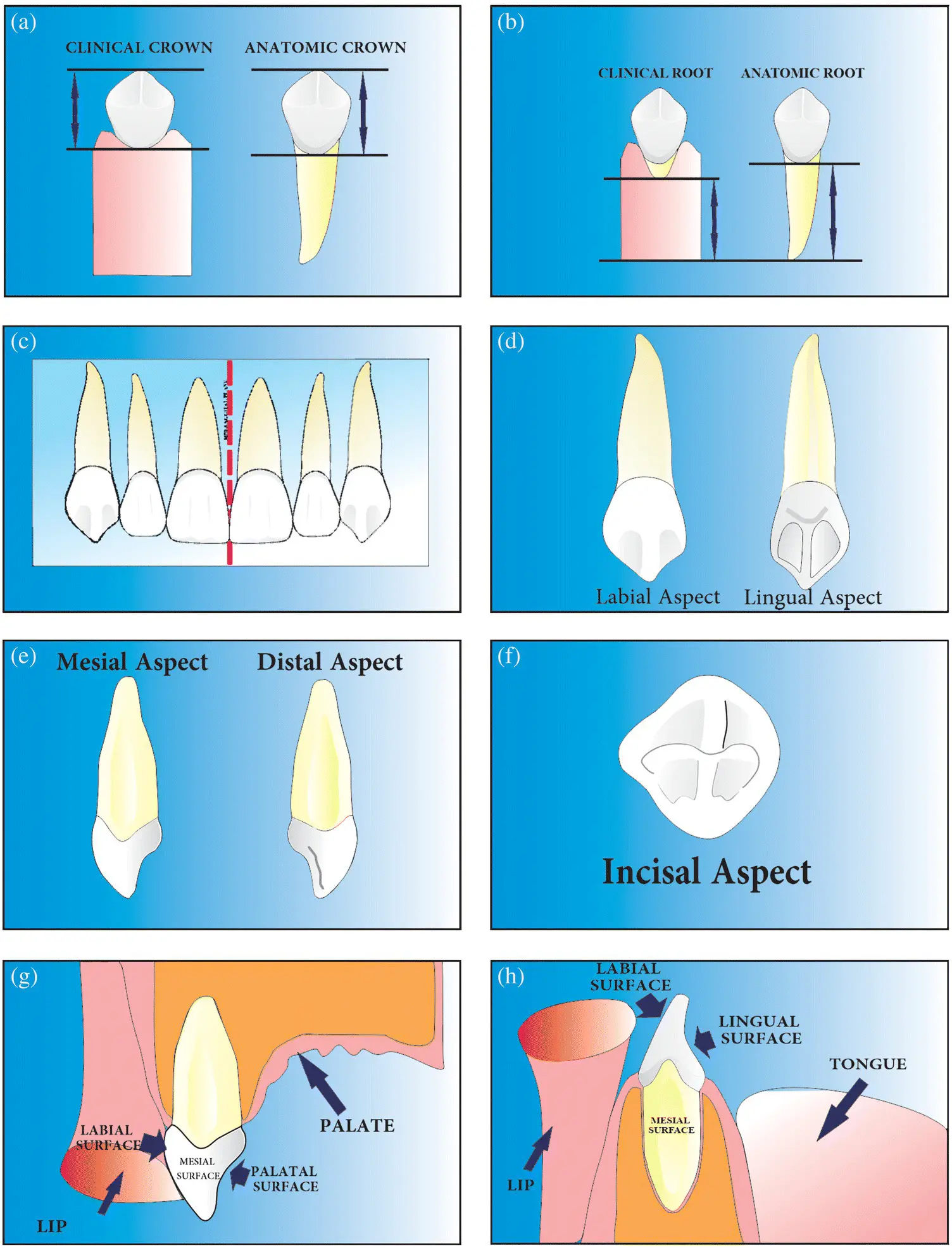
Figure 4.1 (a–h) Anatomical landmarks of human permanent dentition.
Surfaces of posterior teeth (premolars and molars) that contacts teeth of the opposing jaw during occlusion are called occlusal surfaces. These surfaces become incisal edges in case of incisors and cusp tips with respect to canines [1–3].
Mamelons
Rounded elevations or tubercles or protuberances at the incisal portions of incisors, seen on the newly erupted teeth formed from the facial developmental lobes ( Figure 4.2a) [1, 2]. They are usually three in number and the mesial mamelon is usually the tallest [3].
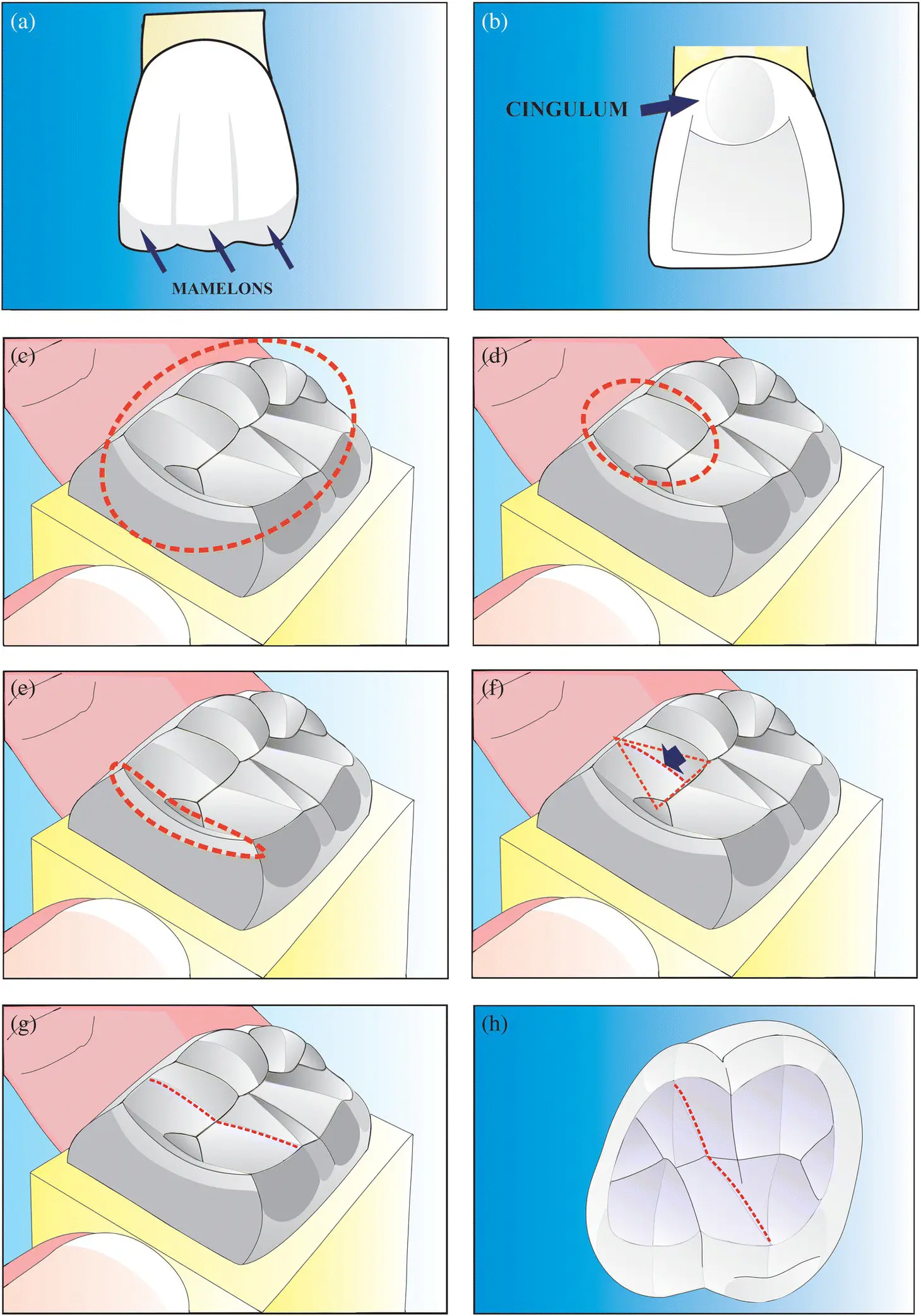
Figure 4.2 (a–h) Anatomical landmarks of human permanent dentition.
Cingulum
In Latin cingulum means girdle [3]. It is the lingual lobe among four developmental lobes of anterior teeth that makes up the bulk of the cervical third of the lingual surface ( Figure 4.2b) [2].
Occlusal Table
The occlusal surface of posterior teeth, bounded by cuspal ridges buccally and lingually, and proximally by marginal ridges ( Figure 4.2c) [1].
Cusp
A triangular or pyramidal elevation that divides the occlusal surface of the crown. Cusps are named according to their location ( Figure 4.2d) [2].
Ridge
The linear elevation on tooth surface [2].
Marginal Ridges
The rounded borders of the enamel that forms margin of a tooth ( Figure 4.2e).
Ex: Mesial and distal marginal ridges on the occlusal surface of a posterior teeth [1, 2].
Triangular Ridge
A linear elevation with slopes on either side, starting from the tip of the cusp descending towards the centre of the occlusal surface of posterior teeth. Named according to the cusp involved ( Figure 4.2f) [1, 2]:
1 Buccal triangular ridge
2 Lingual triangular ridge
Transverse Ridge
A ridge formed by the union of two triangular ridges transversely in the buccolingual direction on the occlusal surfaces of the posterior teeth ( Figure 4.2g).
Ex: buccal and lingual triangular ridges join to form a transverse ridge [1, 2].
Oblique Ridge
A ridge crossing the occlusal surface of the maxillary molar teeth obliquely ( Figure 4.2h).
Formed by the union of the distal cuspal ridge of the mesiopalatal cusp and the triangular ridge of the distobuccal cusp [1–3].
Buccal Ridge
The ridge or elevation that runs cervico‐occlusally on the buccal surface of the posterior teeth ( Figure 4.3a) [1, 2].
Labial Ridge
The ridge or elevation that runs cervico‐occlusally on the labial surface of canines ( Figure 4.3b) [1].
Fossa
An irregular depression or concavity seen on the surface.
Irregular depressions or concavities on the lingual surface of incisors – lingual fossae ( Figure 4.3c).
The concavity formed by the convergence of the ridges at the centre of occlusal surface of posterior teeth where the groove terminal unites is the central fossa ( Figure 4.3d) [1, 2].
Triangular fossae are triangular irregular depressions seen on the occlusal surfaces of premolars and molars that are mesial or distal to marginal ridges. They can also be seen on lingual surfaces of maxillary incisors ( Figure 4.3e) [1, 2]. The canine fossa is a broad concavity on the mesial surface of the maxillary first premolar [3].
Pits
These are the pinpoint depressions seen at the union of groove terminals. Pits are enclosed within the fossae ( Figure 4.3f). Mesial and distal pits are enclosed in respective fossae on posterior teeth [1, 2].
Читать дальшеИнтервал:
Закладка:
Похожие книги на «An Illustrated Atlas of Tooth Carving and Wax-Up Techniques»
Представляем Вашему вниманию похожие книги на «An Illustrated Atlas of Tooth Carving and Wax-Up Techniques» списком для выбора. Мы отобрали схожую по названию и смыслу литературу в надежде предоставить читателям больше вариантов отыскать новые, интересные, ещё непрочитанные произведения.


Обсуждение, отзывы о книге «An Illustrated Atlas of Tooth Carving and Wax-Up Techniques» и просто собственные мнения читателей. Оставьте ваши комментарии, напишите, что Вы думаете о произведении, его смысле или главных героях. Укажите что конкретно понравилось, а что нет, и почему Вы так считаете.