Siegfried Siegesmund - Monument Future
Здесь есть возможность читать онлайн «Siegfried Siegesmund - Monument Future» — ознакомительный отрывок электронной книги совершенно бесплатно, а после прочтения отрывка купить полную версию. В некоторых случаях можно слушать аудио, скачать через торрент в формате fb2 и присутствует краткое содержание. Жанр: unrecognised, на немецком языке. Описание произведения, (предисловие) а так же отзывы посетителей доступны на портале библиотеки ЛибКат.
- Название:Monument Future
- Автор:
- Жанр:
- Год:неизвестен
- ISBN:нет данных
- Рейтинг книги:4 / 5. Голосов: 1
-
Избранное:Добавить в избранное
- Отзывы:
-
Ваша оценка:
Monument Future: краткое содержание, описание и аннотация
Предлагаем к чтению аннотацию, описание, краткое содержание или предисловие (зависит от того, что написал сам автор книги «Monument Future»). Если вы не нашли необходимую информацию о книге — напишите в комментариях, мы постараемся отыскать её.
Alle vier Jahre treffen sich auf einer internationalen Tagung Experten, die sich mit den entsprechenden Sachfragen beschäftigen. Der „14th International Congress on the Deterioration and Conservation of Stone“ findet im September 2020 in Göttingen statt. Er ist die wichtigste Veranstaltung zur Verbreitung des Wissens von Praktikern und Forschern, die im Bereich der Steinkonservierung zur Erhaltung des baulichen Kulturerbes arbeiten: Geowissenschaftler, Architekten, Bauspezialisten, Ingenieure, Restauratoren, Denkmalpfleger und Bauherren.
Der Tagungsband mit über 150 wissenschaftlichen Beiträgen repräsentiert und erfasst den neuesten Stand der Technik auf diesem Gebiet.
Themen sind:
– Charakterisierung von Schadensphänomenen von Steinen und verwandten Baumaterialien (Stuck, Putz, Mörtel usw.)
– Methoden zur Untersuchung des Steinverfalls in situ und zerstörungsfreie Prüfung
– Langzeitüberwachung von Steindenkmälern und Gebäuden
– Simulation und Modellierung des Zerfalls
– Technologien und Entwicklung verbesserter Bearbeitung und Verwendung von Stein in Neubauten
– Bewertung der Langzeitwirkung von Bearbeitungstechniken
– Auswirkungen des Klimawandels auf die Steinverwitterung des Kulturerbes
– Berichte zur Steinkonservierung: Fallstudien und Projekte
– Digitalisierung und Dokumentation von Steinkonservierung
–
The 14th International Congress on the Deterioration and Conservation of Stone, entitled MONUMENT FUTURE: DECAY AND CONSERVATION OF STONE is a quadrennial event that brings together a world-wide community of geoscientists, architects, building specialists, engineers, conservators, restorators, monument curators and building owners who are concerned about the conservation of cultural stone structures and objects. Since antiquity, the weathering and deterioration of historical buildings, masonry, monuments, sculptures etc. using natural stones has been a very well-known problem.
This conference is the main gathering for the dissemination of knowledge in the field of stone deterioration issues. It represents and captures the state-of-the-art in the field of stone conservation and cultural heritage conservation with regards to the following topics:
– Characterisation of damage phenomena of stone and related building materials (plaster, rendering, mortar etc.)
– Methods for the investigation of stone decay; in-situ and non-destructive testing
– Long-term monitoring of stone monuments and buildings
– Simulation and modelling of decay
– Technology and development of improved treatments and use of stone in new buildings
– Assessment of long-term effects of treatments
– Impact of climate change on stone decay of Cultural Heritage
– Reports about stone conservation: case studies and projects
– Digitalization and documentation in stone conservation
Monument Future — читать онлайн ознакомительный отрывок
Ниже представлен текст книги, разбитый по страницам. Система сохранения места последней прочитанной страницы, позволяет с удобством читать онлайн бесплатно книгу «Monument Future», без необходимости каждый раз заново искать на чём Вы остановились. Поставьте закладку, и сможете в любой момент перейти на страницу, на которой закончили чтение.
Интервал:
Закладка:
Calcite decomposition in the same range of temperature is evident in the DSC curves through the 81presence of an endothermic peak. A slight endothermic peak at about 80° C is also present, along with a larger one at 550 °C. Both are better shown in the DSC curves of the insoluble residue. TG/DTG curves (Fig. 6) of the insoluble fraction from the yellow-beige levels show a first mass reduction, with a peak in the DTG curve at 84 °C. In the R sample this mass loss is shifted at 95 °C and it is less pronounced. These thermal variations are consistent with dehydration due to the evaporation of the adsorptively bound water from the specimen (Földvári 2011). A second mass loss with a peak in the DTG curve at 287 °C is observed in the sample from the yellow-beige stone, which can be attributed to the goethite dehydroxylation (Földvári 2011). This peak is absent in the DTG curve of the R sample according to the XRD findings, which did not detect goethite in this sample, but hematite as a product of its transformation. For temperatures higher than 400 °C, the pattern of the DSC curve evidences an endothermic-exothermic process. It corresponds to a solid-phase structural decomposition of organic matter and clay minerals, which is more pronounced in the Y sample compared to the R one. It is followed by a crystallization of new phases, whose evidence is given by a subsequent exothermic bump.
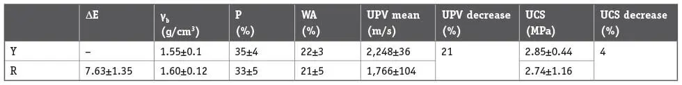
Colour changes, bulk density, porosity accessible to water and water absorption measured for the stone from the yellow-beige and red levels within the blocks are reported in Table 1.
A strong colour variation was recorded in the red level, which may be attributed to the transition of goethite to hematite detected through the mineralogical and thermal analyses. High porosity and water absorption, as well, were measured in the yellow level and not significant decreases of 6 % and 5 %, respectively, were measured in the red level. Also the bulk density showed a slight reduction, namely 3 %. These variations are comparable with those reported for limestones with high porosity and notably lower than the decreases recorded for compact stones (Gomez-Heras et al., Brotons et al. 2013). They were in the range of variability of the measurements, thus they could be due to the intrinsic stone heterogeneity.
However, a decrease of UPV in the samples from the red level was recorded. It was 21 % ( Table 1).
UPV decrease as an effect of the heating is reported in the literature (Yavuz et al. 2010, Andriani 2014), although at entities depending on the temperature and stone structure, as well. It mainly relates to a thermal micro fissuring, which causes the reduction of the propagation velocities.
The microfissuring detected by the UPV test slightly affected the above mentioned physical parameters measured by stauration and buoyancy techniques. This finding suggests that the recorded decreases of the wave velocities may be relevant to the generation of a microporosity which has no effect on the water penetration, as reported in previous studies (Franzoni et al., 2013; Freire-Lista et al., 2016). Microfissuring recorded by UPV had a negligible effect also on the mechanical performance of the stone. Very close values of the compressive strength were measured in both yellow and red levels, corresponding to a strenght loss of 4 % in the discoloured level ( Table 1). Similar entities of decrease have been recorded for porous limestones by Franzoni et al., 2013.
Conclusions
Macroscopic evidences of a fire in the calcarenites employed in an historic building were confirmed by mineralogical changes, which reflects 82on strong color changes. In particular, the change from yellow-beige to reddish color of the stone is consistent with the thermally induced transformation of goethite to hematite. This transition phase indicates that temperatures around 300 °C were reached in the red stone levels during the fire. Effects on the stone microstructure were not visible under optical microscope. Nor the measurement of physical properties showed meaningful variations in this regard. On the contrary, UPV detected a decrease of the propagation velocities, which probably denotes a stone microfissuring. Nonetheless, its entity did not compromise the mechanical resistance of the stone, which remained almost unchanged. High porosity may account for a slight microstructural damage recorded for the investigated calcarenite, where pores likely behave as free spaces for expansion of calcite grain preventing in this way an extensive damage.
Table 1:Color change (ΔE), bulk density (γb), porosity accessible to water (P), water absorption (WA), ultrasonic pulse velocity (UPV) and uniaxial compressive strength (UCS) measured for the stone from the yellow-beige and red levels.
References
Andriani G. F., Germinario L. 2014. Thermal decay of carbonate dimension stones: fabric, physical and mechanical changes. Environ Earth Science 72: 2523–2539.
ASTM D2845-05 2005. Standard test method for laboratory determination of pulse velocities and ultrasonic elastic constants of rock. American Society for Testing Materials.
Brotóns V., Tomás R., Ivorra S., Alarcón J. C. 2013. Temperature influence on the physical and mechanical properties of a porous rock: San Julian’s calcarenite. Engineering Geology 167: 117–127.
Calia A., Colangiuli D., Lettieri M., Quarta G., Masieri M. 2016. Microscopic techniques and a multi-analytical approach to study the fire damage of the painted stuccoes from the Petruzzelli Theatre (Bari, Southern Italy). Microchemical Journal. 126: 42–53.
Földvári M. 2011. Handbook of thermogravimetric system of minerals and its use in geological practice. In: Geological institute of Hungary. Budapest. isbn 978-963-671-288-4.
Franzoni E., Sassoni E., Scherer G.W, Naidu S. 2013. Artificial weathering of stone by heating. Journal of Cultural Heritage. 14: 85–93.
Freire-Lista D. M., Fort R., and Varas-Muriel M. J. 2016. Thermal stress-induced microcracking in building granite. Engineering Geology 206:83–93.
Gomez-Heras M., Alvarez de Buergo M., Fort R., Hajpál M., Török A., Varas M. J. 2006. Evolution of porosity in Hungarian building stones after simulated burning. In: Heritage Weathering and Conservation HWC-2006, Taylor & Francis, Rotterdam, 513–519.
ISRM. Rock characterization testing and monitoring. In: Brown ET, editor. ISRM suggested methods, Oxford:Pergamon Press; 1981.p. 135–40.
Kompaníková Z., Gomez-Heras M., Michňová J., Durmeková T., Vlčko J. 2014. Sandstone alterations triggered by fire-related temperatures. Environmental Earth Science 72: 2569–2581.
Martinho E., Dionísio A. 2018. Assessment Techniques for Studying the Effects of Fire on Stone Materials: A Literature Review. International Journal of Architectural Heritage. 00 1–25.
Siegesmund S., Ullemeyer K., Weiss T., Tschegg E. K. 2000. Physical weathering of marbles caused by anisotropic thermal expansion, Int. J. Earth Sci. 89: 170–182. doi: 10.1007/s005310050324.
Sippel J., Siegesmund S., Weiss T., Nitsch K. H., Korzen M. 2007. In: Pikryl, R. & Smith, B. J. (eds) Building Stone Decay: From Diagnosis to Conservation. Geological Society, London, Special Publications 271: 139–151.
UNI EN 772-1 2011. Methods of test for masonry units – Part 1: Determination of compressive strength.
Yavuz H., Demirdag S., Caran S. 2010. Thermal effect on the physical properties of carbonate rocks. International Journal of Rock Mechanics & MiningSciences 47: 94–103.
Vázquez P., Shushakova V., Gómez-Heras M. 2015. Influence of mineralogy on granite decay induced by temperature increase: Experimental observations and stress simulation. Eng. Geol. 189: 58–67. doi: 10.1016/j.enggeo.2015.01.026
Читать дальшеИнтервал:
Закладка:
Похожие книги на «Monument Future»
Представляем Вашему вниманию похожие книги на «Monument Future» списком для выбора. Мы отобрали схожую по названию и смыслу литературу в надежде предоставить читателям больше вариантов отыскать новые, интересные, ещё непрочитанные произведения.


Обсуждение, отзывы о книге «Monument Future» и просто собственные мнения читателей. Оставьте ваши комментарии, напишите, что Вы думаете о произведении, его смысле или главных героях. Укажите что конкретно понравилось, а что нет, и почему Вы так считаете.