Georgios E. Romanos - Advanced Laser Surgery in Dentistry

Здесь есть возможность читать онлайн «Georgios E. Romanos - Advanced Laser Surgery in Dentistry» — ознакомительный отрывок электронной книги совершенно бесплатно, а после прочтения отрывка купить полную версию. В некоторых случаях можно слушать аудио, скачать через торрент в формате fb2 и присутствует краткое содержание. Жанр: unrecognised, на английском языке. Описание произведения, (предисловие) а так же отзывы посетителей доступны на портале библиотеки ЛибКат.

Advanced Laser Surgery in Dentistry: краткое содержание, описание и аннотация

Предлагаем к чтению аннотацию, описание, краткое содержание или предисловие (зависит от того, что написал сам автор книги «Advanced Laser Surgery in Dentistry»). Если вы не нашли необходимую информацию о книге — напишите в комментариях, мы постараемся отыскать её.

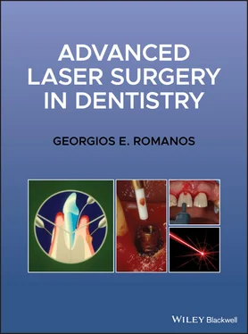
Advanced Laser Surgery in Dentistry  The book includes high-quality, color photographs throughout to support the text and add visual information to the covered topics, which include wound healing, oral surgery, periodontology, implant dentistry, and laser fundamentals and safety considerations. 
provides readers with a step-by-step guide for using lasers in dental practice and discusses likely new directions and possible future treatments in the rapidly advancing field of laser dentistry. Readers will also benefit from a wide variety of subjects, including: 
A thorough introduction to the fundamentals of lasers, including the beam, the laser cavity, active mediums, lenses, resonators, and delivery systems An exploration of lasers and wound healing, including soft tissue and bone healing, as well as laser-assisted excisions and osteotomies An analysis of lasers in periodontology, including laser-assisted bacteria reduction in the periodontal tissues and the removal of subgingival dental calculus A discussion of lasers in implant dentistry and treatment for peri-implantitis Perfect for oral and maxillofacial surgeons, periodontists, and implant dentists, as well as general dentists, 
 will also earn a place in the libraries of dental students and residents seeking to improve their understanding of laser-based oral and dental procedures with a carefully organized reference guide.
Advanced Laser Surgery in Dentistry — читать онлайн ознакомительный отрывок

Ниже представлен текст книги, разбитый по страницам. Система сохранения места последней прочитанной страницы, позволяет с удобством читать онлайн бесплатно книгу «Advanced Laser Surgery in Dentistry», без необходимости каждый раз заново искать на чём Вы остановились. Поставьте закладку, и сможете в любой момент перейти на страницу, на которой закончили чтение.

Тёмная тема
Сбросить

Интервал:

Закладка:

Сделать

2 Chapter 2Figure 2.1 Incision quality during laser irradiation of the bovine mucosa sh...Figure 2.2 Temperature changes of the tissue during laser irradiation. The l...Figure 2.3 Wound healing after scalpel incision (a); 1.75 W power Nd:YAG (b)...Figure 2.4 Wound healing after scalpel incision (a); 1.75 W power Nd:YAG (b)...Figure 2.5 Wound healing after scalpel incision (a); 1.75 W power Nd:YAG (b)...Figure 2.6 Incision area three days after 5W diode laser (980 nm) incision p...Figure 2.7 Incision area 14 days after 10W diode laser (980 nm) presenting c...Figure 2.8 Incision area 14 days after 15W diode (980 nm) laser presenting m...

3 Chapter 3Figure 3.1 Differences in penetration depths between CO 2laser and Nd:YAG la...Figure 3.2 Perpendicular irradiation using a focused CO 2laser (noncontact) ...Figure 3.3 Superficial irradiation of the tissue after excision to create a ...Figure 3.4 Hyperkeratosis of the mandible (a); the pathological tissue was e...Figure 3.5 Necessary instruments for conventional laser‐assisted surgical pr...Figure 3.6 Intraoperative burn of the lower lip of a patient during laser‐as...Figure 3.7 Coagulation of the blood vessel tumor in the papilla #6–7 (a) usi...Figure 3.8 Scar tissue formation due to the use of high‐power diode laser (9...Figure 3.9 Excision of a soft tissue tumor in the maxilla (a) using a contin...Figure 3.10 Removal of a soft tissue tumor in the vestibule of the anterior ...Figure 3.11 Irritation fibroma in the vestibular fold before the laser surge...Figure 3.12 Lingual fibroma (a) excised with a focused non‐contact CO 2laser...Figure 3.13 Fibroma at the buccal mucosa (a) removed with contact pulsed Nd:...Figure 3.14 Excision of a fibroma in the tongue (a) using an Er:YAG laser (b...Figure 3.15 Soft tissue tumor of the alveolar ridge (a); excision with the C...Figure 3.16 Soft tissue tumor in the palate (a). The surgical excision occur...Figure 3.17 Palatal soft tissue lesion (a); using an ablative mode, the lesi...Figure 3.18 Excision of soft tissue tumor (a) with a scalpel and a CO 2laser...Figure 3.19 Clinical diagnosis of symmetric fibromas (a). The surgical proce...Figure 3.20 Pyogenic granuloma of the lower lip (a). The tumor was excised w...Figure 3.21 Fibroma in the buccal mucosa (a). The excision was performed usi...Figure 3.22 Fibroma of the buccal mucosa (a) excised with a CO 2laser with a...Figure 3.23 Surgical excision of the hyperplastic gingiva underneath the bar...Figure 3.24 Hyperplastic and inflamed peri‐implant mucosa with bleeding on p...Figure 3.25 Clinical and radiographical examination of a patient with an oss...Figure 3.26 Soft tissue tumor of the gingiva associated with some bleeding (...Figure 3.27 Soft tissue tumor of the lip (a) removed using an 810 nm diode l...Figure 3.28 Small tumor of the upper lip (a). An Nd:YAG laser with contact f...Figure 3.29 Soft tissue tumor of the buccal mucosa without symptoms (a). The...Figure 3.30 Soft tissue asymptomatic tumor of the tongue (a). Clinical exami...Figure 3.31 Soft tissue tumor of the gingiva (a); excision of the tumor usin...Figure 3.32 Soft tissue tumor in the mouth floor (a); a sialolith was not de...Figure 3.33 Epulis in the interdental area #2–3 (a). A gingivectomy was carr...Figure 3.34 A gingival tumor in a buccal aspect and occlusal view (a, b). Di...Figure 3.35 Mucocele of the low lip (a); a half‐moon‐shaped incision was per...Figure 3.36 Mucus retention cyst of the lower lip (a). After topical anesthe...Figure 3.37 Extravasation cyst of the sublingual gland (a). A plastic cathet...Figure 3.38 Swelling of the cheek (a) and sensitivity to palpation for at le...Figure 3.39 Excision of the lingual frenum (a) with a CO 2laser (4 W; contin...Figure 3.40 The excisions of the two frenula (a, b) in the maxilla and the m...Figure 3.41 Tight frenum in the anterior maxilla (a). Excision with the CO 2...Figure 3.42 Hypertrophic papilla creating esthetic problems for the patient ...Figure 3.43 Excision of the labial frenulum (a) with a focused 810 nm diode ...Figure 3.44 Excision of the labial frenum (a) using a focused 810 nm diode l...Figure 3.45 Insufficient vestibule in the maxilla (a) associated with lack o...Figure 3.46 Lack of vestibule depth before surgery (a); the incision was per...Figure 3.47 Insufficient depth of the vestibule in the mandible (a). The muc...Figure 3.48 A frenectomy (a) was performed using a pulsed Nd:YAG laser (2.0 ...Figure 3.49 Vestibuloplasty with an Er:YAG laser. Observe the insufficient b...Figure 3.50 Insufficient vestibule depth in the maxilla including soft tissu...Figure 3.51 Insufficient vestibule depth in the mandible due to the presence... Figure 3.52 Similar to the previous clinical case, the soft tissue tumor was...Figure 3.53 Oral leukoplakia in the alveolar ridge of the maxilla before exc...Figure 3.54 Oral leukoplakia of the tongue (a) and mouth floor (b); charring...Figure 3.55 Leukoplakia of the gingiva (a); removal of the lesion using a COFigure 3.56 Leukoplakia of the gingiva (a); coagulation after laser excision...Figure 3.57 Oral leukoplakia in the interdental papilla (a); excision with a...Figure 3.58 Oral leukoplakia at the right side of the tongue (a); excision u...Figure 3.59 Soft tissue tumor in a patient with von Willebrand disease befor...Figure 3.60 Schematic illustration of the ice cube technique in conjunction ...Figure 3.61 Vascular lesion of the buccal mucosa (a); coagulation using the ...Figure 3.62 Hemangiomas of the lower lip (a); the lesions were coagulated us...Figure 3.63 Multiple telangiectasias on the skin before (a, b) and after (c,...Figure 3.64 Hemangioma of the lip (a) in a size larger than the width of a t...Figure 3.65 Conventional method of coagulation of the hemangioma of the tong...Figure 3.66 Large hemangioma on the tongue (a) coagulated with the intralumi...Figure 3.67 Hemangioma of the oral mucosa (a) irradiated with the ice cube t...Figure 3.68 Hemangioma of the lower lip (a, b) removed with a diode laser an... Figure 3.69 Initial clinical demonstration of a large hemangioma of the oral... Figure 3.70 Before surgery (a) and laser irradiation (b); three weeks post (... Figure 3.71 Before, during, and after treatment (five months post‐op). Figure 3.72 Hemangioma of the upper lip associated with esthetic concerns (a...Figure 3.73 Amalgam tattoo removal (a) using a pulsed Nd:YAG laser (b); the ...Figure 3.74 Removal of a nevus in the upper lip (a) using an Nd:YAG laser (b...Figure 3.75 Pericoronitis around tooth#17 (a); excision of the mucosa with a...Figure 3.76 Unerupted tooth (a) exposed with a CO 2laser (b) after placement... Figure 3.77 CO2 laser‐assisted surgical exposure of an impacted tooth for or...Figure 3.78 Surgical exposure of an impacted tooth for orthodontic reasons u...Figure 3.79 Surgical exposure of an impacted tooth for orthodontic reasons u...Figure 3.80 A deeply localized salivary stone was palpated in the submandibu...

4 Chapter 4Figure 4.1 Comparison of the osteotomies performed by Lindemann bur, piezosu...Figure 4.2 Electron microscopic analysis of an osteotomy using Er:YAG laser ...Figure 4.3 Electron microscopic analysis of an osteotomy using Er,Cr:YSGG la... Figure 4.4a Bone removal using an Er:YAG laser (Hoya, Japan).Figure 4.4b Osteotomy site presenting a smooth surface after exostosis remov...Figure 4.4c Bone specimen removed with the Er:YAG laser presenting areas of ... Figure 4.5a Preparation of an exostosis (tori mandibularis) after elevation ...Figure 4.5b Osteotomy procedure for removal of a lingual exostosis using an ...Figure 4.6 Crown lengthening using an Er:YAG laser for osseous reduction and...Figure 4.7 Window preparation for sinus augmentation using an Er,Cr:YSGG las...Figure 4.8 Preparation of the cortical layer of the alveolar ridge using an ...Figure 4.9 Minimal invasive removal of an implant using an Er:YAG laser (Hoy...Figure 4.10 Block section using an Er:YAG laser (Hoya Conbio, Versawave, Tok...Figure 4.11 Bone splitting using the Er,Cr:YSGG laser (Waterlase ®MD) w...

Читать дальше
Тёмная тема
Сбросить

Интервал:

Закладка:

Сделать

Похожие книги на «Advanced Laser Surgery in Dentistry»

Представляем Вашему вниманию похожие книги на «Advanced Laser Surgery in Dentistry» списком для выбора. Мы отобрали схожую по названию и смыслу литературу в надежде предоставить читателям больше вариантов отыскать новые, интересные, ещё непрочитанные произведения.


Отзывы о книге «Advanced Laser Surgery in Dentistry»

Обсуждение, отзывы о книге «Advanced Laser Surgery in Dentistry» и просто собственные мнения читателей. Оставьте ваши комментарии, напишите, что Вы думаете о произведении, его смысле или главных героях. Укажите что конкретно понравилось, а что нет, и почему Вы так считаете.

x