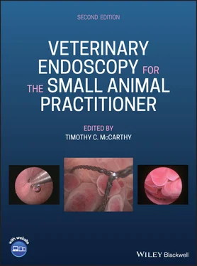
Veterinary Endoscopy for the Small Animal Practitioner
Здесь есть возможность читать онлайн «Veterinary Endoscopy for the Small Animal Practitioner» — ознакомительный отрывок электронной книги совершенно бесплатно, а после прочтения отрывка купить полную версию. В некоторых случаях можно слушать аудио, скачать через торрент в формате fb2 и присутствует краткое содержание. Жанр: unrecognised, на английском языке. Описание произведения, (предисловие) а так же отзывы посетителей доступны на портале библиотеки ЛибКат.
- Название:Veterinary Endoscopy for the Small Animal Practitioner
- Автор:
- Жанр:
- Год:неизвестен
- ISBN:нет данных
- Рейтинг книги:4 / 5. Голосов: 1
-
Избранное:Добавить в избранное
- Отзывы:
-
Ваша оценка:
- 80
- 1
- 2
- 3
- 4
- 5
Veterinary Endoscopy for the Small Animal Practitioner: краткое содержание, описание и аннотация
Предлагаем к чтению аннотацию, описание, краткое содержание или предисловие (зависит от того, что написал сам автор книги «Veterinary Endoscopy for the Small Animal Practitioner»). Если вы не нашли необходимую информацию о книге — напишите в комментариях, мы постараемся отыскать её.
Covers diagnostic endoscopy, interventional endoscopy, and minimally invasive soft tissue surgery Includes thousands of images to illustrate endoscopy concepts for veterinarians Provides a clinically oriented reference book for using rigid and flexible endoscopy in a small animal practice Supports veterinarians who are seeking to increase their services and enhance their revenue streams Any practitioner who is using or preparing to use endoscopic techniques will find
an essential practice resource.
Veterinary Endoscopy for the Small Animal Practitioner — читать онлайн ознакомительный отрывок
Ниже представлен текст книги, разбитый по страницам. Система сохранения места последней прочитанной страницы, позволяет с удобством читать онлайн бесплатно книгу «Veterinary Endoscopy for the Small Animal Practitioner», без необходимости каждый раз заново искать на чём Вы остановились. Поставьте закладку, и сможете в любой момент перейти на страницу, на которой закончили чтение.
Интервал:
Закладка:
9 Chapter 9Figure 9.1 Reusable trocar–cannulas used for thoracoscopy in small animals: ...Figure 9.2 Disposable plastic cannulas used for thoracoscopy in small animal...Figure 9.3 Adequate lung collapse for thoracoscopy in a patient using the se...Figure 9.4 Noncompliant lungs in a dog with pericardial effusion that did no...Figure 9.5 Pleural insufflation used in a dog for a right caudal lung lobect...Figure 9.6 Paraxiphoid telescope portal with the patient in dorsal recumbenc...Figure 9.7 The ventral mediastinum is visible as a complete or incomplete cu...Figure 9.8 When there is a complete mediastinum the pneumothorax produced is...Figure 9.9 A patient with a complete mediastinum that is shifted to the left...Figure 9.10 A natural fenestration visible in the ventral mediastinum that a...Figure 9.11 Cutting a hole in a complete mediastinum to provide access to th...Figure 9.12 Cutting a hole in an incomplete mediastinum without a visible fe...Figure 9.13 A thickened ventral mediastinum covered with reactive pleural ti...Figure 9.14 Cutting a normal mediastinum off of the sternum using sharp diss...Figure 9.15 Cutting through a normal ventral mediastinum where there is a bl...Figure 9.16 A ventral mediastinum with increased number and size of blood ve...Figure 9.17 Cutting the mediastinum off of the sternum in the patient in Fig...Figure 9.18 The mediastinum is cut away from the sternum as far cranially as...Figure 9.19 When indicated the mediastinum is cut off the sternum caudally t...Figure 9.20 Completion of taking the mediastinum down from the sternum provi...Figure 9.21 Placing a lateral chest wall operative portal using a sharp troc...Figure 9.22 Establishing a lateral chest wall portal using blunt dissection ...Figure 9.23 Establishing a lateral chest wall portal by passing a scalpel bl...Figure 9.24 Placing a plastic disposable laparoscopy cannula through the che...Figure 9.25 Placing an EndoTIP cannula through a sharp chest wall incision w...Figure 9.26 Final position of a threaded EndoTIP cannula placed as a lateral...Figure 9.27 A chest tube is placed through the paraxiphoid telescope portal ...Figure 9.28 Adequate chest tube position is confirmed visually with the tele...Figure 9.29 After chest tube placement with this technique the paraxiphoid t...Figure 9.30 Chest tube placement is done with standard open surgical techniq...Figure 9.31 The normal appearance of the right chest wall seen using thoraco...Figure 9.32 The sympathetic trunk is seen running from cranial to caudal acr...Figure 9.33 The thoracic duct is seen as a single clear linear vessel in the...Figure 9.34 A branching thoracic duct is seen as a clear linear vessel in th...Figure 9.35 Smaller lymphatic ducts seen in a 3‐year‐old male English Spring...Figure 9.36 The diaphragm is visible as a convex muscular dome‐shaped struct...Figure 9.37 The dome‐shaped central tendon portion of the diaphragm is a tra...Figure 9.38 The peripheral attachment of the diaphragm meets the chest wall ...Figure 9.39 In the ventral thorax the enlarged costochondral junctions are v...Figure 9.40 The azygos vein is seen in the right dorsal thorax running from ...Figure 9.41 The left pulmonary ligament is seen as the translucent sheet of ...Figure 9.42 The cranial vena cava is visible in the cranial mediastinum with...Figure 9.43 The caudal vena cava seen entering the thorax through the caval ...Figure 9.44 Another view of the caudal vena cava seen in Figure 9.43 showing...Figure 9.45 The caudal vena cava seen from a paraxiphoid telescope portal wi...Figure 9.46 The phrenic nerve is seen as it angles dorsally from the cranial...Figure 9.47 Continuation of the phrenic nerve is seen on the lateral surface...Figure 9.48 The cranial surface of the pericardium is visualized well with t...Figure 9.49 An obese patient with the pericardium mostly covered with fat an...Figure 9.50 Normal lung visible with the patient in dorsal recumbency from a...Figure 9.51 Close‐up examination of lung tissue allows individual alveoli to...Figure 9.52 Normal cranial mediastinal anatomy seen with the patient in dors...Figure 9.53 The sternal lymph node is visible in this picture as the pink ov...Figure 9.54 Removing residual pleural space fluid, in this case, bloody flui...Figure 9.55 Removing residual pericardial fluid that was not removed with pr...Figure 9.56 Subpleural bleeding in the chest wall of a ten‐year‐old neutered...Figure 9.57 A lung lesion with a puncture injury and surrounding contusion s...Figure 9.58 A pericardial puncture lesion seen from the inside of the perica...Figure 9.59 A large chest wall mass originating from a rib in the left crani...Figure 9.60 A primary chondrosarcoma originating in the costal cartilage in ...Figure 9.61 A porcupine quill penetrating the left chest wall from the outsi...Figure 9.62 Secondary chest wall reaction due to a migrating plant material ...Figure 9.63 Pyogranulomatous pleuritis involving the parietal pleura on the ...Figure 9.64 An extensive adhesion from an area of abnormal lung to the chest...Figure 9.65 Thickened pleura obscuring visualization of thoracic wall anatom...Figure 9.66 Pleural thickening due to chylothorax in the left caudal and dor...Figure 9.67 A small number of submacroscopic pleural nodules in the parietal...Figure 9.68 Extensive white mesothelioma nodules on the chest wall pleura an...Figure 9.69 One of several large red irregular pleural masses on the chest w...Figure 9.70 A pleural mass attached to the chest wall of a 12‐year‐old neute...Figure 9.71 Additional smaller discrete nodules on another area of the chest...Figure 9.72 Widely spread submacroscopic nodules on the diaphragm and chest ...Figure 9.73 In this same patient a lesion comprised of a collection of nodul...Figure 9.74 The pulmonary mass in the same patient shown in the Figures 9.70...Figure 9.75 Pyogranulomatous pleuritis and pneumonia with adhesion to and in...Figure 9.76 Adhesion of the right caudal lung lobe to the diaphragm due to c...Figure 9.77 A deformed liver lobe in the chest of a large 12‐year‐old spayed...Figure 9.78 Pyogranulomatous pleuritis secondary to recurrent pyothorax due ...Figure 9.79 Prominent mediastinal blood vessels in a dog with vascular conge...Figure 9.80 A small thymic carcinoma in the cranial mediastinum of a 13‐year...Figure 9.81 A medium‐sized lymphocyte rich thymoma in a 12‐year‐old neutered...Figure 9.82 A large lymphocyte rich thymoma with limited vascularity filling...Figure 9.83 A large lymphocyte rich vascular thymoma in the cranial thorax o...Figure 9.84 A non‐thymic lymphoma mass in the cranial mediastinum of an 8‐ye...Figure 9.85 An ectopic thyroid carcinoma in the cranial mediastinum of a 12‐...Figure 9.86 A poorly differentiated carcinoma of undetermined origin in the ...Figure 9.87 A large neuroendocrine tumor in the cranial mediastinum of a 7‐y...Figure 9.88 A large thin walled cranial mediastinal cyst in a 16‐year‐old sp...Figure 9.89 A cranial mediastinal accumulation of chyle is visible in a 9‐ye...Figure 9.90 A large inflammatory caudal mediastinal mass in a three‐year‐old...Figure 9.91 A partially decomposed grass awn found free in the pleural space...Figure 9.92 A group of porcupine quills in the cranial thorax of a 5‐year‐ol...Figure 9.93 A large primary bronchogenic adenocarcinoma in the right cranial...Figure 9.94 The dorsomedial surface of the same mass as seen in Figure 9.93 ...Figure 9.95 A large adenocarcinoma in the cranial end of the left caudal lun...Figure 9.96 Malignant histiocytosis lesions in the left cranial lung lobe of...Figure 9.97 A massive right caudal lung lobe primary lung tumor in a 10‐year...Figure 9.98 A small primary lung tumor visible on the surface of the right c...Figure 9.99 Superficial changes in the surface of the caudal part of the lef...Figure 9.100 Two small flat red dots representing metastatic lesions on the ...Figure 9.101 Multiple small flat red metastatic hemangiosarcoma lesions were...Figure 9.102 Small raised red hemangiosarcoma metastases in a 10‐year‐old sp...Figure 9.103 A small raised purple metastatic lesion on the ventral margin o...Figure 9.104 A small purple polypoid metastatic lesion on the medial surface...Figure 9.105 A metastatic adenocarcinoma of undetermined, suspected mammary,...Figure 9.106 Massive infiltration of all lung lobes with an uncountable numb...Figure 9.107 An enlarged hilar lymph node in a 6‐year‐old spayed female Mini...Figure 9.108 An air‐filled bulla in the foreground and an empty collapsed bu...Figure 9.109 An air‐filled bulla on the caudal margin of the right cranial l...Figure 9.110 An air‐filled bulla on the dorsal surface of the left cranial l...Figure 9.111 A large air‐filled bulla in the right cranial lung lobe on the ...Figure 9.112 Multiple air‐filled bullae on the caudoventral corner of the ri...Figure 9.113 The bulla seen in Figure 9.109 that is collapsed during the exh...Figure 9.114 An obvious hole in a distended bulla with visible air flow seen...Figure 9.115 The bullae on the dorsal surface of the left cranial lung lobe ...Figure 9.116 Chronic pulmonary fibrosis in a 10‐year‐old spayed female Domes...Figure 9.117 Atelectatic non‐aerated lung in a 13‐year‐old spayed female Ger...Figure 9.118 Diffuse pulmonary hemorrhage and atelectasis seen in the crania...Figure 9.119 Chronic fibrosing pneumonia in the right caudal lung lobe in a ...Figure 9.120 Underdeveloped right cranial and middle lung lobes in an 8‐mont...Figure 9.121 Histiocytic pneumonia in the right cranial lung lobe of an eigh...Figure 9.122 Lung lobe torsion in a 4‐year‐old spayed female Whippet seen af...Figure 9.123 Small white mesothelioma nodules in the cranial mediastinum of ...Figure 9.124 Small white mesothelioma nodules on the caudal vena cava and on...Figure 9.125 Moderate pleural thickening in a 9‐year‐old neutered male Domes...Figure 9.126 Cranial mediastinal subpleural chyle accumulation in the same p...Figure 9.127 In the same patient as seen in Figure 9.125 and 9.126 the thora...Figure 9.128 The same patient after dissection to expose the thoracic duct a...Figure 9.129 The cautery blade from a standard monopolar radiofrequency elec...Figure 9.130 Completion of the cautery process with a dry field and oblitera...Figure 9.131 Marked pleural thickening in a cat with chylothorax of short du...Figure 9.132 Constrictive pleuritis causing collapse of the lungs in the sam...Figure 9.133 Minimal pleural reaction and thickening in an 8‐year‐old neuter...Figure 9.134 In the same patient as Figure 9.133 streaks of fibrin accumulat...Figure 9.135 Marked pleural thickening due to chylothorax in a 9‐year‐old sp...Figure 9.136 Constrictive pleuritis compressing the lung lobes on the right ...Figure 9.137 A large fluid filled pericardium in an 8‐year‐old neutered male...Figure 9.138 A heart base mass visible through the pericardium prior to crea...Figure 9.139 A large heart base tumor visible as a mass distending the peric...Figure 9.140 A normal appearing pericardium in a dog with pericardial effusi...Figure 9.141 Mild pericardial thickening in an 11‐year‐old neutered male lar...Figure 9.142 Moderate pericardial thickening secondary to a heart base tumor...Figure 9.143 Marked pericardial thickening in a 12‐year‐old spayed female La...Figure 9.144 A pericardium with mesothelial reaction suspicious of mesotheli...Figure 9.145 A metastatic thyroid carcinoma lesion on the base of the aorta ...Figure 9.146 Inflammatory pericarditis in a spayed female Whippet of unknown...Figure 9.147 Mesothelioma nodules on the internal surface of the pericardium...Figure 9.148 A solid sheet of mesothelioma lining the pericardium in a neute...Figure 9.149 A completed pericardial window in a patient with a heart base t...Figure 9.150 Elevation of the pericardium looking cranially from the paraxip...Figure 9.151 Using a 30° telescope with the angle directed down in the same ...Figure 9.152 Additional elevation with two instruments and the 30° telescope...Figure 9.153 A large dark purple heart base tumor in a ten‐year‐old neutered...Figure 9.154 A heart base tumor with areas of multiple different appearances...Figure 9.155 A heart base tumor with the appearance of blood clots of differ...Figure 9.156 Biopsy of a pleural mesothelioma nodule using 5 mm diameter opp...Figure 9.157 Biopsy of a large mesothelioma mass on the parietal pleura in a...Figure 9.158 Biopsy of a pleural lesion in a dog with chylothorax. The lesio...Figure 9.159 A metastatic mass on the parietal pleura of a 12‐year‐old neute...Figure 9.160 An inoperable cranial mediastinal mass biopsied with the patien...Figure 9.161 A primary osteosarcoma in the chest wall of a 16‐month‐old neut...Figure 9.162 Biopsy of an enlarged hilar lymph node in a 6‐year‐old spayed f...Figure 9.163 Placement of 5 mm diameter opposing cup or clam shell biopsy fo...Figure 9.164 Placement of 5 mm biopsy forceps at the margin of a focal lung ...Figure 9.165 A defect in the margin of normal appearing lung tissue after a ...Figure 9.166 The biopsy site at the side of the focal lung lesion seen in Fi...Figure 9.167 Abnormal lung tissue in the cranial portion of the left cranial...Figure 9.168 The cut end of the resected left cranial lung lobe after applic...Figure 9.169 Abnormal lung tissue involving the right cranial and middle lun...Figure 9.170 An articulated EndoGIA linear stapler is positioned across the ...Figure 9.171 The lobectomy resection site after removal of the stapler and r...Figure 9.172 This is the authors preference for operating room setup and pat...Figure 9.173 An alternative operating room setup, patient positioning, and p...Figure 9.174 The authors preferred portal placement for pericardial window s...Figure 9.175 An alternative for portal placement for pericardial window surg...Figure 9.176 A pericardial window can also be created with the patient in le...Figure 9.177 Pericardial window surgery portal placement when the patient is...Figure 9.178 Grasping the pericardium with 5 mm diameter aggressive rat toot...Figure 9.179 Excessive fat covering the pericardium and preventing access to...Figure 9.180 Reactive loose pleura on the pericardium interfering with visua...Figure 9.181 Grasping forceps elevating tissue in a patient where it cannot ...Figure 9.182 Grasping forceps with a grip that has clearly elevated the peri...Figure 9.183 Dissecting pleura and fat off of the pericardium to provide acc...Figure 9.184 Metzenbaum scissors positioned to cut into the pericardium in F...Figure 9.185 Cutting pericardium with scissors after initial penetration and...Figure 9.186 Elevation of the pericardium is maintained with the grasping fo...Figure 9.187 The procedure is continued until a patch of pericardium approxi...Figure 9.188 In this picture there is loss of pericardial elevation during c...Figure 9.189 The same patient as in Figure 9.188 with the grasping forceps r...Figure 9.190 This image demonstrates grasping forceps with a position that d...Figure 9.191 Thickened pericardial tissue may require more robust scissors t...Figure 9.192 Using a 5 mm vessel sealing device to create a pericardial wind...Figure 9.193 A thickened highly vascular pericardium in an 8‐year‐old neuter...Figure 9.194 Removing pericardial window tissue through a 6 mm EndoTIP cannu...Figure 9.195 Extracting pericardial window tissue with a significant adipose...Figure 9.196 Performing a subtotal pericardiectomy for treatment of chylotho...Figure 9.197 Subtotal pericardiectomy being performed in a 3‐year‐old neuter...Figure 9.198 Completed subtotal pericardiectomy in a dog with no visible per...Figure 9.199 Completed subtotal pericardiectomy in a cat with no visible per...Figure 9.200 Abnormal lung tissue in the ventral margin of the caudal portio...Figure 9.201 Positioning an articulating EndoGIA stapling device across the ...Figure 9.202 The stapling cartridge in Figure 9.201 was too short leaving a ...Figure 9.203 Completion of transection of lung tissue for this partial lung ...Figure 9.204 The resection line of a partial lobectomy of the right cranial ...Figure 9.205 Positioning of an EndoGIA stapling device on the ventral margin...Figure 9.206 The sealed staple line following partial lung lobectomy in the ...Figure 9.207 Running saline over a partial lung lobectomy site done with an ...Figure 9.208 Operating room arrangement for total lobectomy of a left crania...Figure 9.209 Operating room arrangement for total lobectomy of a left caudal...Figure 9.210 Portal placement for total lobectomy of the right caudal lung l...Figure 9.211 Portal placement for total lobectomy of the right cranial lung ...Figure 9.212 A 5 mm diameter vessel sealing device in position demonstrating...Figure 9.213 Cutting an avascular dorsal pulmonary ligament of the left caud...Figure 9.214 The activated vessel sealing device on the dorsal pulmonary lig...Figure 9.215 An EndoGIA stapler placed across the hilus of the left caudal l...Figure 9.216 A second EndoGIA stapler placed across the untransected portion...Figure 9.217 A tissue retrieval bag being used to remove a resected lung lob...Figure 9.218 The right cranial lung lobe elevated to expose the hilus in a p...Figure 9.219 The hilar resection site in the right cranial lung lobe seen in...Figure 9.220 A stapling device on the hilus of the cranial portion of the le...Figure 9.221 The hilar resection site in the left cranial portion of the lef...Figure 9.222 Operating room setup for combined thoracic duct occlusion and s...Figure 9.223 Portal placement for thoracic duct occlusion and subtotal peric...Figure 9.224 Pleural reaction with thickening in a 9‐year‐old spayed female ...Figure 9.225 Pleural reaction with thickening in a three‐year‐old neutered m...Figure 9.226 Incising the thickened pleura in the dog in Figure 9.224 expose...Figure 9.227 Incised thickened pleura in the cat in Figure 9.226 with expose...Figure 9.228 Clips applied after dissection of multiple thoracic duct branch...Figure 9.229 Dissected periaortic tissue in the cat seen in Figures 9.225 an...Figure 9.230 Portal placement for correction of persistent right aortic arch...Figure 9.231 Exposure and identification of the ligamentum arteriosum by sha...Figure 9.232 The ligamentum arteriosum is isolated from the esophagus and su...Figure 9.233 Patency of the ligamentum arteriosum in many patients requires ...Figure 9.234 Dissection over the constricted area of the esophagus is contin...Figure 9.235 Dissection of the cranial mediastinal mass shown in Figure 9.83...Figure 9.236 Completed dissection of the mass in Figure 9.235 with removal t...Figure 9.237 Dissection of a cranial mediastinal ectopic thyroid carcinoma i...Figure 9.238 The tumor site after complete resection of the ectopic thyroid ...Figure 9.239 Resecting a large vascular lymphocyte rich thymoma from the cra...Figure 9.240 Completed resection of the cranial mediastinal mass in Figure 9...Figure 9.241 The reactive ventral mediastinum in a five‐year‐old intact male...Figure 9.242 Transection of reactive caudal mediastinal tissue in the same p...Figure 9.243 Completed resection of the caudal mediastinal mass seen in Figu...Figure 9.244 Removing one of 45 porcupine quills seen in Figure 9.92 from th...Figure 9.245 Removing the quill from the patient seen in Figure 9.61 using 5...Figure 9.246 The quill in Figure 9.245 after it was withdrawn from its posit...Figure 9.247 The lung lesion caused by the porcupine quill shown being remov...Figure 9.248 A porcupine quill protruding from the heart of a dog with multi...Figure 9.249 A lung penetration with air leakage caused during portal placem...
Читать дальшеИнтервал:
Закладка:
Похожие книги на «Veterinary Endoscopy for the Small Animal Practitioner»
Представляем Вашему вниманию похожие книги на «Veterinary Endoscopy for the Small Animal Practitioner» списком для выбора. Мы отобрали схожую по названию и смыслу литературу в надежде предоставить читателям больше вариантов отыскать новые, интересные, ещё непрочитанные произведения.
Обсуждение, отзывы о книге «Veterinary Endoscopy for the Small Animal Practitioner» и просто собственные мнения читателей. Оставьте ваши комментарии, напишите, что Вы думаете о произведении, его смысле или главных героях. Укажите что конкретно понравилось, а что нет, и почему Вы так считаете.


