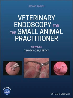
Veterinary Endoscopy for the Small Animal Practitioner
Здесь есть возможность читать онлайн «Veterinary Endoscopy for the Small Animal Practitioner» — ознакомительный отрывок электронной книги совершенно бесплатно, а после прочтения отрывка купить полную версию. В некоторых случаях можно слушать аудио, скачать через торрент в формате fb2 и присутствует краткое содержание. Жанр: unrecognised, на английском языке. Описание произведения, (предисловие) а так же отзывы посетителей доступны на портале библиотеки ЛибКат.
- Название:Veterinary Endoscopy for the Small Animal Practitioner
- Автор:
- Жанр:
- Год:неизвестен
- ISBN:нет данных
- Рейтинг книги:4 / 5. Голосов: 1
-
Избранное:Добавить в избранное
- Отзывы:
-
Ваша оценка:
- 80
- 1
- 2
- 3
- 4
- 5
Veterinary Endoscopy for the Small Animal Practitioner: краткое содержание, описание и аннотация
Предлагаем к чтению аннотацию, описание, краткое содержание или предисловие (зависит от того, что написал сам автор книги «Veterinary Endoscopy for the Small Animal Practitioner»). Если вы не нашли необходимую информацию о книге — напишите в комментариях, мы постараемся отыскать её.
Covers diagnostic endoscopy, interventional endoscopy, and minimally invasive soft tissue surgery Includes thousands of images to illustrate endoscopy concepts for veterinarians Provides a clinically oriented reference book for using rigid and flexible endoscopy in a small animal practice Supports veterinarians who are seeking to increase their services and enhance their revenue streams Any practitioner who is using or preparing to use endoscopic techniques will find
an essential practice resource.
Veterinary Endoscopy for the Small Animal Practitioner — читать онлайн ознакомительный отрывок
Ниже представлен текст книги, разбитый по страницам. Система сохранения места последней прочитанной страницы, позволяет с удобством читать онлайн бесплатно книгу «Veterinary Endoscopy for the Small Animal Practitioner», без необходимости каждый раз заново искать на чём Вы остановились. Поставьте закладку, и сможете в любой момент перейти на страницу, на которой закончили чтение.
Интервал:
Закладка:
4 Chapter 4Figure 4.1 Direct visual and physical access to the nasal cavity is achieved...Figure 4.2 The working tips of the two rigid biopsy forceps most commonly us...Figure 4.3 Rigid biopsy and operating forceps for turbinectomy are larger an...Figure 4.4 Five millimeter diameter Ferris‐Smith arthroscopy rongeurs with 5...Figure 4.5 Lateral radiograph of a normal dog showing the area of the skull ...Figure 4.6 Positioning for lateral radiographic projection of the nasal cavi...Figure 4.7 Lateral radiograph of a normal dog showing the area of the skull ...Figure 4.8 Positioning for ventral 20° rostral dorsocaudal oblique open mout...Figure 4.9 Ventral 20° rostral dorsocaudal oblique open mouth projection of ...Figure 4.10 Positioning for rostrocaudal projection of the skull for frontal...Figure 4.11 Rostrocaudal projection of the skull demonstrating normal fronta...Figure 4.12 HemaBlock ®placed in the nasal cavity to arrest an otherwis...Figure 4.13 Nasal tumor biopsy using the 3.0 mm rigid biopsy forceps passed ...Figure 4.14 The 3.0 mm rigid biopsy forceps positioned for biopsy of a nasop...Figure 4.15 Five French flexible biopsy forceps passed through the working c...Figure 4.16 A 1000 μm laser fiber used to vaporize tissue and control bleedi...Figure 4.17 A small quantity of mucus in the nasal cavity that is a normal f...Figure 4.18 Nasal mucosal appearance through air with a bright red color. Hi...Figure 4.19 Nasal mucosal appearance through liquid at the same location and...Figure 4.20 Turbinates are not well developed in the rostral nasal cavity ap...Figure 4.21 Normal pigment extending into the rostral nasal passage.Figure 4.22 Gently curving smooth turbinates in the rostral nasal cavity tha...Figure 4.23 Branching of the ventral nasal conchae seen from the middle meat...Figure 4.24 Ethmoidal turbinates with the normal crumpled appearance with mi...Figure 4.25 Thick ethmoidal turbinates approaching the upper end of normal....Figure 4.26 Ethmoidal turbinates at the thin end of normal with midrange int...Figure 4.27 Minimal interturbinate space in the ethmoidal turbinates at the ...Figure 4.28 Ethmoidal turbinates with midrange interturbinate space.Figure 4.29 Wide ethmoidal interturbinate space at the upper limit of normal...Figure 4.30 The same area of the nasal cavity as in Figure 4.22 with more in...Figure 4.31 The area on the nasal septum caudally that is normally roughened...Figure 4.32 The normal curved caudal margin of the nasal septum.Figure 4.33 Normal nasolacrimal duct opening in the rostral nasal cavity.Figure 4.34 Air bubbles seen during examination of a normal nasal cavity. Th...Figure 4.35 The normal nasopharynx seen with the telescope passed from the n...Figure 4.36 In this image, the endotracheal tube is seen in the right side o...Figure 4.37 A normal nasopharynx with subtle vasculature visible in the muco...Figure 4.38 Prominent vasculature visible in the mucosa of a normal nasophar...Figure 4.39 The opening of a eustachian tube in the nasopharynx at the appro...Figure 4.40 A small hard protrusion that is a common finding on the dorsal w...Figure 4.41 A wider view of the nasopharyngeal protrusion seen in the Figure...Figure 4.42 An air–liquid interface interfering with visualization of the na...Figure 4.43 The smooth discolored, greenish‐brown appearance of the mucosa i...Figure 4.44 Roughening of the mucosal surface in the discolored area of the ...Figure 4.45 An area of disrupted mucosal discoloration due to contact with t...Figure 4.46 An area of the nasal cavity with mild roughening of the mucosal ...Figure 4.47 Small random blood vessels visible in the normal nasal mucosa.Figure 4.48 Larger visible blood vessels at the margin of the olfactory orga...Figure 4.49 A normal frontal sinus lined with a thin transparent membrane an...Figure 4.50 A bone ridge extending into a normal frontal sinus. Highlights i...Figure 4.51 An air–water interface in a normal frontal sinus interfering wit...Figure 4.52 Active bleeding encountered at the beginning of rhinoscopy in a ...Figure 4.53 Fresh blood adhered to the surface of a visible tumor in the nas...Figure 4.54 A fresh blood clot from a recent bleeding episode that has not u...Figure 4.55 Abnormal material in the nasal cavity of a 12‐year‐old Brittany ...Figure 4.56 Clotted blood from a recent bleeding episode that formed into a ...Figure 4.57 Clotted blood in the same patient as Figure 4.56 in a different ...Figure 4.58 A smooth mass of clotted blood that has changed from red to a br...Figure 4.59 This clotted blood has changed from red to purple forming a larg...Figure 4.60 A purple blood clot that has formed a lobulated structure.Figure 4.61 A small blood clot‐like structure protruding between turbinates ...Figure 4.62 An organized blood clot‐like structure that appears encapsulated...Figure 4.63 A dark blood clot‐like structure with a ragged surface and stran...Figure 4.64 Ragged vascular appearing tissue that is mostly white structure ...Figure 4.65 Blood clot‐like material with three different appearances in one...Figure 4.66 The internal appearance of a blood clot‐like structure following...Figure 4.67 The internal appearance of a blood clot‐like mass with organized...Figure 4.68 Free mucopurulent exudate obscuring visibility of tumor surface ...Figure 4.69 Free bloody exudate in the nasal cavity of a dog with a nasal ma...Figure 4.70 Mucopurulent exudate that is adherent to the surface of a neopla...Figure 4.71 Large numerous blood vessels visible on the surface of a smooth ...Figure 4.72 Numerous small irregular blood vessels on the surface of a solid...Figure 4.73 Sparse small blood vessels in a solid smooth mass in the nasal c...Figure 4.74 Red coloration of a solid smooth nasal mass indicating significa...Figure 4.75 A smooth solid avascular‐appearing neoplastic mass in the nose o...Figure 4.76 A solid smooth avascular‐appearing neoplastic nasal mass in a do...Figure 4.77 An irregular solid nasal mass with an avascular appearance.Figure 4.78 A pink irregular solid neoplastic mass in the nasal cavity of a ...Figure 4.79 A dense red solid irregular mass in the nasal cavity of a dog.Figure 4.80 An irregular neoplastic nasal mass in a dog with sparse small bl...Figure 4.81 Numerous small irregular blood vessels in a solid irregular nasa...Figure 4.82 Numerous large prominent blood vessels in an irregular solid neo...Figure 4.83 A lobulated avascular appearing neoplastic nasal mass in a 14‐ye...Figure 4.84 The roughened surface of a neoplastic nasal mass.Figure 4.85 An unusual striated surface of a neoplastic mass in the nasal ca...Figure 4.86 A ragged tumor surface of a squamous cell carcinoma immediately ...Figure 4.87 A large neoplastic nasal mass with a red polypoid surface. Also ...Figure 4.88 A small polypoid neoplastic mass in the nasal cavity of an eight...Figure 4.89 A fimbriated neoplastic nasal mass with large blood vessels in t...Figure 4.90 A fimbriated neoplastic nasal mass with small blood vessels in t...Figure 4.91 A smooth cystic avascular neoplastic nasal mass with clear appea...Figure 4.92 A smooth cystic avascular neoplastic nasal mass with blue colora...Figure 4.93 A close‐up image of an avascular cystic mass showing that there ...Figure 4.94 A small avascular clear cystic projection from a nasal neoplasti...Figure 4.95 A sheet of avascular blue cystic‐appearing neoplastic tissue in ...Figure 4.96 A turbinate‐shaped avascular blue cystic structure in a dog with...Figure 4.97 A white avascular‐appearing cystic neoplastic lesion in the nasa...Figure 4.98 A pink cystic avascular‐appearing structure in the nasal cavity ...Figure 4.99 A red neoplastic cyst filled with fresh blood in the nasal cavit...Figure 4.100 Small sparse blood vessels in the wall of a neoplastic cyst in ...Figure 4.101 Numerous larger blood vessels visible in a neoplastic nasal cys...Figure 4.102 Large prominent blood vessels in the wall of a cystic neoplasti...Figure 4.103 Nasal neoplasia with solid vascular and solid avascular portion...Figure 4.104 Cystic avascular and solid vascular neoplastic tissue adjacent ...Figure 4.105 Solid vascular and solid avascular tissue in the same neoplasti...Figure 4.106 Cystic vascular tissue, solid irregular vascular tissue, and ro...Figure 4.107 Solid smooth avascular‐appearing neoplastic nasal tissue adjace...Figure 4.108 A cystic avascular‐appearing nasal tumor with an area of cyst w...Figure 4.109 Greenish coloration of a neuroendocrine tumor in the area of th...Figure 4.110 An area of gray tumor appearing in an area of normally pigmente...Figure 4.111 A nasal tumor with an unusual speckled surface.Figure 4.112 Internal tumor tissue following biopsy of a mass revealing a fr...Figure 4.113 Solid white internal tumor tissue exposed with biopsy of a nasa...Figure 4.114 A nasal tumor that has ruptured prior to rhinoscopy exposing in...Figure 4.115 A smooth vascular red‐appearing nasopharyngeal polyp in a one‐y...Figure 4.116 A smooth avascular white appearing nasopharyngeal polyp in a fo...Figure 4.117 An irregular vascular purple‐appearing nasopharyngeal polyp in ...Figure 4.118 A multicolored nasopharyngeal polyp visible in the nasopharynx ...Figure 4.119 A nasopharyngeal polyp attached to the stalk where it exits the...Figure 4.120 Two‐millimeter biopsy forceps passed from the nasopharynx into ...Figure 4.121 The eustachian tube after removal of the nasopharyngeal portion...Figure 4.122 The middle ear portion of a nasopharyngeal polyp seen with a 1....Figure 4.123 A small nasal tumor that is extending between turbinates with l...Figure 4.124 The site or origin of a nasal tumor, or invasion of tumor into ...Figure 4.125 A unilateral nasal tumor extending caudally into and completely...Figure 4.126 Extension of unilateral nasal neoplasia caudal to the septum as...Figure 4.127 A solid irregular avascular‐appearing unilateral mass extending...Figure 4.128 A cystic avascular bluish extension of a unilateral neoplasia c...Figure 4.129 A smooth neoplastic mass extending caudal to the nasal septum t...Figure 4.130 Adhesions of turbinates to the nasal septum in the contralatera...Figure 4.131 Displacement of the nasal septum to the contralateral side by a...Figure 4.132 A view of the contralateral nasal cavity with invasion of the n...Figure 4.133 Penetration of the nasal septum into the contralateral nasal ca...Figure 4.134 Penetration of the nasal septum into the contralateral nasal ca...Figure 4.135 Biopsy forceps placed to remove solid tumor tissue under endosc...Figure 4.136 Biopsy forceps placed to remove an area of cystic tumor tissue ...Figure 4.137 Tumor tissue is removed with biopsy forceps or rongeurs until t...Figure 4.138 Using the diode laser to control bleeding during tumor debulkin...Figure 4.139 The diode laser being used to vaporize tissue as part of the tu...Figure 4.140 Completed resection of a nasal tumor with a narrow attachment a...Figure 4.141 The diode laser has been used to provide hemostasis and vaporiz...Figure 4.142 Using biopsy forceps to remove additional tissue following diod...Figure 4.143 A large nasal septal defect created by debulking a tumor that h...Figure 4.144 An adequate airway has been re‐established by removal of tumor ...Figure 4.145 Completion of nasal tumor debulking when adequate neoplastic ti...Figure 4.146 Completion of nasal tumor debulking when a total turbinectomy h...Figure 4.147 Completion of a nasal tumor debulking procedure due to inabilit...Figure 4.148 Continued active bleeding after extensive tumor debulking and t...Figure 4.149 Application of a hemostatic powder achieved immediate hemostasi...Figure 4.150 A neoplastic clear‐walled irregular avascular‐appearing cyst wi...Figure 4.151 Rapid drainage of the cyst following laser penetration. The cys...Figure 4.152 Drainage of the cyst allows the cyst wall to shrink changing fr...Figure 4.153 A solid smooth nasal mass with varied coloration but no visible...Figure 4.154 At reoperation three weeks after the initial procedure in the c...Figure 4.155 Reoperation of a nasal tumor that at initial operation was an e...Figure 4.156 A large quantity of mucopurulent exudate in the nasal cavity of...Figure 4.157 Thick viscous mucopurulent exudate seen between nasal turbinate...Figure 4.158 Mucopurulent exudate covering and adhering to a fungal colony o...Figure 4.159 Thick viscous mucopurulent exudate extending caudal to the nasa...Figure 4.160 Decreased turbinate thickness with increased interturbinate spa...Figure 4.161 Early mild change in turbinate shape due to mycotic rhinitis in...Figure 4.162 Mild loss of turbinate height with turbinate thickening in a do...Figure 4.163 Mild loss of turbinate bulk with loss of both height and thickn...Figure 4.164 Marked turbinate distortion in a dog with mycotic rhinitis.Figure 4.165 Marked distortion of the ethmoidal turbinates in a dog with an Figure 4.166 Turbinate destruction and loss of turbinate support from remain...Figure 4.167 An end‐stage nasal cavity with a nasal fungal infection and alm...Figure 4.168 Marked hyperemia of the nasal turbinate mucosa and mild turbina...Figure 4.169 Fine roughening of the nasal turbinate mucosa in response to a ...Figure 4.170 A patient with a nasal Aspergillus infection showing coarse rou...Figure 4.171 Variable mucosal roughening on the nasal septum of the dog seen...Figure 4.172 Fine uniform roughening of the nasal septum mucosa of a dog wit...Figure 4.173 Increased vascularity of the turbinate mucosa in a dog with myc...Figure 4.174 An increased visible vascular pattern in the nasopharynx of a d...Figure 4.175 Replacement of nasal mucosa with granulation tissue in direct c...Figure 4.176 An adhesion between a nasal turbinate and the nasal septum in a...Figure 4.177 Individual small white nodules representing inflammatory polyps...Figure 4.178 Inflammatory polyps on the dorsal wall of the nasopharynx appea...Figure 4.179 White nodular inflammatory polyps coalesced into a solid layer ...Figure 4.180 Two small irregular inflammatory polyps in the nasal cavity of ...Figure 4.181 A cluster of small irregular polyps in a dog with a nasal Asper...Figure 4.182 Large inflammatory polyps coalescing into a solid irregular mas...Figure 4.183 A tumor‐like mass in the nasal cavity of a dog with a nasal fun...Figure 4.184 A larger tumor‐like mass in the nasal cavity secondary to a nas...Figure 4.185 An area of septal destruction due to a nasal fungal infection. ...Figure 4.186 The bright iridescent metallic appearance of a small fungal col...Figure 4.187 A medium‐sized fungal colony showing the bright iridescent meta...Figure 4.188 A large fungal colony with areas of the bright iridescent metal...Figure 4.189 A small spherical dull fungal colony sitting directly on the mu...Figure 4.190 A small white smooth shiny fungal colony sitting on a bed of gr...Figure 4.191 A small white fuzzy fungal colony in the frontal sinus of a dog...Figure 4.192 An upright spherical nasal fungal colony partially obscured wit...Figure 4.193 An irregular fungal colony with a smooth surface in the frontal...Figure 4.194 A fuzzy fungal colony in the nasal cavity of the dog in Figure ...Figure 4.195 A fungal colony in the nasal cavity of a dog with fuzzy white a...Figure 4.196 A large black Aspergillus niger colony in the nasal cavity of a...Figure 4.197 Mucopurulent drainage in the caudodorsal nasal cavity of a dog ...Figure 4.198 Mucopurulent drainage from the frontal sinus of a dog with a fu...Figure 4.199 Thickening of the lining membrane and increased vascular patter...Figure 4.200 Removing fungal colony material from the nasal cavity of a dog ...Figure 4.201 Using a stone basket to remove a fungal colony from the frontal...Figure 4.202 A frontal sinus after complete removal of a fungal colony with ...Figure 4.203 Correct placement of nasopharynx occlusion catheters for use wi...Figure 4.204 Foreign material in the nasal cavity of a dog with an Aspergill...Figure 4.205 Fungal material surrounding a tooth foreign body in the nasal c...Figure 4.206 Thickening of the lymphatic tissue on the dorsal wall of the na...Figure 4.207 A nasopharyngeal mass partially filling the nasopharynx in a ca...Figure 4.208 A cryptococcal mass completely filling the nasopharynx of a cat...Figure 4.209 A nasal cavity cryptococcal mass in a cat with chronic nasal di...Figure 4.210 Mucopurulent nasal discharge in the nasal cavity of a cat with ...Figure 4.211 Turbinate distortion and loss of turbinate mass in a cat with a...Figure 4.212 Mucopurulent exudate in the nasal cavity of a dog with allergic...Figure 4.213 Hyperemic turbinate mucosa in a case of allergic rhinitis. Ther...Figure 4.214 Roughening of the turbinate mucosa in a case of allergic rhinit...Figure 4.215 A case of allergic rhinitis with roughening of the septa mucosa...Figure 4.216 Roughening of the dorsal surface of the nasopharynx in a dog wi...Figure 4.217 Friable mucosa damaged by minimal endoscope contact during rhin...Figure 4.218 Turbinate swelling with loss of interturbinate air space in a d...Figure 4.219 Mild turbinate distortion in a dog with allergic rhinitis.Figure 4.220 An inflammatory nodule on a turbinate in a dog with allergic rh...Figure 4.221 Multiple small inflammatory nodules on the dorsal wall of the n...Figure 4.222 Inflammatory nodules coalesced into a solid sheet on the dorsal...Figure 4.223 A solitary irregular inflammatory polyp in the nasal cavity of ...Figure 4.224 A cluster of small irregular inflammatory polyps in the nasal c...Figure 4.225 Inflammatory polyps appearing as large contiguous masses.Figure 4.226 A grass awn foreign body in the nasal cavity of a dog.Figure 4.227 A blade of grass in the nasal cavity of a dog. There is thick i...Figure 4.228 A metallic bullet fragment in the nasal cavity of a dog.Figure 4.229 An Oregon grape leaf in the nasal cavity of a dog.Figure 4.230 Unidentified amorphous organic material in the nasal cavity of ...Figure 4.231 Unidentifiable inorganic material in the nose of a dog.Figure 4.232 Unidentifiable friable inorganic material in the nasal cavity o...Figure 4.233 Thick inspissated exudate completely hiding the foreign body in...Figure 4.234 Mucopurulent exudate containing blood surrounding a nasal forei...Figure 4.235 Removing a nasal foreign body with standard alligator forceps. ...Figure 4.236 Undisturbed exudate adjacent to an abscessed tooth root in a do...Figure 4.237 Cyst‐like structures in the ventrolateral nasal cavity over inv...Figure 4.238 A fractured upper fourth premolar tooth associated with the cys...Figure 4.239 White turbinates in a cat with turbinate infarction and complet...Figure 4.240 Pale turbinates in a cat with decreased vascular supply due to ...Figure 4.241 White distorted turbinates in a cat with chronic turbinate infa...Figure 4.242 Turbinate destruction in a case of turbinate infarction with a ...Figure 4.243 Biopsy of infarcted turbinates produces minimal or no bleeding....Figure 4.244 Mild turbinate damage to an infarcted turbinate caused by conta...Figure 4.245 No bleeding even with significant iatrogenic turbinate trauma c...Figure 4.246 Mucopurulent exudate in the nasal cavity of a cat with turbinat...Figure 4.247 White avascular turbinates on one side of the nasal cavity in a...Figure 4.248 Normally vascularized turbinate tissue in the contralateral nas...Figure 4.249 An area of completely infarcted turbinate in the foreground wit...Figure 4.250 An area of white completely infarcted tissue on the right and a...Figure 4.251 Turbinate distortion and destruction in a chronic case of turbi...Figure 4.252 Inflammatory nodule formation in a cat with chronic turbinate i...Figure 4.253 A large irregular inflammatory polyp in a cat with chronic turb...Figure 4.254 An area of turbinates with loss of cartilage support in a cat w...Figure 4.255 A nasal airway stricture immediately caudal to the nares in an ...Figure 4.256 Laser correction of the stricture seen in Figure 4.255.Figure 4.257 Turbinate inflammation and distortion visible through the corre...Figure 4.258 Recurrence of the nasal stricture in Figures 4.255–4.257 two we...Figure 4.259 Laser ablation of the recurrent stricture in Figures 4.255–4.25...Figure 4.260 Turbinate inflammation, distortion, and destruction visible cau...Figure 4.261 Fibrosis and inflammation in the nasal cavity at the level of a...Figure 4.262 Inflammatory nodules in the nasal cavity of the dog with the in...Figure 4.263 Turbinate distortion and loss of turbinate mass in a dog with a...Figure 4.264 A grass awn in the deep horizontal ear canal that has penetrate...Figure 4.265 Marked hyperemia of the ipsilateral wall of the nasopharynx in ...Figure 4.266 An eight‐month‐old DSH cat with a middle ear polyp visible thro...Figure 4.267 The normal middle ear visible through a normal translucent tymp...Figure 4.268 A typical nasopharyngeal polyp seen in an 11‐year‐old DLH cat t...Figure 4.269 An atypical nasopharyngeal polyp visible in the ear canal of a ...Figure 4.270 A carcinoma in the ear canal of an eight‐year‐old DSH cat with ...Figure 4.271 Removing the external ear canal portion of a nasopharyngeal pol...Figure 4.272 A middle ear cavity that has had a polyp debrided with biopsy f...Figure 4.273 Three parts of a nasopharyngeal polyp removed from a cat. Rhino...Figure 4.274 An unusual extension of a malignant middle ear canal tumor thro...Figure 4.275 A small inflammatory lesion protruding from the eustachian tube...Figure 4.276 A close-up endoscopic view of the dorsal view of the nasal mite...Figure 4.277 A close-up lateral endoscopic view of the nasal mite “Pneumony...Figure 4.278 A solitary nasal mite in the nasal cavity of an eight-year-old...Figure 4.279 A solitary nasal mite in the frontal sinus of a dog.Figure 4.280 A solitary nasal mite in the nasopharynx of a dog.Figure 4.281 A herd of nasal mites in the nasopharynx of a three-year-old Go...Figure 4.282 An inflammatory reaction of the lymphatic tissue on the dorsal...Figure 4.283 Reactive hyperemic lymphoid tissue on the dorsal wall of the na...Figure 4.284 Solid thickened lymphatic tissue on the dorsal wall of the naso...Figure 4.285 A large mass originating on the dorsal wall of the nasopharynx ...Figure 4.286 An area of hyperemic lymphatic tissue representing the “felenoi...Figure 4.287 Thickened lymphatic tissue on the dorsal wall of the nasopharyn...Figure 4.288 A smooth mass on the dorsal wall of the nasopharynx of a 13‐yea...Figure 4.289 An irregular mass on the dorsal wall of the nasopharynx in a 11...Figure 4.290 An irregular enlarged nasal turbinate that histopathology revea...Figure 4.291 Septal thickening in the nasal cavity of the dog in Figure 4.29...Figure 4.292 Large hamartoma ridges extending from the right nasal cavity in...Figure 4.293 A nasopharyngeal hamartoma in a cat.Figure 4.294 Vascular dysplasia in a 14‐year‐old neutered male West Highland...Figure 4.295 Vascular dysplasia in a 12‐year‐old neutered male Schipperke. T...Figure 4.296 A close‐up of the vascular appearance in a free‐floating turbin...Figure 4.297 Mucosal hyperemia in a patient with rhinitis of undetermined or...Figure 4.298 Roughened nasal mucosa in a Great Dane dog with rhinitis with a...Figure 4.299 Enlarged prominent blood vessels in the nasal turbinate mucosa ...Figure 4.300 Enlarged blood vessels in the nasopharynx of a dog with rhiniti...Figure 4.301 Swollen thickened turbinates in a dog with rhinitis of undeterm...Figure 4.302 Numerous small inflammatory nodules.Figure 4.303 Pedunculated polyps in the nasal cavity of a dog with rhinitis ...Figure 4.304 An irregular cluster of inflammatory tissue.Figure 4.305 A large inflammatory tumor‐like mass in the nasal cavity of a d...Figure 4.306 Thickened turbinates seen in a rhinitis case with no documented...Figure 4.307 An area of decreased turbinate bulk in the same case as Figure ...Figure 4.308 A white mass‐like lesion in the nasal cavity of the same case a...Figure 4.309 A red mass‐like lesion in the nasal cavity of the same case as ...Figure 4.310 An area of mucosal hyperemia in the nasal cavity of the case in...Figure 4.311 Hyperemia and thickening of the caudal portion of the nasal sep...Figure 4.312 Inflammatory nodules scattered over the area of the “catenoid” ...Figure 4.313 An increased number of enlarged blood vessels on the lateral wa...Figure 4.314 A hamartoma like mass in the nasopharynx of this same case as i...Figure 4.315 Contact between rostral turbinates in a brachiocephalic dog see...Figure 4.316 Contact between rostral turbinates in a brachiocephalic dog see...Figure 4.317 An area of turbinates in a brachiocephalic dog where there is a...Figure 4.318 The nasopharynx of a French Bulldog with extension of the ethmo...Figure 4.319 The ethmoidal turbinates are seen bending around the dorsal mar...Figure 4.320 The normal nasopharynx of the above case caudal to the abnormal...Figure 4.321 A completed partial turbinectomy in the French Bulldog seen in ...Figure 4.322 A nasopharyngeal stricture in an eight‐year‐old DSH cat present...Figure 4.323 A long tapering nasopharyngeal stenosis ending in a blind pouch...Figure 4.324 A small open communication in the center of a thin wall of tiss...Figure 4.325 A large residual lumen in a nasopharyngeal stenosis with reflec...Figure 4.326 A smaller residual lumen in a nasopharyngeal stricture obscured...Figure 4.327 Unidentified material occluding the residual lumen of a nasopha...Figure 4.328 Inflammatory nodules in the nasopharynx caudal to a nasopharyng...Figure 4.329 A cluster of inflammatory polyps in the nasopharynx rostral to ...Figure 4.330 Preparing to pass a 5 Fr biopsy forceps through a small residua...Figure 4.331 The dilated stricture from Figure 4.330 after passage of the bi...Figure 4.332 A 5 Fr red rubber catheter passed through the working channel o...Figure 4.333 Positioning of a nasopharyngeal stent using rhinoscopy with the...Figure 4.334 In this image, the stent has been retracted until it is fully w...Figure 4.335 The inflated balloon after expansion of the stent. The balloon ...Figure 4.336 A fully expanded nasopharyngeal stent sitting tightly against t...Figure 4.337 A nasopharyngeal stent in a cat two years after placement that ...Figure 4.338 Removal of the exudate in the case shown in Figure 4.337 reveal...Figure 4.339 The central portion of the stent in Figures 4.337 and 4.338 was...Figure 4.340 Multiple white raised avascular appearing masses in the nasopha...Figure 4.341 Multiple small white raised avascular appearing elongated nodul...Figure 4.342 A mass in the nasal cavity partially covered with exudate.Figure 4.343 Another mass in a different area of the same nasal cavity.Figure 4.344 An irregular deformed turbinate in this case of lymphoid hyperp...Figure 4.345 Irregular turbinates with loss of bulk and support.Figure 4.346 Thickened swollen puffy irregular turbinates.Figure 4.347 Roughened mucosa on the surface of an otherwise unchanged turbi...Figure 4.348 The fractured internal structure of one of the masses after bio...Figure 4.349 An area of white solid tissue in the nasal cavity of an eight‐y...Figure 4.350 Another area of the same case that has a cystic avascular appea...Figure 4.351 Completion of the debridement procedure with minimal residual a...Figure 4.352 Rostral turbinates with mild loss of bulk and distortion in the...Figure 4.353 Extensive mucopurulent exudate in the nasal cavity of the same ...Figure 4.354 A white lobulated avascular appearing mass in the caudolateral ...Figure 4.355 Debulking the recurrent mass from the nasal cavity of the same ...Figure 4.356 Completion of the debulking process with laser charred tissue o...Figure 4.357 Completion of the scheduled debulking two weeks after the previ...Figure 4.358 A nasal mass protruding from the right nares of an eight‐year‐o...Figure 4.359 An elongated pink solid mass in the right nasal cavity in the p...Figure 4.360 A white avascular cystic or edematous mass in another part of t...Figure 4.361 A dark cystic mass and an irregular solid mass in the same pati...Figure 4.362 Another area of the same nasal cavity with long vascular floati...Figure 4.363 The tissue displaced from the nasopharynx into the oral cavity ...
Читать дальшеИнтервал:
Закладка:
Похожие книги на «Veterinary Endoscopy for the Small Animal Practitioner»
Представляем Вашему вниманию похожие книги на «Veterinary Endoscopy for the Small Animal Practitioner» списком для выбора. Мы отобрали схожую по названию и смыслу литературу в надежде предоставить читателям больше вариантов отыскать новые, интересные, ещё непрочитанные произведения.
Обсуждение, отзывы о книге «Veterinary Endoscopy for the Small Animal Practitioner» и просто собственные мнения читателей. Оставьте ваши комментарии, напишите, что Вы думаете о произведении, его смысле или главных героях. Укажите что конкретно понравилось, а что нет, и почему Вы так считаете.


