Jörg Flock - X-Ray Fluorescence Spectroscopy for Laboratory Applications
Здесь есть возможность читать онлайн «Jörg Flock - X-Ray Fluorescence Spectroscopy for Laboratory Applications» — ознакомительный отрывок электронной книги совершенно бесплатно, а после прочтения отрывка купить полную версию. В некоторых случаях можно слушать аудио, скачать через торрент в формате fb2 и присутствует краткое содержание. Жанр: unrecognised, на английском языке. Описание произведения, (предисловие) а так же отзывы посетителей доступны на портале библиотеки ЛибКат.
- Название:X-Ray Fluorescence Spectroscopy for Laboratory Applications
- Автор:
- Жанр:
- Год:неизвестен
- ISBN:нет данных
- Рейтинг книги:5 / 5. Голосов: 1
-
Избранное:Добавить в избранное
- Отзывы:
-
Ваша оценка:
X-Ray Fluorescence Spectroscopy for Laboratory Applications: краткое содержание, описание и аннотация
Предлагаем к чтению аннотацию, описание, краткое содержание или предисловие (зависит от того, что написал сам автор книги «X-Ray Fluorescence Spectroscopy for Laboratory Applications»). Если вы не нашли необходимую информацию о книге — напишите в комментариях, мы постараемся отыскать её.
on using X-ray fluorescence for laboratory applications
This book focuses on the practical aspects of X-ray fluorescence (XRF) spectroscopy and discusses the requirements for a successful sample analysis, such as sample preparation, measurement techniques and calibration, as well as the quality of the analysis results.
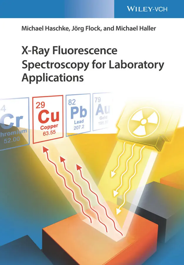
X-Ray Fluorescence Spectroscopy for Laboratory Applications
X
An important resource for the analytical chemist, providing concrete guidelines and support for everyday analyses Focuses on daily laboratory work with commercially available devices Offers a unique compilation of knowledge and best practices from equipment manufacturers and users Covers the entire work process: sample preparation, the actual measurement, data processing, assessment of uncertainty, and accuracy of the obtained results
appeals to analytical chemists, analytical laboratories, materials scientists, environmental chemists, chemical engineers, biotechnologists, and pharma engineers.
X-Ray Fluorescence Spectroscopy for Laboratory Applications — читать онлайн ознакомительный отрывок
Ниже представлен текст книги, разбитый по страницам. Система сохранения места последней прочитанной страницы, позволяет с удобством читать онлайн бесплатно книгу «X-Ray Fluorescence Spectroscopy for Laboratory Applications», без необходимости каждый раз заново искать на чём Вы остановились. Поставьте закладку, и сможете в любой момент перейти на страницу, на которой закончили чтение.
Интервал:
Закладка:
8 Chapter 9Table 9.1 Mean deviations for a cast iron calibration.Table 9.2 Typical analytical performance of WD spectrometers for low-alloy st...Table 9.3 Analytical performance of WD spectrometers for stainless steel.Table 9.4 Analytical performance of WD spectrometers for tool steel.Table 9.5 Analytical performance of WD spectrometers for the analysis of Al a...Table 9.6 Composition of eutectics of soft solders.Table 9.7 Analytical performance for the analyses of soft solders.Table 9.8 Accuracies for the analysis of gold alloys.Table 9.9 Relative standard deviations of repeated measurements depending on ...Table 9.10 Typical composition and measurement uncertainties of flat glasses.Table 9.11 Polymers and their approximate composition and mass fractions in w...Table 9.12 Correlation coefficients R 2for different energy resolutions.Table 9.13 Mass fractions of hydrocarbon groups and H, N, and O for PUR and A...
9 Chapter 10Table 10.1 Detection limits for some heavy elements using K-radiation.Table 10.2 Maximum loads of soils and sewage sludge.Table 10.3 Typical compositions of components for the cement production in wt...Table 10.4 Relative standard deviations of repeated measurements for samples ...Table 10.5 Concentrations of typical ferroalloys in wt%.Table 10.6 Digestion procedures for different Ferroalloys.Table 10.7 Analyses results of ferro-silicon using powder samples in wt%.Table 10.8 Relative standard deviation σ relof repeated measurements of s...Table 10.9 Mass fractions ranges of refractory materials.Table 10.10 Results of repeated measurements on a refractory.Table 10.11 Average limits for toxic elements in animal food.Table 10.12 Comparison between calibration results and limits of detection fo...Table 10.13 Maximal amount of intake of toxic elements per day in micrograms.Table 10.14 Reference values for the content of heavy metals in secondary fue...Table 10.15 Typical line overlaps for toxic elements.Table 10.16 Results of repeated measurements for secondary fuels.Table 10.17 Reproducibility and trueness of the analyses of toxic liquids.
10 Chapter 11Table 11.1 Limit of detection for S in fuels depending on the measuring method...Table 11.2 Repeatability and reproducibility limits for the detection of sulf...Table 11.3 Requirements according to ASTM D 4927-05 and ASTM D6481.Table 11.4 Analytical performance for additives with a WD spectrometer.
11 Chapter 12Table 12.1 Preparation procedures for TXRF.Table 12.2 Concentration limits of elements in drinking water.Table 12.3 Quantification results of the fresh water sample (NIST SRM 1640).Table 12.4 TXRF results of blood serum compared with certified values.
12 Chapter 13Table 13.1 Properties of energy-dispersive detectors.
13 Chapter 14Table 14.1 Detector influence on analytical results.Table 14.2 Coating thickness results of an Au/Ni/Cu layer system evaluated in...Table 14.3 Absolute and relative standard deviations of 10 repeated measureme...Table 14.4 Selection of reference standards and their uncertainties (95%).Table 14.5 Measurement results of a CIGS layer system.Table 14.6 Characterization of a SnPb/Ni/AgPb/ceramic system.
14 Chapter 15Table 15.1 Maximum concentrations for toxic substances according to RoHS.Table 15.2 Maximum loads according to Toy Regulation (BGBl., II, Nr. 203/2011...Table 15.3 Maximum soluble migrated element in ppm (mg/kg) for surface coatin...
15 Chapter 16Table 16.1 Concentrations at the measuring points in wt%.
16 Chapter 19Table 19.1 Parameters in dependence of the task for checking.Table 19.2 Determination of the parameter range of application, linearity, an...
17 Appendix A Table A.1 Periodic table of elements with atomic number, atomic weight, and d...Table A.2 Atomic weight and density (in g/cm 3) of all elements.Table A.3 Energies of absorption edges (keV).Table A.4 K-line energies (keV).Table A.5 L-line energies (keV).Table A.6 M-line energies (keV).Table A.7 Wavelengths of K-lines (nm).Table A.8 Wavelengths of L-lines (nm).Table A.9 Wavelengths of M-lines (nm).Table A.10 Common line interferences, energies in keV.Table A.11 Fluorescence yields.Table A.12 Transfer factors between elements and oxides.
List of Illustrations
1 Chapter 2 Figure 2.1 Parts of the continuous spectrum of an X-ray tube. Figure 2.2 Line energy as a function of the atomic number. Figure 2.3 Electron transitions with the corresponding line names. Figure 2.4 Fluorescence yield as a function of the atomic number. Figure 2.5 Contributions to the attenuation coefficient for X-radiation of (... Figure 2.6 Elastic and inelastic scattering coefficients for selected elemen... Figure 2.7 (a) Influence of the sample type on the intensity of the scattere... Figure 2.8 Spectra of PMMA, Al, Ti, and Ni demonstrating the different scatt... Figure 2.9 PW 1540 from Philips. Figure 2.10 Analyst 0700 from KEVEX. Figure 2.11 Steps for an analytical procedure.
2 Chapter 3 Figure 3.1 Information depth of fluorescence radiation. Figure 3.2 Information depth for different matrices. Figure 3.3 Cd intensities measured on polymer samples of different thickness... Figure 3.4 Analyzed volume limited by the measurement geometry. Figure 3.5 Specimen generated by sampling from a melt. Figure 3.6 Influence of surface roughness on the measured fluorescence inten... Figure 3.7 Dependence of fluorescence intensity on grinding time. Figure 3.8 Dependence of fluorescence intensity on the morphology of the sam... Figure 3.9 Press die for to produce pellets from small-sized materials. Figure 3.10 Pressed pellet (a) and cross section of a pressed pellet in bind... Figure 3.11 Influence of grain size distribution and different hardness of s... Figure 3.12 Calibration curves for the same powder material prepared as pres... Figure 3.13 Arrangement of layers of melting agent and sample in the fusion ... Figure 3.14 (a) Pt crucibles over a gas burner. (b) Temperature profile of t... Figure 3.15 Cooling curve to produce glass pellets. Figure 3.16 Glassy (a) and milky, too quickly cooled fusion bead (b). Figure 3.17 Sample cup for the analysis of liquids. Figure 3.18 Transmission of low-energy radiation of sample cup films.
3 Chapter 4 Figure 4.1 Energy resolution of different ED spectrometer (blue) and WD spec... Figure 4.2 Spectrum of stainless steel, measured by WDS and EDS: (a) low-ene... Figure 4.3 Influence of the selected lattice plane (a) and the collimator si... Figure 4.4 Spectra of a gold alloy measured with a proportional counter and ... Figure 4.5 Energy dependence of energy resolution of ED-detectors (FWHM, ful... Figure 4.6 Detection efficiency of prop-counter and scintillation counter. Figure 4.7 Detection efficiency of ED-detectors. Figure 4.8 Throughput of ED-detectors and their influence on the detector re...Figure 4.9 Peak shape changes for very high count rates of WD spectrometers....Figure 4.10 Effect of the count rate on the peak-to-background ratios.Figure 4.11 Comparison of the ratios of the relative standard deviation (rel...Figure 4.12 Low-energy spectral background due to a Compton-induced escape e...Figure 4.13 Escape and pile-up peak of Ti radiation ( y -axis in square root s...Figure 4.14 Pt spectrum measured without and with Al filter ( y -axis in squar...Figure 4.15 Shelf and tail contributions to a Cu line ( y -axis in square root...Figure 4.16 Principle layout of EDS instruments (MCA, multichannel analyzer)...Figure 4.17 Handheld instruments from various manufacturers: (a) XL5, Thermo...Figure 4.18 Portable instruments of different manufacturers: (a) FXL, Thermo...Figure 4.19 Various energy-dispersive spectrometers: (a) Epsilon 3, Panalyti...Figure 4.20 Principle layout of WDS instruments.Figure 4.21 A selection of wavelength-dispersive X-ray spectrometers: (a) Ze...Figure 4.22 A selection of multichannel X-ray spectrometers: (a) S8 Lion, Br...Figure 4.23 Principle setup of a TXRF instrument (MCA, multichannel analyzer...Figure 4.24 Total reflection X-ray spectrometers: (a) TXRF-310, Rigaku Corp....Figure 4.25 Layout of an X-ray spectrometer with monoenergetic excitation.Figure 4.26 Spectrum of an oil sample excited with Mo radiation.Figure 4.27 Measurement geometry for the excitation with polarized radiation...Figure 4.28 Energy-dispersive instruments using polarized radiation: (a) XEP...Figure 4.29 Basic layouts of position-sensitive X-ray spectrometers with a c...Figure 4.30 Coating thickness instruments with collimators: (a) XDAL, Helmut...Figure 4.31 Instruments utilizing X-ray optics for beam focusing: (a) M4 Tor...Figure 4.32 Macro-X-ray fluorescence spectrometer for the investigation of l...Figure 4.33 Confocal geometry for the investigation of 3D elemental distribu...Figure 4.34 Grazing incident measurement geometry.Figure 4.35 Grazing exit measurement geometry.
Читать дальшеИнтервал:
Закладка:
Похожие книги на «X-Ray Fluorescence Spectroscopy for Laboratory Applications»
Представляем Вашему вниманию похожие книги на «X-Ray Fluorescence Spectroscopy for Laboratory Applications» списком для выбора. Мы отобрали схожую по названию и смыслу литературу в надежде предоставить читателям больше вариантов отыскать новые, интересные, ещё непрочитанные произведения.


Обсуждение, отзывы о книге «X-Ray Fluorescence Spectroscopy for Laboratory Applications» и просто собственные мнения читателей. Оставьте ваши комментарии, напишите, что Вы думаете о произведении, его смысле или главных героях. Укажите что конкретно понравилось, а что нет, и почему Вы так считаете.