Philias R. Garant - Oral Cells and Tissues
Здесь есть возможность читать онлайн «Philias R. Garant - Oral Cells and Tissues» — ознакомительный отрывок электронной книги совершенно бесплатно, а после прочтения отрывка купить полную версию. В некоторых случаях можно слушать аудио, скачать через торрент в формате fb2 и присутствует краткое содержание. Жанр: unrecognised, на английском языке. Описание произведения, (предисловие) а так же отзывы посетителей доступны на портале библиотеки ЛибКат.

- Название:Oral Cells and Tissues
- Автор:
- Жанр:
- Год:неизвестен
- ISBN:нет данных
- Рейтинг книги:3 / 5. Голосов: 1
-
Избранное:Добавить в избранное
- Отзывы:
-
Ваша оценка:
Oral Cells and Tissues: краткое содержание, описание и аннотация
Предлагаем к чтению аннотацию, описание, краткое содержание или предисловие (зависит от того, что написал сам автор книги «Oral Cells and Tissues»). Если вы не нашли необходимую информацию о книге — напишите в комментариях, мы постараемся отыскать её.
Oral Cells and Tissues — читать онлайн ознакомительный отрывок
Ниже представлен текст книги, разбитый по страницам. Система сохранения места последней прочитанной страницы, позволяет с удобством читать онлайн бесплатно книгу «Oral Cells and Tissues», без необходимости каждый раз заново искать на чём Вы остановились. Поставьте закладку, и сможете в любой момент перейти на страницу, на которой закончили чтение.
Интервал:
Закладка:
It has been calculated that the lipid phase of the plasma membrane of a fibroblast turns over in about 50 minutes. Some intramembrane proteins are caught up in this flow, while others remain in place because of their association with the internal cytoskeleton or with extracellular substrates.
Protrusion of lamellae and filopodia at the leading edge is driven by rapid polymerization of actin filaments (see chapter 11for a discussion of actin filament formation). Assembly of linear actin bundles may push the membrane outward or cause an increase in local hydrostatic pressure to deform the membrane outward at the leading edge. Because calcium triggers actin polymerization, it has been proposed that filopodial formation at the leading edge might be regulated by the entry of calcium ions through cell membrane channels.
Another explanation for the forward extension of the plasma membrane is the assembly of new membrane via exocytosis at the leading edge and the simultaneous endocytosis toward the middle and rear of a migrating cell. Polarized exocytosis-endocytosis cycles have been observed in migrating fibroblasts and neurite growth cones.
To develop traction and forward movement, cells must form attachments between their leading edge and the substratum. Cells migrating in vitro on glass cover slips make close contacts and focal contacts with the surface of the glass. 82At close contacts, the cell membrane is separated from the substratum by a space of 20 to 30 nm. Close contacts represent the initial association of specific cell membrane attachment proteins to the extracellular matrix. Close contacts are typically found at the very leading edge of lamellae and filopodia.
In contrast, focal contacts typically occur just distal to the outer zone of the leading edge ( Figs 1-16and 1-17). In focal contacts, the cell membrane is only 10 to 15 nm from the surface of the substrate. The focal contact is the product of the maturation of the close contact by recruitment of integrin receptors and other membrane-associated proteins. Along with the integrins, actin, vinculin, and talin are rapidly associated with the initial site of attachment to form a focal contact or focal adhesion. Thus, the integrins mediate transmembrane linkage of the cytoskeletal proteins to the extracellular matrix. 83
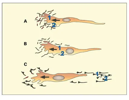
Fig 1-17Hypothesis proposed by Harris (1973) to explain how the forward movement of cells is coordinated to the development of stable cell-to-matrix contacts associated with actin and myosin filament bundles. (A) Focal contacts (1 and 2) established at the leading edge remain in position as (B) new membrane and cytosol advance in the continued protrusion of the leading lamella. (C) With time, the focal contacts, first established at A, become located at the trailing end of the cell, and will eventually be ruptured as the tail is pulled forward. The detached focal contacts with bits of cytoplasm remain attached to the substratum. Contraction of actin and myosin in the cell body propels cytosol forward to the leading lamella. In the process, matrix molecules become aligned parallel to the direction of cell migration. (Adapted from Hay. 88)
The integrin dimer α5β1 represents one type of integrin fibronectin receptor. Fibronectin participates as the extracellular component of the close contact in migrating fibroblasts and neural crest cells. 84Motile cells make cell-to-matrix attachment interactions of a transient nature (close contacts). Fibronectin receptors tend to be more dispersed over the surface of migrating cells.
Cell-to-cell attachments and stable cell-to-matrix adhesions (focal adhesions) assume greater importance in stabilizing nonmotile cells at their final destination. In stationary cells, the fibronectin receptors are clustered in alignment with extracellular fibronectin fibrils. 85 , 86When cells are attached to matrix fibrils, which are under tension, the cells develop large focal adhesions (fibronexus) associated with cytoplasmic actin and myosin bundles (stress fibers). The fibronexus junction is described in chapter 6.
Specific extracellular matrix molecules, organized into three-dimensional scaffolds, provide pathways for the selective migration of certain cell types. Neural crest cells migrate in defined tracks rich in fibronectin and hyaluronic acid. The same is true for the migration of fibroblasts into the primary corneal stroma. The basal lamina, or substances associated with it, can also act as a substrate for the preferential migration of cells in vivo. Certain types of neural crest cells end their migration when they encounter regions rich in tenascin, a large extracellular attachment molecule.
Several environmental stimuli cause cells to undergo directed migration. Cells can move along a concentration gradient of an extracellular matrix molecule (haptotaxis). In an electrical field, cells migrate toward the cathode (galvanotaxis). Fibronectin fragments induce directed migration of fibroblasts, a stimulus likely to be important in wound healing. 87
Cells also tend to move outward from a cell mass. Cells on the perimeter of the cell mass continue to form leading lamellae and filopodia along their free surface and thus are able to move away from the cell mass. Within the cell mass, however, cells are contact inhibited; a state of reduced membrane ruffling and filopodial extension occurs along the adjacent surfaces of juxtaposed cells. Directed migrations of neural crest cells within the extracellular matrix scaffold proceed from areas of high to low cell density because of contact inhibition.
Extracellular matrix molecules may undergo reorganization following interaction with the cell surface of a migrating cell (see Fig 1-17). 84 , 88 – 90Traction transmitted to the extracellular matrix by migrating (contracting) cells also exerts an organizational influence over matrix molecules. As fibroblasts migrate through a collagen gel in vitro, they cause the extracellular collagen fibrils to become aligned parallel to the long axis of the fibroblasts and the gel to contract. Fibronectin fibrils increase in size and organization toward the trailing edge of migrating fibroblasts. The role of cell polarity and migration in determining the organization of collagen in the periodontal ligament is discussed in chapter 6.
Cell and substrate adhesion molecules
Calcium-dependent cadherins, integrins, selectins, plasma membrane proteoglycans, and members of the immunoglobulin superfamily, such as neural cell adhesion molecule, participate in forming cell-to-cell and cell-to-matrix adhesions. 91Members of these transmembrane proteins play essential roles in the cellular organization of tissues and organs and in the migration of cells in embryonic and adult tissues. 91 – 93The cadherins, components of desmosomes, are discussed in chapter 4, and the selectins, adhesion molecules that regulate leukocyte emigration from blood vessels, are described in chapters 13 and 14.
Integrins The integrins are a family of cell surface transmembrane proteins that developed very early in evolution 91 – 94( Figs 1-18and 1-19). Integrins are heterodimers made up of α and β subunits. At least 14 α and eight β subunits have been identified. Figure 1-19contains a chart of the subunits and ligands of the very late activation–type integrins.
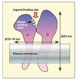
Fig 1-18Integrin-type receptors. The α and β integrin transmembrane proteins form a dimer with a shared ligand-binding site. Metal-binding sites on the α subunit are needed for receptor function.
Читать дальшеИнтервал:
Закладка:
Похожие книги на «Oral Cells and Tissues»
Представляем Вашему вниманию похожие книги на «Oral Cells and Tissues» списком для выбора. Мы отобрали схожую по названию и смыслу литературу в надежде предоставить читателям больше вариантов отыскать новые, интересные, ещё непрочитанные произведения.


Обсуждение, отзывы о книге «Oral Cells and Tissues» и просто собственные мнения читателей. Оставьте ваши комментарии, напишите, что Вы думаете о произведении, его смысле или главных героях. Укажите что конкретно понравилось, а что нет, и почему Вы так считаете.