Philias R. Garant - Oral Cells and Tissues
Здесь есть возможность читать онлайн «Philias R. Garant - Oral Cells and Tissues» — ознакомительный отрывок электронной книги совершенно бесплатно, а после прочтения отрывка купить полную версию. В некоторых случаях можно слушать аудио, скачать через торрент в формате fb2 и присутствует краткое содержание. Жанр: unrecognised, на английском языке. Описание произведения, (предисловие) а так же отзывы посетителей доступны на портале библиотеки ЛибКат.

- Название:Oral Cells and Tissues
- Автор:
- Жанр:
- Год:неизвестен
- ISBN:нет данных
- Рейтинг книги:3 / 5. Голосов: 1
-
Избранное:Добавить в избранное
- Отзывы:
-
Ваша оценка:
Oral Cells and Tissues: краткое содержание, описание и аннотация
Предлагаем к чтению аннотацию, описание, краткое содержание или предисловие (зависит от того, что написал сам автор книги «Oral Cells and Tissues»). Если вы не нашли необходимую информацию о книге — напишите в комментариях, мы постараемся отыскать её.
Oral Cells and Tissues — читать онлайн ознакомительный отрывок
Ниже представлен текст книги, разбитый по страницам. Система сохранения места последней прочитанной страницы, позволяет с удобством читать онлайн бесплатно книгу «Oral Cells and Tissues», без необходимости каждый раз заново искать на чём Вы остановились. Поставьте закладку, и сможете в любой момент перейти на страницу, на которой закончили чтение.
Интервал:
Закладка:
I am also grateful to all the scientists and publishers who graciously permitted me to adapt and/or reproduce their illustrations for this book.
Finally, I am most grateful to my spouse and best friend, Jeanne. Without her help and support I would not have been able to complete my professional education, nor lead a life in academia. I am also grateful to my six children for the love they have shown me.
Early Tooth Development
Teeth are formed from oral epithelium, in the form of a dental lamina, and neural crest ectomesenchyme of the maxillary and mandibular processes ( Fig 1-1). The oral epithelium contributes the enamel component, and the ectomesenchyme contributes the dentin and cementum components of the fully formed tooth. Although the initiating events that trigger downgrowth of the oral epithelium to form a dental lamina are incompletely understood, it is known that neural crest ectomesenchyme is necessary. 1 , 2Early reciprocal inductive interactions between the oral epithelium and the underlying ectomesenchyme, and subsequent interactions between the enamel organ and dental papilla, coordinate the sequential events of tooth development. 3 – 5
Efforts to understand the instructional signals that originate in each of these interacting tissues have been ongoing for more than 50 years. 6 , 7Most investigations have been performed with dental tissues obtained from embryonic mice and rats or with the continuously growing incisor teeth of adult mice and rats. Organ culture techniques have been perfected to study the growth of dental tissues in chemically defined media, to observe the results of various epithelial-mesenchymal combinations, and to examine the effects of various growth factors on odontogenesis. Thus, nearly all current insight into the regulatory mechanisms of tooth development has come from studies of animal models, often from tooth buds grown in organ culture.
This chapter contains a discussion of the initiation of tooth formation and the histodifferentiation of the enamel organ and dental papilla. Subsequent chapters will examine the cytodifferentiation of dentin-and enamel-forming cells and the secretion and mineralization of their respective matrices.
Role of the Neural Crest
Early in embryogenesis, soon after the neural tube forms by invagination of the overlying ectoderm, migratory pluripotent neuroepithelial cells, the neural crest cells, migrate from the dorsal midline region of the neural tube. 8In exiting from the neural tube, neural crest cells lose their epithelioid characteristics and assume a mesenchymal phenotype capable of directed cell migration. Cranial neural crest cells invade the developing branchial arches and, in a series of reciprocal inductive interactions with early oral epithelium, form tooth primordia ( Figs 1-1and 1-2). When the movement of dye-injected neural crest cells was traced in organ cultures of developing dental arches, it was shown that neural crest cells from the posterior midbrain, and to a lesser extent from the anterior hindbrain, form dental ectomesenchyme. 9The failure of neural crest ectomesenchymal cells to migrate normally to appropriate sites during craniofacial development leads to serious developmental defects, including the absence of teeth (anodontia) and underdeveloped jawbones (micrognathia).
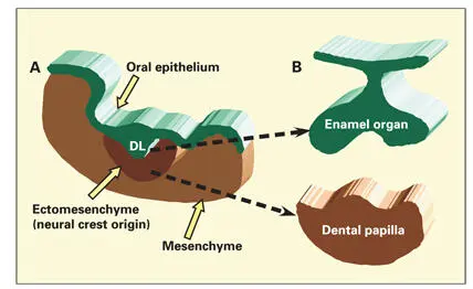
Fig 1-1Stages in the development of a tooth bud. (A) Oral epithelium and the underlying ectomesenchyme and mesenchyme during the development of the dental lamina (DL). (B) The enamel organ arises from a genetically determined site of the dental lamina by cell proliferation. The dental papilla develops from ectomesenchymal cells of neural crest origin.
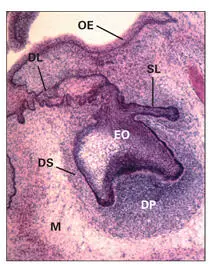
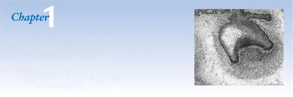
Fig 1-2Histologic section of a developing tooth at early bell stage. (DL) Dental lamina; (DP) dental papilla; (DS) dental sac; (EO) enamel organ; (M) mesenchyme; (OE) oral epithelium; (SL) successional lamina. (Hematoxylin-eosin stain. Original magnification × 220.)
Subsets of cranial neural crest cells give rise to chondrocytes, osteoblasts, periodontal ligament fibroblasts, cementoblasts, and odontoblasts. Final phenotype differentiation is regulated by interaction of the ectomesenchymal cells with extrinsic factors, such as growth factors, and substrate adhesion molecules in the local microenvironment. 10It has been suggested that there may be separate populations of neural crest cells for each tooth type. The molecular code for each tooth type appears to reside in specific sets of homeobox genes. 11 , 12
Development of the Dental Lamina, Enamel Organ, and Dental Papilla
The first evidence of tooth formation in humans is observed as a thickening of the oral epithelium in the mandibular, maxillary, and medial nasal processes in the 1-month-old fetus ( Figs 1-3to 1-5). It has been suggested that the zone of epithelial thickening (the dental plate or placode) contains the genetic determinants for the initiating signals that regulate the number and position of the future tooth buds. Experiments with epithelial-mesenchymal tissue recombination have shown that early-stage oral epithelium is capable of inducing tooth development in non-oral ectomesenchyme. 13 – 15When non-oral epithelium is used in the recombination, only bone and cartilage form in the ectomesenchyme. Mouse oral epithelium has been shown to induce biochemical markers of early tooth development in chick oral ectomesenchyme, a tissue thought to have lost its ability to form teeth. 16The results of these studies suggest that the oral ectoderm contains instructional signals for tooth development and perhaps the prepattern for the entire dentition. Weiss et al 17suggested that a very early signaling system (prior to neural crest migration) involving Shh and Pax6 genes might form the basis of epithelial patterning mechanisms for tooth development.
Formation of the dental lamina
At a slightly later stage of development (11- to 14-mm embryos), the epithelium invaginates into the underlying mesenchyme to form the dental lamina. This process begins in the distal (molar) region and later in the midline. In 15- to 20-mm human embryos, the dental lamina shows signs of additional differential growth, reflecting the determination of incisor, canine, and molar domains (see Figs 1-4and 1-5). Deep notches in the dental lamina are present between the incisor and canine domains, especially in the mandible. Continued site-specific enlargement of the dental lamina, along with condensation of neural crest ectomesenchyme, gives rise to the individual tooth buds.
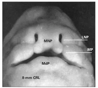
Fig 1-3Facial region of a human embryo. (LNP) Lateral nasal process; (MNP) medial nasal process; (MP) maxillary process; (MdP) mandibular process; (CRL) crown-rump length. (Adapted from Ooe 74with permission.)
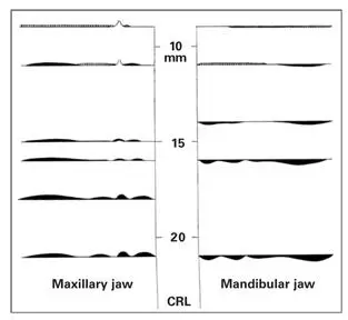
Fig 1-4Degree of oral epithelial thickening in various human embryos ranging from 10- to 20-mm crown-rump length (CRL). Note the undulating character of the undersurface of the epithelium. (Adapted from Ooe 74with permission.)
Читать дальшеИнтервал:
Закладка:
Похожие книги на «Oral Cells and Tissues»
Представляем Вашему вниманию похожие книги на «Oral Cells and Tissues» списком для выбора. Мы отобрали схожую по названию и смыслу литературу в надежде предоставить читателям больше вариантов отыскать новые, интересные, ещё непрочитанные произведения.


Обсуждение, отзывы о книге «Oral Cells and Tissues» и просто собственные мнения читателей. Оставьте ваши комментарии, напишите, что Вы думаете о произведении, его смысле или главных героях. Укажите что конкретно понравилось, а что нет, и почему Вы так считаете.