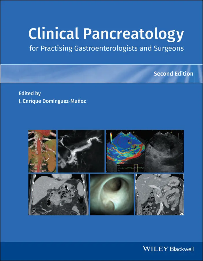
Clinical Pancreatology for Practising Gastroenterologists and Surgeons
Здесь есть возможность читать онлайн «Clinical Pancreatology for Practising Gastroenterologists and Surgeons» — ознакомительный отрывок электронной книги совершенно бесплатно, а после прочтения отрывка купить полную версию. В некоторых случаях можно слушать аудио, скачать через торрент в формате fb2 и присутствует краткое содержание. Жанр: unrecognised, на английском языке. Описание произведения, (предисловие) а так же отзывы посетителей доступны на портале библиотеки ЛибКат.
- Название:Clinical Pancreatology for Practising Gastroenterologists and Surgeons
- Автор:
- Жанр:
- Год:неизвестен
- ISBN:нет данных
- Рейтинг книги:4 / 5. Голосов: 1
-
Избранное:Добавить в избранное
- Отзывы:
-
Ваша оценка:
Clinical Pancreatology for Practising Gastroenterologists and Surgeons: краткое содержание, описание и аннотация
Предлагаем к чтению аннотацию, описание, краткое содержание или предисловие (зависит от того, что написал сам автор книги «Clinical Pancreatology for Practising Gastroenterologists and Surgeons»). Если вы не нашли необходимую информацию о книге — напишите в комментариях, мы постараемся отыскать её.
was first published sixteen years ago, the knowledge and clinical management of pancreatic diseases have developed markedly. Thanks to the development of translational research and the “from bench to bedside” concept, much progress from the lab has been applied to clinical practice. Additionally, several highly relevant clinical trials published over the last decade have resulted in the updating and optimisation of clinical guidelines.
A new and validated classification of the severity and complications of acute pancreatitis that is firmly rooted in clinical practice has become the basis for the development of minimally invasive approaches to pancreatic necrosis. The etiopathogenic knowledge of chronic pancreatitis and other pancreatopaties, like that associated with diabetes mellitus, has developed significantly. Increased study of cystic pancreatic tumours, which has been reflected in the publication of several guidelines and consensus reports over the last few years, is especially important. Most research efforts have focused on pancreatic cancer, which have led and will further lead to a significant increase in the therapeutic armamentarium against this devastating disease. Finally, many newly published studies have changed the concept, causes, clinical relevance, diagnosis and treatment of exocrine pancreatic insufficiency. Updates based on these developments and more are included in the new edition of
.
This new edition of
is a result of the collaboration between the world's leading experts in each area of clinical pancreatology, with the aim of facilitating gastroenterologists, surgeons, oncologists, internists, nutritionists, diabetologists, paediatricians, radiologists, pathologists and other specialists in their daily clinical practice. This book is an indispensable update providing leading knowledge in clinical pancreatology.
Clinical Pancreatology for Practising Gastroenterologists and Surgeons — читать онлайн ознакомительный отрывок
Ниже представлен текст книги, разбитый по страницам. Система сохранения места последней прочитанной страницы, позволяет с удобством читать онлайн бесплатно книгу «Clinical Pancreatology for Practising Gastroenterologists and Surgeons», без необходимости каждый раз заново искать на чём Вы остановились. Поставьте закладку, и сможете в любой момент перейти на страницу, на которой закончили чтение.
Интервал:
Закладка:
107 107 Sadr‐Azodi O, Oskarsson V, Discacciati A, et al. Pancreatic cancer following acute pancreatitis: a population‐based matched cohort study. Am J Gastroenterol 2018; 113:1711–1719.
108 108 Kirkegard J, Cronin‐Fenton D, Heide‐Jorgensen U, Mortensen FV. Acute pancreatitis and pancreatic cancer risk: a nationwide matched‐cohort study in Denmark. Gastroenterology 2018; 154:1729–1736.
109 109Yadav D, Lowenfels AB. The epidemiology of pancreatitis and pancreatic cancer. Gastroenterology 2013; 144:1252–1261.
2 How to Deal with the Etiological Diagnosis of Acute Pancreatitis in Clinical Practice?
Soumya Jagannath and Pramod Kumar Garg
Department of Gastroenterology, All India Institute of Medical Sciences, New Delhi, India
Introduction
Acute pancreatitis (AP) is an acute inflammatory condition of the pancreas with significant morbidity and mortality. Global incidence of AP is 34 per 100 000 person‐years with a mortality of 1.6 per 100 000 person‐years [1]. Most cases of AP are mild but severe pancreatitis occurs in 10–20% of patients, who present with organ failure with a high mortality of up to 40% [2,3]. Around 25% of patients with AP develop recurrence of pancreatitis and a variable proportion of them progress to chronic pancreatitis over time [4].
AP occurs due to a variety of causes. Making the etiological diagnosis of AP is important and possible in most cases. Two of the most common causes of AP are biliary (40–70%) and alcohol (25–35%) [3,5,6]. Other causes of AP are enumerated in Table 2.1. Although the etiology has no major bearing on outcome of AP [7], its determination is important to prevent the recurrence and progression to chronic pancreatitis since most of the causes are either modifiable or treatable. The annual rate of relapse after first attack of AP is 5.3 per 100 per year in patients with alcoholic etiology and 1.5 per 100 per year in those with biliary etiology [5]. Hence, identification of the etiology dictates further management after the acute episode resolves. In this chapter we discuss the various causes of AP ( Table 2.1) and a diagnostic algorithm for etiological diagnosis of AP.
Etiological Diagnosis
Gallstone‐induced Pancreatitis
Gallstones have been suspected to be a cause of pancreatitis since the seventeenth century [9]. Claude Bernard [10] first demonstrated in 1856 that injection of bile into the pancreatic duct caused pancreatitis in an experimental model. In 1901, Eugene Opie postulated that pancreatic outflow obstruction due to a gallstone caused pancreatitis following his observation of this phenomenon in an autopsy case (duct obstruction hypothesis) [11]. In the same year, he also postulated the “common channel hypothesis” which states that impaction of gallstones at the papilla of Vater creates a common channel between bile duct and pancreatic duct allowing bile to reflux into the pancreatic duct [12]. However, anatomical studies have shown that the common channel is less than 6 mm which is too short to permit reflux of bile into the pancreas, and a stone at the papilla of Vater is likely to obstruct both the ducts [13]. Also, pressure in the pancreatic duct is higher than that in the bile duct, which should preclude bile reflux into the pancreas [14]. Experiments performed on American opossum, an animal model well suited to test the common channel hypothesis, showed that obstruction of the pancreas rather than bile reflux resulted in pancreatitis [15]. In an elegant study, Acosta and Ledesma [16] showed that small stones could be recovered by sieving the stool samples of patients with gallstone pancreatitis. This observation suggested that small stones migrating from the gallbladder to the biliary tract transiently obstructed the papilla during their onward journey to the duodenum and thus caused pancreatitis.
Rise in liver enzymes within the first 24 hours of onset of AP is suggestive of a biliary etiology, with a sensitivity of 85–90% [17,18]. Elevations of alanine aminotransferase (ALT) levels to threefold or more alone has a positive predictive value of 95% as shown in a meta‐analysis and increases to 100% if combined with abdominal ultrasound [19,20]. However, elevation of ALT also occurs in alcoholism, nonalcoholic steatohepatitis, and viral hepatitis. Hence, we recommend monitoring hepatic transaminase levels to document its rapid decline to baseline within a few days for predicting biliary pancreatitis.
Table 2.1 Causes of acute pancreatitis.
| Obstructive Gallstone aTumor Benign: ampullary adenoma, ampullary GIST Malignant: periampullary carcinoma, pancreatic ductal adenocarcinoma, other pancreatic neoplasms Biliary parasites Ampullary stenosis Toxin and drugs Alcohol aScorpion venom Organophosphorus poisoning Tobacco (smoking) bDrug (list is not exhaustive) [8] Definite : L‐asparaginase, azathioprine, codeine, carbamazepine, cisplatin, co‐trimoxazole, cytarabine, dapsone, didanosine, enalapril, erythromycin, estrogens, furosemide, hydrochlorothiazide, interferon alfa‐2b, itraconazole, lamivudine, mercaptopurine, mesalamine, methyldopa, olanzapine, opiates, pentamidine, simvastatin, steroids, sulfasalazine, sulindac, tamoxifen, tetracycline, valproic acid Probable : didanosine, doxycycline, ifosfamide, imatinib, liraglutide, orlistat, oxaliplatin, rifampicin, secnidazole, sitagliptin, sorafenib, tigecycline, vildagliptin Metabolic aHypertriglyceridemia Hypercalcemia Diabetes mellitus bTraumatic aPost‐ERCP aBalloon enteroscopy Hereditary/familial/genetic bInfections Viruses, e.g. mumps, hepatitis B and E, coxsackievirus, cytomegalovirus, varicella‐zoster virus, SARS‐CoV2 Bacteria, e.g. Salmonella , Mycoplasma pneumoniae , Legionella Parasites: ascariasis, Clonorchis sinensis Vascular disorders Vasculitis: microscopic polyangiitis, Wegener’s granulomatosis Hypotension/ischemia Embolic occlusion Congenital anomalies bAnnular pancreas Choledochocele Pancreas divisum Anomalous pancreatobiliary union Uncertain causes: sphincter of Oddi dysfunction Idiopathic a |
aCommon causes of acute pancreatitis.
bMay be a cofactor rather than cause.
ERCP, endoscopic retrograde cholangiopancreatography; GIST, gastrointestinal stromal tumor.
Abdominal ultrasonography (USG) should be performed in all patients with AP to look for gallstones [21]. A biliary etiology should be suspected in patients with AP who have gallstones and a dilated bile duct with or without choledocholithiasis on abdominal USG. Abdominal USG has a very high sensitivity and specificity for cholecystolithiasis, but a lower sensitivity for choledocholithiasis [22–24]. Magnetic resonance cholangiopancreatography (MRCP) and endoscopic ultrasound (EUS) have similar high sensitivity (91–100%) and specificity (85–98%) for detection of common bile duct stones [23,25,26]. In a prospective study which considered endoscopic extraction of a biliary stone as the gold standard, the sensitivity of abdominal USG, computed tomography (CT), MRCP, endoscopic retrograde cholangiopancreatography (ERCP) and intraductal endosonography was 20, 40, 80, 90, and 95%, respectively, for detecting bile duct stones [24]. The presence of gallstones alone might suggest a biliary etiology, but it is not conclusive. Biochemical and radiological investigations are needed to differentiate it from other etiologies. If there is suspicion of a common bile duct stone in view of persistently deranged liver function tests, then either MRCP or EUS should be done to look for bile duct stones. If suspicion of biliary pancreatitis remains, repeat USG is recommended after recovery [27,28].
Microlithiasis
Microliths are gallstones less than 3 mm in size and which cannot be detected on abdominal USG. Because of the lithogenic bile, microliths generally grow in size. Patients with suspected microlithiasis have been shown to develop gallstones on follow‐up abdominal USG in up to 75% of cases [29,30]. Microliths are different from biliary sludge. Biliary sludge is seen as low‐amplitude echoes in the gallbladder without acoustic shadowing on USG and layer in the dependent part. Microliths are best diagnosed on EUS with a sensitivity of 96% [35].
Читать дальшеИнтервал:
Закладка:
Похожие книги на «Clinical Pancreatology for Practising Gastroenterologists and Surgeons»
Представляем Вашему вниманию похожие книги на «Clinical Pancreatology for Practising Gastroenterologists and Surgeons» списком для выбора. Мы отобрали схожую по названию и смыслу литературу в надежде предоставить читателям больше вариантов отыскать новые, интересные, ещё непрочитанные произведения.


Обсуждение, отзывы о книге «Clinical Pancreatology for Practising Gastroenterologists and Surgeons» и просто собственные мнения читателей. Оставьте ваши комментарии, напишите, что Вы думаете о произведении, его смысле или главных героях. Укажите что конкретно понравилось, а что нет, и почему Вы так считаете.