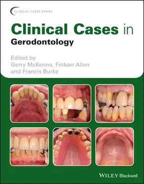
Clinical Cases in Gerodontology
Здесь есть возможность читать онлайн «Clinical Cases in Gerodontology» — ознакомительный отрывок электронной книги совершенно бесплатно, а после прочтения отрывка купить полную версию. В некоторых случаях можно слушать аудио, скачать через торрент в формате fb2 и присутствует краткое содержание. Жанр: unrecognised, на английском языке. Описание произведения, (предисловие) а так же отзывы посетителей доступны на портале библиотеки ЛибКат.
- Название:Clinical Cases in Gerodontology
- Автор:
- Жанр:
- Год:неизвестен
- ISBN:нет данных
- Рейтинг книги:3 / 5. Голосов: 1
-
Избранное:Добавить в избранное
- Отзывы:
-
Ваша оценка:
- 60
- 1
- 2
- 3
- 4
- 5
Clinical Cases in Gerodontology: краткое содержание, описание и аннотация
Предлагаем к чтению аннотацию, описание, краткое содержание или предисловие (зависит от того, что написал сам автор книги «Clinical Cases in Gerodontology»). Если вы не нашли необходимую информацию о книге — напишите в комментариях, мы постараемся отыскать её.
Gerodontology
Clinical Cases in
Gerodontology
Offers a case-based guide to geriatric dental care Includes the thinking behind clinical decision making Fosters independent learning and prepares for case-based examinations Contains review questions and relevant literature citations Written for graduate and undergraduate dental students and professionals,
offers an instructive case-based guide to the oral health of older adults.
Clinical Cases in Gerodontology — читать онлайн ознакомительный отрывок
Ниже представлен текст книги, разбитый по страницам. Система сохранения места последней прочитанной страницы, позволяет с удобством читать онлайн бесплатно книгу «Clinical Cases in Gerodontology», без необходимости каждый раз заново искать на чём Вы остановились. Поставьте закладку, и сможете в любой момент перейти на страницу, на которой закончили чтение.
Интервал:
Закладка:
Table of Contents
1 Cover
2 Series Page
3 Title Page
4 Copyright Page
5 List of Contributors
6 Introduction Epidemiology of the Ageing Population The oral health of older adults The importance of oral health for older adults: links between oral disease and systemic well‐being References
7 Chapter 1: Management of Chronic Dental Disease Case 1 Management of Root Caries Case 2 Caries Management in a Long‐Term Care Facility Using Atraumatic Restorative Treatment (ART) Case 3 Non‐surgical Periodontal Treatment (NSPT) for Periodontally Involved Lower Incisors Case 4 Splinting and Maintenance of Periodontally InvolvedLower Incisors Case 5 Management of Toothwear Using Direct Composite Restorations
8 Chapter 2: Replacement of Missing Teeth Case 6 Fabrication of Complete Conventional Dentures for edentate patients Case 7 Fabrication of New Complete Replacement Dentures Using a Copy Technique Case 8 Provision of Upper and Lower Implant‐Retained Overdentures for an Older Patient Case 9 Use of a Removable Partial Denture to Replace Missing Teeth Case 10 Integrating Fixed and Removable Prosthodontics Case 11 Utilising Upper and Lower Overdentures for a Partially Dentate Patient Case 12 Single Tooth Replacement Using Adhesive Bridgework Case 13 Tooth Replacement According to the Principles of the Shortened Dental Arch Case 14 Use of a Natural Pontic to Replace a Lower Incisor Lost Due to Periodontal Disease Case 15 The Use of Dental Implants to Provide Fixed Prosthodontics in a Partially Dentate Older Patient
9 Chapter 3: Management of Failing Restorations Case 16 Endodontic Treatment for a Fractured Tooth and Conversion to an Overdenture Abutment Case 17 Managing the Failing Restored Dentition: Replacement of Failing Crowns Case 18 Removal and Replacement of Heavily Restored Anterior Teeth Case 19 Dismantling a Long‐Span Fixed Bridge and Replacement with a Removable Partial Denture Case 20 Replacement of a Failing Implant Bridge for a Patient with Missing Lower Teeth
10 Chapter 4: Management of Malignancy and Other Oral Conditions Case 21 Managing Malignant Oral Disease Case 22 Maintenance of Multiple Overdenture Abutments for a Patient Following Head and Neck Radiotherapy Case 23 Management of Primary Sjögren’s Syndrome in a Partially Dentate Patient Case 24 Management of Drug‐Induced Gingival Overgrowth Case 25 Vital Bleaching to Improve the Aestheticsof Natural Teeth
11 Index
12 End User License Agreement
List of Illustrations
1 Chapter 1 Figure 1.1.1 Circumferential root caries lesions affecting the remaining low... Figure 1.1.2 Glass ionomer cement (Fuji IX GP Extra™, GC Corporation, Japan)... Figure 1.1.3 Patient at three‐month review. Figure 1.2.1 Carious lesion in an upper molar tooth. Figure 1.2.2 Carious lesion in an upper lateral incisor. Figure 1.2.3 Basic atraumatic restorative technique instrument kit used for ... Figure 1.2.4 Cavity in upper molar after caries removal using hand instrumen... Figure 1.2.5 Cavity in upper canine after caries removal using hand instrume... Figure 1.2.6 Glass ionomer cement (Fuji IX GP Extra™, GC Corporation, Japan)...Figure 1.3.1 Clinical presentation of patient before treatment.Figure 1.3.2 Calculus deposits on lower anterior teeth.Figure 1.3.3 Radiographic findings at initial presentation.Figure 1.3.4 Patient after completion of non‐surgical periodontal treatment....Figure 1.3.5 Lower incisors after removal of supragingival calculus deposits...Figure 1.3.6 Radiographic review after 1 year illustrating no progression of...Figure 1.4.1 Lower incisor teeth at initial presentation.Figure 1.4.2 Putty matrix fabricated for placement of the periodontal splint...Figure 1.4.3 Lower incisor teeth isolated using rubber dam.Figure 1.4.4 Periodontal splint in situ.Figure 1.4.5 (a) and (b) Checking of periodontal splint for occlusal interfe...Figure 1.5.1 Appearance of patient’s teeth at initial presentation.Figure 1.5.2 Intraoral appearance of patient at initial presentation.Figure 1.5.3 Upper and lower models mounted on semi‐adjustable articulator. ...Figure 1.5.4 Right lateral view of wax‐up. Note the disclusion of the molars...Figure 1.5.5 Anterior view of putty matrix which was made from wax‐up. The m...Figures 1.5.6 (a) and (b) Polishing of restorations.Figure 1.5.7 Palatal view of maxillary teeth showing occlusal contact marked...Figure 1.5.8 Lingual view of mandibular teeth showing occlusal contact marke...Figure 1.5.9 Anterior view after treatment.
2 Chapter 2Figure 2.6.1 Set of complete replacement dentures which the patient was wear...Figure 2.6.2 Open tray over ‘flabby’ anterior maxillary ridge.Figure 2.6.3 Master impression recorded using selective pressure impression ...Figure 2.6.4 Record blocks with wax rims and permanent bases.Figure 2.6.5 (a) and (b) Incremental build‐up of occlusal surfaces of comple...Figure 2.6.6 Pre‐treatment view of patient with historical dentures in situ....Figure 2.6.7 Post‐treatment view with new dentures in situ.Figure 2.7.1 Patient at initial presentation.Figure 2.7.2 (a) and (b) Putty impression of existing upper denture (Lab‐Put...Figure 2.7.3 Denture try‐in.Figure 2.7.4 Completed full lower denture.Figure 2.8.1 Pre‐treatment view of complete replacement dentures.Figure 2.8.2 (a) Right tuberosity region: note the extensive bulk of soft ti...Figure 2.8.3 Pre‐treatment orthopantomograph (OPG). Note the lack of bone in...Figure 2.8.4 Post‐surgical orthopantomograph (OPG) showing the implants plac...Figure 2.8.5 Locator TM(Zest Dental Systems, USA) abutments in situ , favoura...Figure 2.8.6 Locator TM(Zest Dental Systems, USA) abutments in situ. Note th...Figure 2.8.7 (a) Fitting surface of new complete replacement denture prior t...Figure 2.8.8 Lateral views (a) pre treatment; (b) post treatment.Figure 2.9.1 Note the first point of contact (RCP) between 14 and 45. This i...Figure 2.9.2 Bite‐raising appliance (splint) in centric relation.Figure 2.9.3 Anterior view of definitive overlay maxillary removable partial...Figure 2.9.4 Occlusal view of maxillary overlay prosthesis. Note the metal b...Figure 2.9.5 Index of desired anterior tooth position. This is used on the m...Figure 2.9.6 Putty index made on diagnostic wax‐up cast, used to guide posit...Figure 2.10.1 The patient’s appearance at initial presentation.Figure 2.10.2 The patient’s worn lower removable partial denture.Figure 2.10.3 Palatal view of upper teeth.Figure 2.10.4 Residual lower dentition.Figure 2.10.5 The patient’s occlusion.Figure 2.10.6 Root canal treatment completed on the lower second molar.Figure 2.10.7 (a) and (b) Gold crowns fabricated with milled features for re...Figure 2.10.8 Upper arch with crowns fitted to aid denture retention.Figure 2.10.9 Upper denture inserted.Figure 2.10.10 Lower denture inserted.Figure 2.10.11 Final result at review.Figure 2.11.1 The patient at initial presentation, including his upper and l...Figure 2.11.2 Intraoral photographs at initial presentation.Figure 2.11.3 Intraoral periapical radiographs (IOPAs) at initial presentati...Figure 2.11.4 Temporary acrylic upper and lower partial dentures were provid...Figure 2.11.5 Intraoral picture after initial stabilisation treatment.Figure 2.11.6 Use of root cap overdenture abutments, to help retain new pros...Figure 2.11.7 Construction of the final removable prostheses (note the fitti...Figure 2.11.8 Occlusal scheme on final removable prostheses.Figure 2.12.1 The patient at initial presentation demonstrating his missing ...Figure 2.12.2 Missing upper lateral incisor and failing restorations on uppe...Figure 2.12.3 (a) and (b) Removal of failing restorations on upper left cani...Figure 2.12.4 Adhesive bridge after cementation.Figure 2.12.5 Checking dynamic occlusion to ensure that the pontic is not in...Figure 2.12.6 The patient at review.Figure 2.13.1 The patient’s teeth at the initial consultation appointment.Figure 2.13.2 Upper teeth, including unrestorable upper lateral incisors.Figure 2.13.3 Lower teeth, including retained roots.Figure 2.13.4 Upper arch after caries management, removal of unrestorable te...Figure 2.13.5 Adhesive bridges placed in lower arch to provide the patient w...Figure 2.13.6 Final result incorporating upper removable partial denture and...Figure 2.14.1 Lower incisor teeth at initial presentation.Figure 2.14.2 Extracted tooth with portion of root removed and glass fibre r...Figure 2.14.3 Natural pontic seated using composite resin (G‐aenial, GC Corp...Figure 2.14.4 Natural pontic in place after careful consideration of the occ...Figure 2.15.1 Orthopantomogram (OPG) of the patient’s dentition. Note the im...Figure 2.15.2 (a)–(e): Series of intraoral photographs demonstrating a heavi...
Читать дальшеИнтервал:
Закладка:
Похожие книги на «Clinical Cases in Gerodontology»
Представляем Вашему вниманию похожие книги на «Clinical Cases in Gerodontology» списком для выбора. Мы отобрали схожую по названию и смыслу литературу в надежде предоставить читателям больше вариантов отыскать новые, интересные, ещё непрочитанные произведения.
Обсуждение, отзывы о книге «Clinical Cases in Gerodontology» и просто собственные мнения читателей. Оставьте ваши комментарии, напишите, что Вы думаете о произведении, его смысле или главных героях. Укажите что конкретно понравилось, а что нет, и почему Вы так считаете.


